Author contributions: H.Y.Y., J.Y. B. performed the experiments. G.S. per- formed the statistical analysis. Y.S.K and J.H.K analyzed the data. J.Y.B. and J.H.L. wrote the manuscript. J.H.L. designed overall study and edited the manuscript.
This is an Open Access article distributed under the terms of the Creative Commons Attribution Non-Commercial License, which permits unrestricted non-commercial use, distribution, and reproduction in any medium, provided the original work is properly cited.
Copyright © Korean J Physiol Pharmacol, pISSN 1226-4512, eISSN 2093-3827
INTRODUCTION
BIS (BCL-2 interacting cell death suppressor), also called BAG3, has been originally identified as an anti-apoptotic protein and co- chaperone which binds to BCL-2 and HSP (heat shock protein)70, respectively [1,2]. Evidence subsequently showed that BIS was involved in diverse cellular functions including anti-apoptotic, anti-stress, migration and invasion, autophagy, development, and cell senescence, which might be mediated by its various binding partner proteins [3]. Clinically, BIS has been implicated in several pathological states including human cancers of various origins, which was supported by its pro-survival function [4,5]. BIS has
been also studied in myopathy and neurodegeneration, which is related with its protein quality control ability, targeting z-disk composing proteins or aggregation-prone proteins [6-11]. There- fore, the regulation of BIS expression might be critical for cellular resistance to various stresses or the maintenance of integrity of cytoskeletal organization, as well as protein homeostasis under physiological and pathological conditions.
While BIS is expressed constitutively at high levels in skeletal and cardiac muscles as well as in various human cancers, BIS has been reported to be induced by stress such as heat shock, heavy metals, serum deprivation, electrophile stress, and oxida- tive stress [12]. Moreover, BIS expression is considerably elevated
Original Article
Effect of BIS depletion on HSF1-dependent transcriptional activa- tion in A549 non-small cell lung cancer cells
Hye Hyeon Yun
1,2,#, Ji-Ye Baek
1,2,#, Gwanwoo Seo
2,3, Yong Sam Kim
4,5, Jeong-Heon Ko
4,5, and Jeong-Hwa Lee
1,2,*
1Department of Biochemistry, College of Medicine, The Catholic University of Korea, Seoul 06591, 2The Institute for Aging and Metabolic Diseases, College of Medicine, The Catholic University of Korea, Seoul 06591, 3Laboratory of Genomic Instability and Cancer Therapeutics, Cancer Mutation Research Center, Chosun University School of medicine, Gwangju 61452, 4Genome Editing Research Center, KRIBB, Daejeon 34141, 5Department of Biomolecular Science, Korea University of Science and Technology, Daejeon 34113, Korea
ARTICLE INFO Received April 10, 2018 Revised May 1, 2018 Accepted May 1, 2018
*Correspondence Jeong-Hwa Lee
E-mail: [email protected] Key Words
A549
BCL-2 interacting cell death suppressor Heat shock factor1
Heat shock protein
#These authors contributed equally to this study.
ABSTRACT The expression of BCL-2 interacting cell death suppressor (BIS), an anti-
stress or anti-apoptotic protein, has been shown to be regulated at the transcription- al level by heat shock factor 1 (HSF1) upon various stresses. Recently, HSF1 was also shown to bind to BIS, but the significance of these protein-protein interactions on HSF1 activity has not been fully defined. In the present study, we observed that com- plete depletion of BIS using a CRISPR/Cas9 system in A549 non-small cell lung cancer did not affect the induction of heat shock protein (HSP) 70 and HSP27 mRNAs under various stress conditions such as heat shock, proteotoxic stress, and oxidative stress.
The lack of a functional association of BIS with HSF1 activity was also demonstrated
by transient downregulation of BIS by siRNA in A549 and U87 glioblastoma cells. En-
dogenous BIS mRNA levels were significantly suppressed in BIS knockout (KO) A549
cells compared to BIS wild type (WT) A549 cells at the constitutive and inducible
levels. The promoter activities of BIS and HSP70 as well as the degradation rate of BIS
mRNA were not influenced by depletion of BIS. In addition, the expression levels of
the mutant BIS construct, in which 14 bp were deleted as in BIS-KO A549 cells, were
not different from those of the WT BIS construct, indicating that mRNA stability was
not the mechanism for autoregulation of BIS. Our results suggested that BIS was not
required for HSF1 activity, but was required for its own expression, which involved an
HSF1-independent pathway.
upon proteasome inhibition, viral infection, as well as in response to fibroblast growth factor 2 (FGF-2) [13-15]. Recently, the 3’- UTR of BIS mRNA was shown to be the target for microRNA (MiR)-371a-5p, MiR143, or MiR-217-5p, resulting in upregulation of BIS in cardiomyocytes and downregulation in glioblastoma and colorectal cells, respectively [16-18]. The expression of BIS, however, is mainly regulated at the transcriptional level by activa- tion of several transcription factors [19-21]. Among those, heat shock factor 1(HSF1) is the representative transcription factor that regulates BIS expression by interacting with heat shock- responsive elements (HSEs) located in the promoter region of BIS, similar to other stress-responsive genes such as HSP70 [22-25].
Furthermore, ectopic expression of BIS was shown to activate its own promoter as well as HSP70, through a positive feedback loop involving HSF1 [26,27].
In addition to a main transcription regulator of BIS expression, HSF1 was recently identified as a binding protein that binds to BIS protein, as demonstrated by a BIS interactom analysis based on quantitative immunoprecipitation combined with knockdown (QUICK) and human proteome microarrays [28]. Recent evi- dences suggest that HSF1 is constitutively active in cancer cells in contrast to the rapid inactivation upon removal of stress in physiological state, but the molecular mechanism for its persistent activation in cancers is not poorly understood [29,30]. Therefore, it is possible that interaction of HSF1 and BIS might modulate the protein stability or localization of HSF1, thereby resulting in ac- cumulation of HSP70 and HSP27 which might confer tumor cells resistant against proteotoxic stress and consequent expansion of tumor cells. However, the significance or requirement of the interaction of HSF1 and BIS on their respective functions is not clearly defined yet.
Therefore, the present study is designed to investigate the consequence of BIS depletion on the HSF1 activity as a transcrip- tional factor using BIS depletion strategy. Our results showed that activation of HSF1 by several stresses was not affected by BIS knockout or knockdown as determined by the endogenous HSP70 and HSP27 mRNAs as well as HSP70 promoter activity.
However, the expression of the BIS gene that also contains HSEs in its promoter region was reduced specifically in the BIS knock- out (KO) cells. Our results suggested that BIS was not critically required for HSF1 activity, but was required for its own expres- sion, which involved a HSF1-independent pathway.
METHODS
Cells and treatment
A549 human lung cancer cells and U87 glioblastoma cells were obtained from American Type Culture collection and maintained in DMEM supplemented with 10% feat-inactivated fetal bovine serum (Biowest, Nuaillé, France). The complete depletion of BIS
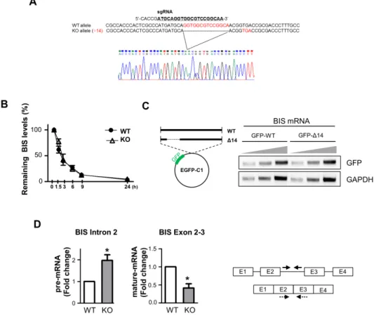
protein was achieved by CRISPR/Cas9 system as previously de- scribed [31]. DNA sequencing revealed that only single type of deletion (14 bp) was found in BIS-knockout (KO) clone used in this study while mixed genotypes were observed in the BIS clone used in previous study [31]. Transient knockdown of BIS or HSF1 expression was performed by transfection of 200 nM of specific small interfering RNA (siRNA) for BIS (5’-AAGGUUCAGAC- CAUCUUGGAA-3’) or HSF1 (5’-CUGAAGAGUGAAGACAU- AAAGA-3’) using G-fectin (Genolution, Seoul, Korea). To induce stress response, cells were exposed to heat shock at 43°C for 30 min and further incubated at 37°C for recovery as indicated.
Cells were also treated with MG132 (Sigma-aldrich, St. Louis, MO, USA), a proteasome inhibitor, as proteotoxic stress. As an oxidative stress, cells were exposed to glucose free and serum free condition for 3 h and then and incubated in the normal media for 3 h. For the determination of BIS mRNA stability, 200 ng/ml of actinomycin D (Sigma-aldrich) was added to cells for indicated times prior to the quantification of BIS mRNA.
Quantitative real time PCR (qRT-PCR)
For gene expression analyses, total RNA was isolated with an RNA extraction kit AcuZol (Bioneer, Daejeon, Korea) and cDNA was prepared with AccuPower
®Customized RocketScript Cycle RT premix (Bioneer) according to the manufacturer’s protocol.
To determine BIS pre-mRNA levels, DNase I (Thermo scientific, Waltham, MA, USA) was treated to exclude the possible con- tamination of genomic DNA and the reverse transcription was performed using random priming reaction with AccuPower
®RocketScript Cycle RT premix (Bioneer). Then, quantitative real- time PCR (qRT-PCR) was performed using SYBR premix Ex Taq (Takara Biotechnology, Shiga, Japan) with specific primers on CFX96 Connect Real-Time PCR Detection System (Biorad, Hércules, CA, USA). The relative values for BIS, HSP70, HSP27 or HSF1 mRNA were calculated after normalizing the Ct value to β-actin levels from the same sample using the ddCt method. For BIS pre-mRNA, normalization was performed using 18S RNA.
The specific primers for each mRNA are listed in Table S1.
Western blotting assay
Cell protein extracts were prepared and western blotting analy-
sis was carried out as previously described following standard
procedures [32]. The membranes were then incubated overnight
with antiserum against BIS [1], HSP70 (Enzo Life Sciences,
Farmingdale, NY, USA), HSP27 (Santa Cruz, Santa Cruz, CA,
USA) or HSF1 (Santa Cruz) followed by appropriate secondary
antibodies. The immunoreactive proteins were visualized using
enhanced chemiluminescence system (ECL Western Blotting
Substrate, Promega, Madison, WI, USA).
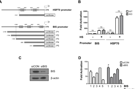
Promoter assay
The promoter activity of BIS or HSP70 was evaluated using luciferase reporter assay. A series of reporter plasmid for BIS promoter with various lengths was prepared by PCR with corre- sponding primer sets from human genomic DNA, and subsequent cloning with KpnI and XhoI sites pGL3 basic plasmid (Promega) as previously described [23]. A 1.04 kb fragment of 5’-franking re- gion of human HSP70 DNA containing two heat shock elements was also amplified with specific primers and subcloned into KpnI and XhoI sites of pGL3 basic plasmid. Following knockdown of BIS expression, A549 cells were transfected with 0.25 μg of indi- cated reporter plasmids with Fugene 6 (Roche Life Science, Basel, Switzerland) for 24 h. Wild type (WT) A549 and BIS-KO A549 cells were transfected with a BIS or HSP70 promoter for 24 h and then exposed to heat shock. The activity reporter was measured using a Dual-Luciferase reporter assay system (Promega). After normalization with renilla activity, the promoter activities of each constructs are presented as fold change of luciferase activities relative to that from pGL3 basic plasmid, which was taken as 1.0.
GFP-BIS plasmid constructs
To construct the open reading frame of WT BIS gene and the corresponding region of BIS gene with 14 bp deletion, PCR was performed with the cDNA from WT A549 and BIS-KO A549 cells as a template, respectively. After verification of the correct sequences, the PCR product was then digested and cloned into XhoI and EcoRI sites of pEGFP-C1 (Promega). After transfection of these constructs for 48 h, total RNA was extracted and cDNA was prepared following treatment of DNase I (Thermo scientific).
The GFP mRNA levels from each constructs were determined by PCR amplification and the PCR products were analyzed by aga- rose electrophoresis.
Statistics
The data are presented as the mean±standard error of the mean (SEM). Statistical significance between two groups was analyzed by Student’s t test. A p-value of <0.05 was considered statistically significant.
RESULTS
Depletion of BIS did not affect HSF1-dependent transcriptional activation in A549 cells
HSF1 is known as a main transcriptional regulator for BIS ex- pression under stress [22,23,25]. Recently, HSF1 was also shown to interact with BIS, but the significance of the physical interac- tion of these proteins on HSF1 activity was not clearly defined.
Previously we established BIS-KO A549 cells by the CRISPR/
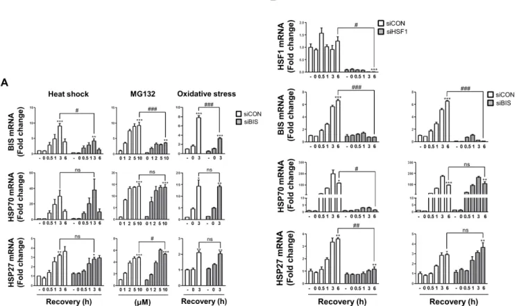
Cas9 system and demonstrated that BIS depletion sensitizes A549 cells to cisplatin via suppressing the stability of MCL-1 [31]. How- ever, the association of BIS depletion on HSF1 activity was not studied using this KO strategy. Therefore, we investigated here if BIS depletion affected the transcriptional activation of HSF1 tar- get genes in response to several stresses. First, we determined the expression profiles of HSP70 and HSP27 mRNA upon heat shock in WT A549 and BIS-KO A549 cells in which 14 bp was deleted in exon 1 of the BIS gene [31]. Quantitative analysis of mRNA in- dicated that the induction patterns of HSP70 and HSP27 mRNA levels in response to heat shock were not significantly different between BIS-KO A549 and WT A549 cells (Fig. 1A). At 1 h of recovery following heat shock, the HSP70 mRNA levels were in- creased to approximately 11-fold both in WT A549 and BIS-KO A549 cells compared to those of WT A549 at normal conditions.
The induction of HSP27 levels was sustained up to 6 h of recovery after heat shock both in WT A549 and BIS-KO A549 cells with 3-fold and 3.4-fold increases, respectively. Western blots also revealed that the expressions of HSP70 and HSP27 proteins were not significantly affected by BIS depletion upon heat shock (Fig.
1B). Moreover, treatment with MG132, a proteasome inhibitor, or recovery from serum free condition, an oxidative stress [33,34], also increased HSP70 and HSP27 mRNA expression in WT A549 cells, which were not significantly repressed in BIS-KO A549 cells (Fig. 1A).
Previously, ectopic expression of BIS was shown to activate its own promoter, involving a 5’-UTR sequence of the BIS gene in glial cells [26]. A subsequent study demonstrated that the positive feedback loop of BIS expression appeared to be a consequence of stress responses generated by the accumulation of BIS protein and subsequently ubiquitination of client proteins, which trig- gered nuclear translocation of HSF1 [27]. These previous studies prompted us to determine the effects of complete depletion of BIS on the expression of its own mRNA. Because the BIS-KO A549 cells have only the 14 bp deletion in exon 1 of the BIS gene lead- ing to a frame shift, the promoter region harboring several HSEs [23,25] should be intact and therefore respond to cellular stresses.
As shown in Fig. 1A, the BIS mRNA levels in WT A549 cells were
increased by 7.5-fold at 1 h recovery following heat shock while
those in the BIS-KO A549 cells were only 2.4-fold increased rela-
tive to those in WT A549 cells under non-stressed conditions. In
response to proteasome inhibition or oxidative stress, BIS induc-
tion was also considerably decreased in BIS-KO A549 cells ; 9.3-
fold induction vs. 4.5-fold after treatment with 5 mM of MG132,
and 6.2-fold induction vs. 2.3-fold at 3 h recovery after glucose
and serum deprivation, respectively. It is noteworthy that, in BIS-
KO cells, the basal levels of BIS mRNA were also decreased by
0.45-fold compared to WT A549 cells. These results indicated
that BIS depletion inhibited the constitutive as well as inducible
expression of the BIS mRNA without alteration of HSP70 and
HSP27 mRNA levels.
To exclude the possibility that our results were due to the cellu- lar features acquired during the selection process of BIS-KO A549 cells, which were not relevant with BIS depletion, we transiently suppressed BIS expression in WT A549 cells using siRNA and examined the induction of HSP70 and HSP27 mRNA. Fig. 2A shows that BIS knockdown neither affected the constitutive nor inducible expression of HSP70 and HSP27 mRNAs in response to heat shock, proteasome inhibition, or serum deprivation in con- ditions in which BIS expression was sufficiently decreased. Thus, these results clearly demonstrated that activation of HSF1 on the HSP27 and HSP70 was not dependent on BIS expression in A549 cells.
Transcriptional activity of HSF1 was not suppressed by siRNA-mediated BIS depletion in U87 glioblastoma cells
Our findings were inconsistent with a previous report showing that BIS knockdown using adenovirus was accompanied by de- creases in HSP70 and HSP27 expression as determined by west- ern blots of T98G glioblastoma cells [27]. Thus, to determine if the lack of a functional association of BIS with HSF1 activity was the specific phenotypes of A549 cells, we suppressed HSF1 or BIS expression in U87 glioblastoma cells and examined the HSP70 and HSP27 mRNA levels upon heat shock stress. As shown in Fig. 2B, HSF1 knockdown substantially decreased HSP70 and HSP27 mRNAs. At 6 h of recovery following heat shock, HSP70
and HSP27 mRNA levels were 116-fold and 3.6-fold increased, re- spectively, compared to unstressed control U87 cells, whereas the levels were 1.4-fold and 1.2-fold in HSF1-knockdown U87cells, re- spectively. In addition, HSF1 knockdown significantly suppressed BIS induction upon heat shock. At 6 h of recovery, BIS mRNA ex- pression was 6.6-fold increase in control cells while it was 0.7-fold increase in HSF1-depleted A549 cells. However, effective suppres- sion of BIS did not cause any significant alteration of induction of these two HSP mRNAs in U87 cells, as observed in A549 cells.
Thus, BIS was the downstream target for HSF1 transcription factor, but BIS had no ability to cross-regulate HSF1 activity espe- cially in a knockdown model, irrespective of cell type.
Promoter activity of HSP70 and BIS was not dependent on BIS expression levels
We next measured the promoter activity of BIS and HSP70 us- ing a luciferase reporter assay in BIS WT and BIS-KO-A549 cells.
The aim of the reporter assay was to ensure that the promoter activity mediated through HSF1-HSE interaction was not influ- enced by BIS depletion, and to further determine if BIS specifi- cally regulated its own promoter without affecting other HSF1- target gene via an HSF1-independent mechanism. Fig. 3 A shows the relative locations of HSEs in the 5’-UTRs of the BIS gene and HSP70 gene. The basal luciferase activity using the HSP70 promoter containing two putative HSEs in WT A549 cells was comparable to that of the KO-A549 cells (Fig. 3B). Furthermore,
Fig. 1. The induction of HSP70 and HSP27 mRNA was not affected by BIS depletion under stress conditions. (A) Wild type (WT) A549 cells and BIS-knockout (KO) A549 cells were exposed to heat shock (43°C) for 30 min and subsequent recovery for up to 6 h, to MG132 for 6 h, or to glucose free and serum free conditions for 3 h followed by 3 h of recovery. The mRNA expression levels at the indicated times or indicated treatment concentra- tions were measured by qRT-PCR analyses. The fold induction was determined as the relative value of each mRNA level compared to the untreated WT A549 cells, which was designated arbitrarily as 1.0. Data are represented as the mean±SEM from three independent experiments. *p≤0.05, **p≤0.01,
***p≤0.001 vs. control in each group; #p≤0.05, ##p≤0.01, ###p≤0.001 vs. WT at the indicated times or concentrations. ns, statistically non-significant. (B) The protein levels of BIS, HSP70, and HSP27 in the WT and BIS-KO A549 were determined by western blotting of samples after 3 h of recovery (+R3) following heat shock and control cell samples. Actin expression was used as a loading control.
even after exposure to heat shock stress, the luciferase activity driven by the HSP70 promoter was not reduced in BIS-KO A549 cells compared to WT A549 cells. Similarly, the activity of the BIS promoter with three HSEs was not significantly different between the WT A549 cells and BIS-KO A549 cells before or after heat shock stress. When a series of reporter constructs of various 5’- UTR lengths of the BIS gene were expressed in control A549 cell or BIS knockdown A549 cells, the luciferase activities from all the constructs tested revealed no significant difference between con- trol and BIS-KD A549 cells, regardless of the presence of any pu- tative HSE or length of the BIS promoter region (Figs. 3C and D).
Therefore, the reporter assay results suggested that the promoter activity driven by the HSF1-HSE axis was not influenced by BIS depletion, correlating with the expression levels of target genes as shown in Figs. 1 and 2. These results also suggest that alteration of promoter activity did not contribute to the autoregulation cir- cuit of BIS expression at the constitutive or inducible level.
Autoregulation of BIS mRNA expression was not attributable to an altered regulation of mRNA stability
It has been shown that an aberrant mRNA with a truncated open reading frame and a long 3’-UTR due to the presence of a premature stop codon was recognized and degraded by specific mRNA degradation systems, called nonsense-mediated decay.
The nonsense-medicated decay pathway is a known translation- coupled mRNA quality control system that prevents cells from producing potentially deleterious truncated proteins, by recruit- ing a set of factors that destabilize a target RNA [35,36]. Therefore, it was possible that the decreases of BIS mRNA in BIS-KO A549 cells were the consequence of nonsense-mediated BIS mRNA decay due to the premature stop codon (Fig. 4A), and were not re- lated with the absence of BIS protein. To confirm this hypothesis, we measured the BIS mRNA stability by an actinomycin D chase assay in WT and BIS-KO A549 cells. As determined by the level of remaining BIS mRNA, determined using qRT-PCR, at the in- dicated times following treatment with actinomycin D, the degra- dation rate of BIS mRNA was not notably different between WT
Fig. 2. BIS knockdown did not affect HSP70 and HSP27 induction by various stress conditions. (A) BIS expression was transiently suppressed by 200 nM of control siRNA (siCON) and BIS specific siRNA (siBIS) for 48 h in A549 cells, and the fold induction was compared for BIS, HSP70, and HSP27 mRNA expression in response to heat shock, MG132, or oxidative stress as described in Fig. 1. (B) U87 cells were transfected with HSF1 siRNA (siHSF1) or BIS siRNA (siBIS) for 48 h and exposed to heat shock. The expression levels of HSF1, BIS, HSP70, and HSP27 mRNA were determined as in Fig. 1.
*p≤0.05, **p≤0.01, ***p≤0.001 vs. untreated control cells in each group; #p≤0.05, ##p≤0.01, ###p≤0.001 vs. siCON at the indicated times or treatment concentrations. ns, statistically non-significant.
A549 cells and BIS-KO A549 cells; t
1/2=2.54 h and 2.44 h in WT and BIS-KO A549 cells, respectively. From these results, we found that the regulation of BIS mRNA stability, regardless of 14-bp deletion, was not an essential mechanism which conferred auto- regulation of BIS expression. In addition, when the WT BIS gene and deletion mutant of the BIS gene (Δ14-BIS) were expressed as a GFP-fusion construct in WT-A549 cells, the GFP mRNA expression levels were not notably different between the two cell types transfected with each construct (Fig. 4C). Taken together, the deletion of 14 nucleotides in the BIS gene was not responsible for the decrease in BIS mRNA levels in BIS-KO cells. Finally, we tested the BIS pre-mRNA levels in WT A549 and BIS-KO A549 cells using specific primers for intron 2. As shown in Fig. 4D, the BIS pre-mRNA levels represented by intron 2 levels were higher in BIS-KO A549 cells than those in WT A549 cells, while BIS ma- ture mRNA levels determined by the primers covering exon2 and 3 were decreased in BIS-KO A549 cells as measured by primers for exon4 (Figs. 1-3). Thus, these findings suggest the possibility that splicing process of BIS pre-mRNA may be retarded by BIS depletion.
DISCUSSION
The expression of BIS, a pro-survival protein, is mainly regu-
lated at the transcriptional level by HSF1 in response to various forms of stress [22-25]. Recent studies have shown that BIS physi- cally binds to HSF1 [28], but the effect of the interaction of these two proteins on each other’s function has not been clearly de- fined. BIS was shown to exert diverse effects on cell fates through interaction with various proteins, which affect their stability, lo- calization and activity [3]. Therefore, it is probable that BIS might modulate either the translocation or the transcriptional activity of HSF1. Supporting this possibility, the effect of BIS on nuclear translocation of HSF1 has been previously reported. For example, oxidative stress-induced phosphorylation of BIS protein weakens its binding ability to HSF1, thereby accelerating its nuclear trans- location in A172 cells [37]. BIS was also shown to co-translocate into the nucleus with HSF1 upon heat shock stress in HeLa cells [38]. In addition, under glioblastoma stem cell like sphere-form- ing conditions, BIS depletion is known to decrease HSF1 protein levels and its nuclear localization [39]. Collectively, these results indicate that BIS might be positively involved in the nuclear trans- location of HSF1. In contrast, the effects of BIS depletion on the transcriptional activity of HSF1 are not consistent. In the present study, we clearly demonstrated that BIS depletion did not affect the activation of HSF1 as evidenced by the expressions of HSP70 and HSP27 mRNA as well as HSP70 promoter activity in A549 lung cancer cells. However, a previous report showed that adeno- virus-mediated suppression of BIS decreased HSP70 and HSP27
Fig. 3. The promoter activities of BIS and HSP70 were not affected by BIS depletion. (A) Schematic diagram for the relative position of heat shock element (HSE) to ATG in the promoter region of the HSP70 and BIS genes. (B) WT A549 and BIS-KO A549 cells were transfected with BIS or HSP70 pro- moter constructs for 24 h and then exposed to heat shock (HS) at 43°C for 30 min, and then further at 37°C for 1 h. Transcriptional activation of the BIS (P3) and HSP70 promoters are presented as fold changes compared to the value of the pGL3 basic construct in untreated conditions in WT and BIS-KO A549 cells. (C) Western blotting shows that BIS protein expression was sufficiently repressed by siRNA. (D) Following suppression of BIS in A549 cells with BIS siRNA (siBIS), the reporter constructs were expressed and compared to the control (siCON). After normalization with renilla activity, the re- sults are presented as fold change compared to the activity from the pGL3 basic vector (mean±SE, n=3). #p≤0.05 vs. siCON for P1. ns, statistically non- significant.
protein expression in T98G glioblastoma cells [27]. On the other hands, HSP70 expression was significantly increased in BIS-KO cardiomyocytes [40]. The molecular basis for this inconsistency between our findings and those of previous reports are uncertain.
The viral transfection method can create a cell environment in which the host cell is more dependent on the BIS in activating HSF1. It is also likely that BIS depletion either caused or aggra- vated the accumulation of protein aggregates, thereby secondarily activating HSF1 in cells vulnerable to proteotoxic stress. These results show that nuclear translocation of HSF1 may be affected by the interaction with BIS, which is not necessarily linked to al- teration of transcriptional activity of HSF1 on the stress-inducible target genes.
A possible explanation for the lack of functional association of BIS with HSF1 activity shown in our study could have resulted from the application of limited forms of stress such as proteotoxic stress and canonical target genes including HSPs. In addition to proteotoxic stress, HSF1 and subsequent heat shock response are known to be influenced by the proliferative and metabolic state
of the cell, which can provide protein homeostasis according to cellular demands [41,42]. Thus, BIS might be involved in the acti- vation of HSF1 under specific conditions other than proteotoxic stress. Furthermore, even in unstressed conditions, HSF1 plays an important role in the diverse biological processes including development, metabolism and carcinogenesis through a tran- scriptional program that is uncoupled from heat shock response [43,44]. Therefore, it is also possible that BIS might participate in an alternative transcriptional program that leads to long-term cell survival, which is supported by the constitutive high expres- sion of HSF1 and BIS in various type of human cancer [3,4,44,45].
However, to investigate the mechanistic network between BIS and HSF1 in the pro-growth program in cancer cells, further studies should be performed under more detailed and diverse experi- mental conditions.
In the present study, we also demonstrated that the induction profile of the BIS gene, which includes HSEs in the promoter, did not parallel that of the other HSF1- target genes in BIS-KO A549 cells. Considering that BIS expression was primarily regulated
Fig. 4. Autoregulation of BIS was not mediated by the alteration of mRNA stability. (A) The sequencing results of genotyping in BIS-KO A549 cells show the deletion in exon 1 of the BIS gene and consequent generation of a premature stop codon. (B) WT and BIS-KO A549 cells were treated with actinomycin D for up to 24 h. At the indicated times, mRNA was extracted, and the remaining BIS mRNA was measured and compared to the initial value with qRT-PCR analysis (mean±SE, n=3). (C) The coding region of the WT BIS and the deletion mutant of the BIS gene, in which 14 bp were deleted (Δ14), were cloned into the pEGFP-C1 construct (left). After transfection for 48 h, the GFP mRNA levels representing WT and Δ14-BIS were determined with PCR amplification and agarose electrophoresis by loading different quantities of PCR products (right). (D) The BIS pre-mRNA levels were determined in WT A549 and BIS-KO A549 cells using qRT-PCR analysis with specific primers for intron 2 (solid arrows). The BIS mature mRNA was levels were also determined by the primers covering exon 2 and 3 (dotted arrows). Reverse transcription was performed as described in Method sec- tion. *p≤0.05 vs. WT.
by HSF1 upon stress or ectopic expression of BIS itself, our find- ings were unexpected. Thus, autoregulation of BIS expression may involve separate mechanisms; an HSF1-dependent positive feedback pathway triggered by BIS overexpression and HSF1- independent suppression of its own transcript mediated by BIS depletion. Our findings show that neither promoter activity nor mRNA stability of BIS was affected by BIS depletion. Therefore, the nuclear or cellular events linking these two processes, includ- ing splicing process, should be further studied to investigate the molecular mechanism by which BIS controls the autoregulatory loop. Targeting the auto-regulatory pathway is an efficient strat- egy to suppress BIS expression, which is aberrantly expressed in most cancers.
ACKNOWLEDGEMENTS
This research was supported by Basic Science Research Pro- grams through the National Research Foundation of Korea (NRF), funded by the Minister of Education, Science and Technology (2017R1A2B2005508) and by a grant of the Korea Health Tech- nology R&D Project through the Korea Health Industry Devel- opment Institute (KHIDI), funded by the Ministry of Health &
Welfare, Republic of Korea (HI17C1257).
CONFLICTS OF INTEREST
The authors declare no conflicts of interest.
SUPPLEMENTARY MATERIALS
Supplementary data including one table can be found with this article online at http://pdf.medrang.co.kr/paper/pdf/Kjpp/
Kjpp022-04-11-s001.pdf.
REFERENCES
1. Lee JH, Takahashi T, Yasuhara N, Inazawa J, Kamada S, Tsujimoto Y.
Bis, a Bcl-2-binding protein that synergizes with Bcl-2 in preventing cell death. Oncogene. 1999;18:6183-6190.
2. Takayama S, Xie Z, Reed JC. An evolutionarily conserved fam- ily of Hsp70/Hsc70 molecular chaperone regulators. J Biol Chem.
1999;274:781-786.
3. Rosati A, Graziano V, De Laurenzi V, Pascale M, Turco MC. BAG3:
a multifaceted protein that regulates major cell pathways. Cell Death Dis. 2011;2:e141.
4. Rosati A, Ammirante M, Gentilella A, Basile A, Festa M, Pascale M, Marzullo L, Belisario MA, Tosco A, Franceschelli S, Moltedo O, Pa- gliuca G, Lerose R, Turco MC. Apoptosis inhibition in cancer cells: a novel molecular pathway that involves BAG3 protein. Int J Biochem
Cell Biol. 2007;39:1337-1342.
5. Zhu H, Liu P, Li J. BAG3: a new therapeutic target of human can- cers? Histol Histopathol. 2012;27:257-261.
6. Selcen D, Muntoni F, Burton BK, Pegoraro E, Sewry C, Bite AV, Engel AG. Mutation in BAG3 causes severe dominant childhood muscular dystrophy. Ann Neurol. 2009;65:83-89.
7. Odgerel Z, Sarkozy A, Lee HS, McKenna C, Rankin J, Straub V, Lochmüller H, Paola F, D’Amico A, Bertini E, Bushby K, Goldfarb LG. Inheritance patterns and phenotypic features of myofibrillar myopathy associated with a BAG3 mutation. Neuromuscul Disord.
2010;20:438-442.
8. Lei Z, Brizzee C, Johnson GV. BAG3 facilitates the clearance of en- dogenous tau in primary neurons. Neurobiol Aging. 2015;36:241- 248.
9. Seidel K, Vinet J, Dunnen WF, Brunt ER, Meister M, Boncoraglio A, Zijlstra MP, Boddeke HW, Rüb U, Kampinga HH, Carra S. The HSPB8-BAG3 chaperone complex is upregulated in astrocytes in the human brain affected by protein aggregation diseases. Neuropathol Appl Neurobiol. 2012;38:39-53.
10. Merabova N, Sariyer IK, Saribas AS, Knezevic T, Gordon J, Turco MC, Rosati A, Weaver M, Landry J, Khalili K. WW domain of BAG3 is required for the induction of autophagy in glioma cells. J Cell Physiol. 2015;230:831-841.
11. Behl C. Breaking BAG: the Co-Chaperone BAG3 in health and dis- ease. Trends Pharmacol Sci. 2016;37:672-688.
12. Rosati A, Basile A, Falco A, d’Avenia M, Festa M, Graziano V, De Laurenzi V, Arra C, Pascale M, Turco MC. Role of BAG3 protein in leukemia cell survival and response to therapy. Biochim Biophys Acta. 2012;1826:365-369.
13. Wang HQ, Liu HM, Zhang HY, Guan Y, Du ZX. Transcriptional upregulation of BAG3 upon proteasome inhibition. Biochem Bio- phys Res Commun. 2008;365:381-385.
14. Rosati A, Leone A, Del Valle L, Amini S, Khalili K, Turco MC.
Evidence for BAG3 modulation of HIV-1 gene transcription. J Cell Physiol. 2007;210:676-683.
15. Gentilella A, Khalili K. BAG3 expression is sustained by FGF2 in neural progenitor cells and impacts cell proliferation. Cell Cycle.
2010;9:4245-4247.
16. Liu J, Qu CB, Xue YX, Li Z, Wang P, Liu YH. MiR-143 enhances the antitumor activity of shikonin by targeting BAG3 expression in human glioblastoma stem cells. Biochem Biophys Res Commun.
2015;468:105-112.
17. Flum M, Kleemann M, Schneider H, Weis B, Fischer S, Handrick R, Otte K. miR-217-5p induces apoptosis by directly targeting PRKCI, BAG3, ITGAV and MAPK1 in colorectal cancer cells. J Cell Com- mun Signal. 2018;12:451-466.
18. d’Avenia M, Citro R, De Marco M, Veronese A, Rosati A, Visone R, Leptidis S, Philippen L, Vitale G, Cavallo A, Silverio A, Prota C, Gravina P, De Cola A, Carletti E, Coppola G, Gallo S, Provenza G, Bossone E, Piscione F, Hahne M, De Windt LJ, Turco MC, De Lau- renzi V. A novel miR-371a-5p-mediated pathway, leading to BAG3 upregulation in cardiomyocytes in response to epinephrine, is lost in Takotsubo cardiomyopathy. Cell Death Dis. 2015;6:e1948.
19. Ben Aicha S, Lessard J, Pelletier M, Fournier A, Calvo E, Labrie C.
Transcriptional profiling of genes that are regulated by the endo- plasmic reticulum-bound transcription factor AIbZIP/CREB3L4 in prostate cells. Physiol Genomics. 2007;31:295-305.
20. Gentilella A, Passiatore G, Deshmane S, Turco MC, Khalili K. Ac- tivation of BAG3 by Egr-1 in response to FGF-2 in neuroblastoma cells. Oncogene. 2008;27:5011-5018.
21. Cesaro E, Montano G, Rosati A, Crescitelli R, Izzo P, Turco MC, Costanzo P. WT1 protein is a transcriptional activator of the anti- apoptotic bag3 gene. Leukemia. 2010;24:1204-1206.
22. Song S, Kole S, Precht P, Pazin MJ, Bernier M. Activation of heat shock factor 1 plays a role in pyrrolidine dithiocarbamate-mediated expression of the co-chaperone BAG3. Int J Biochem Cell Biol.
2010;42:1856-1863.
23. Yoo HJ, Im CN, Youn DY, Yun HH, Lee JH. Bis is induced by oxi- dative stress via activation of HSF1. Korean J Physiol Pharmacol.
2014;18:403-409.
24. Jacobs AT, Marnett LJ. HSF1-mediated BAG3 expression attenuates apoptosis in 4-hydroxynonenal-treated colon cancer cells via stabili- zation of anti-apoptotic Bcl-2 proteins. J Biol Chem. 2009;284:9176- 9183.
25. Franceschelli S, Rosati A, Lerose R, De Nicola S, Turco MC, Pascale M. Bag3 gene expression is regulated by heat shock factor 1. J Cell Physiol. 2008;215:575-577.
26. Gentilella A, Khalili K. Autoregulation of co-chaperone BAG3 gene transcription. J Cell Biochem. 2009;108:1117-1124.
27. Gentilella A, Khalili K. BAG3 expression in glioblastoma cells pro- motes accumulation of ubiquitinated clients in an Hsp70-dependent manner. J Biol Chem. 2011;286:9205-9215.
28. Chen Y, Yang LN, Cheng L, Tu S, Guo SJ, Le HY, Xiong Q, Mo R, Li CY, Jeong JS, Jiang L, Blackshaw S, Bi LJ, Zhu H, Tao SC, Ge F.
Bcl2-associated athanogene 3 interactome analysis reveals a new role in modulating proteasome activity. Mol Cell Proteomics.
2013;12:2804-2819.
29. Dai C, Sampson SB. HSF1: Guardian of proteostasis in can- cer. Trends Cell Biol. 2016;26:17-28.
30. Dai C, Santagata S, Tang Z, Shi J, Cao J, Kwon H, Bronson RT, Whi- tesell L, Lindquist S. Loss of tumor suppressor NF1 activates HSF1 to promote carcinogenesis. J Clin Invest. 2012;122:3742-3754.
31. Cui MN, Yun HH, Lee NE, Kim HY, Im CN, Kim YS, Lee JH. De- pletion of BIS sensitizes A549 cells to treatment with cisplatin. Mol Cell Toxicol. 2016;12:63-71.
32. Baek JY, Yun HH, Im CN, Ko JH, Jeong SM, Lee JH. BIS overexpres- sion does not affect the sensitivity of HEK 293T cells against apop- tosis. Mol Cell Toxicol. 2017;13:95-103.
33. Jelluma N, Yang X, Stokoe D, Evan GI, Dansen TB, Haas-Kogan
DA. Glucose withdrawal induces oxidative stress followed by apop- tosis in glioblastoma cells but not in normal human astrocytes. Mol Cancer Res. 2006;4:319-330.
34. Liu Y, Song XD, Liu W, Zhang TY, Zuo J. Glucose deprivation in- duces mitochondrial dysfunction and oxidative stress in PC12 cell line. J Cell Mol Med. 2003;7:49-56.
35. Schweingruber C, Rufener SC, Zünd D, Yamashita A, Mühlemann O. Nonsense-mediated mRNA decay - mechanisms of substrate mRNA recognition and degradation in mammalian cells. Biochim Biophys Acta. 2013;1829:612-623.
36. Karousis ED, Nasif S, Mühlemann O. Nonsense-mediated mRNA decay: novel mechanistic insights and biological impact. Wiley In- terdiscip Rev RNA. 2016;7:661-682.
37. Kim HY, Kim YS, Yun HH, Im CN, Ko JH, Lee JH. ERK-mediated phosphorylation of BIS regulates nuclear translocation of HSF1 un- der oxidative stress. Exp Mol Med. 2016;48:e260.
38. Jin YH, Ahn SG, Kim SA. BAG3 affects the nucleocytoplasmic shut- tling of HSF1 upon heat stress. Biochem Biophys Res Commun.
2015;464:561-567.
39. Im CN, Yun HH, Lee JH. Heat shock factor 1 depletion sensitizes A172 glioblastoma cells to temozolomide via suppression of cancer stem cell-like properties. Int J Mol Sci. 2017;18:E468.
40. Fang X, Bogomolovas J, Wu T, Zhang W, Liu C, Veevers J, Stroud MJ, Zhang Z, Ma X, Mu Y, Lao DH, Dalton ND, Gu Y, Wang C, Wang M, Liang Y, Lange S, Ouyang K, Peterson KL, Evans SM, Chen J. Loss-of-function mutations in co-chaperone BAG3 de- stabilize small HSPs and cause cardiomyopathy. J Clin Invest.
2017;127:3189-3200.
41. Li J, Labbadia J, Morimoto RI. Rethinking HSF1 in stress, develop- ment, and organismal health. Trends Cell Biol. 2017;27:895-905.
42. Su KH, Dai C. Metabolic control of the proteotoxic stress response:
implications in diabetes mellitus and neurodegenerative disor- ders. Cell Mol Life Sci. 2016;73:4231-4248.
43. Jiang S, Tu K, Fu Q, Schmitt DC, Zhou L, Lu N, Zhao Y. Multifac- eted roles of HSF1 in cancer. Tumour Biol. 2015;36:4923-4931.
44. Mendillo ML, Santagata S, Koeva M, Bell GW, Hu R, Tamimi RM, Fraenkel E, Ince TA, Whitesell L, Lindquist S. HSF1 drives a tran- scriptional program distinct from heat shock to support highly ma- lignant human cancers. Cell. 2012;150:549-562.
45. Dai C, Whitesell L, Rogers AB, Lindquist S. Heat shock factor 1 is a powerful multifaceted modifier of carcinogenesis. Cell.
2007;130:1005-1018.