Expression of polo-like kinase 1 in pre-implantation stage murine somatic cell nuclear transfer embryos JVS
8
0
0
전체 글
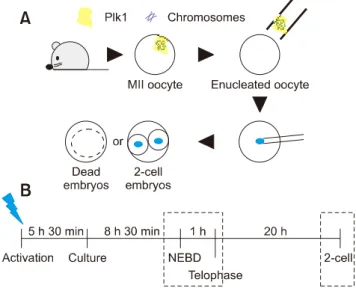
(2) Role of polo-like kinase 1 in nuclear transfer murine embryos. reported to occur frequently in SCNT murine embryos [25]. Thus, the expression and localization patterns of Plk1 in SCNT murine embryos were investigated and compared with in vivo counterparts to demonstrate the correlation between Plk1 expression and poor development of SCNT murine embryos. Although, some studies have demonstrated that Plk1 is an essential factor in mammalian embryos, in SCNT murine embryos, the expression patterns of Plk1 have not been reported. Therefore, this study investigated the Plk1 expression patterns in SCNT murine embryos.. Materials and Methods Materials All organic and inorganic compounds were purchased from Sigma-Aldrich Korea (Korea). Animal use and ethical statement All metaphase II (MII) oocytes and donor cells were acquired from 6 to 8 weeks old C57BL6 X DBA2 F1-hybrid (B6D2F1) female mice (Orient Bio, Korea). Animal experiments were approved under the agreement guidelines of the Institutional Animal Care and Use Committee of Seoul National University (approval No. SNU-130123-5-5). Collection oocytes and preparation of donor cells The 7.5 IU of equine chorionic gonadotropin (eCG; Daesung Microbiology Labs, Korea) were introduced to female B6D2F1 mice by intraperitoneal injection for superovulation. Forty-eight hours later, 7.5 IU of human chorionic gonadotropin (hCG; Daesung Microbiology Labs) were injected into the mice. To obtain in vivo-fertilized embryos, two female mice were mated with a male mouse immediately after hCG injection. Oocytes and in vivo-fertilized embryos were collected 15 h after the hCG injection. Briefly, oviducts of mice were transferred to 2 mL TCM-199 with Earl’s salts medium (TCM-washing) including 300 IU/mL of hyaluronidase to remove cumulus cells. Oocytes were recovered from torn ampullae. After 3 to 4 min of exposure to the hyaluronidase-containing TCM-washing medium, the cumulus-free oocytes were washed five times in Hepes-CZB medium (HCZB) before manipulation. The cumulus cells were suspended in a 10 L TCM-washing droplet mixed with 12% polyvinylpyrrolidone (PVP) for subsequent use as donor cells. Finally, the entire dish was covered with mineral oil. Enucleation Oocytes (12–15) were set in a 10 L droplet of HCZB, including 10 g/mL CB, and were then covered by mineral oil o and kept in a 37 C operation heat chamber on a microscope stage. The zona pellucida of the oocyte was pierced by an enucleation pipette with several piezo pulses using a piezo-actuated micromanipulator (PMM-150FU; Prime Tech,. 3. Japan). The diameter of the enucleation pipette was 6 to 8 m. The MII chromosome-spindle complex was then removed via enucleation pipette (panel A in Fig. 1). The enucleated oocytes were transferred to HCZB, washed three times, and kept for up to 30 min in KSOM medium. Nuclear injection The cumulus cells were collected by using 5 m diameter injection pipettes. The cells were aspirated in and out several times to accomplish membrane destruction. The zona pellucida was drilled out by an injection pipette and piezo pulses. Then, the donor cells in the pipette were injected into the enucleated oocytes by a single piezo pulse. After that SCNT, all of the reconstructed oocytes were washed three times in HCZB and kept for 10 min in room temperature HCZB to stabilize the oocytes. All injection protocols were undertaken at room temperature. After oocyte stabilization, reconstructed oocytes were kept in KSOM in a humidified 37oC incubator. Activation and culture The embryo culture medium was based on KSOM and CZB. Reconstructed SCNT oocytes and MII oocytes were kept separately in calcium-free CZB with 5 g/mL cytochalasin B (CB) and 10 mM SrCl2 (ACZB) for oocyte activation. After 5 h 30 min, activated oocytes were washed in HCZB five times and transferred in KSOM medium to a humidified 37oC incubator. The in vivo-fertilized embryos were similarly cultured in. Fig. 1. Schematic representation of the experiments. (A) Illustration of the sequential experimental process. The blue dots are nuclei from the donor cell. (B) Timeline of the somatic cell nuclear transfer procedure. Immunofluorescence analysis was performed at the times within the boxes with dotted lines. The blue lightning bolt is at the time of activation treatment. Plk1, polo-like kinase 1; MII, metaphase II; NEBD, nuclear envelop breakdown. www.vetsci.org.
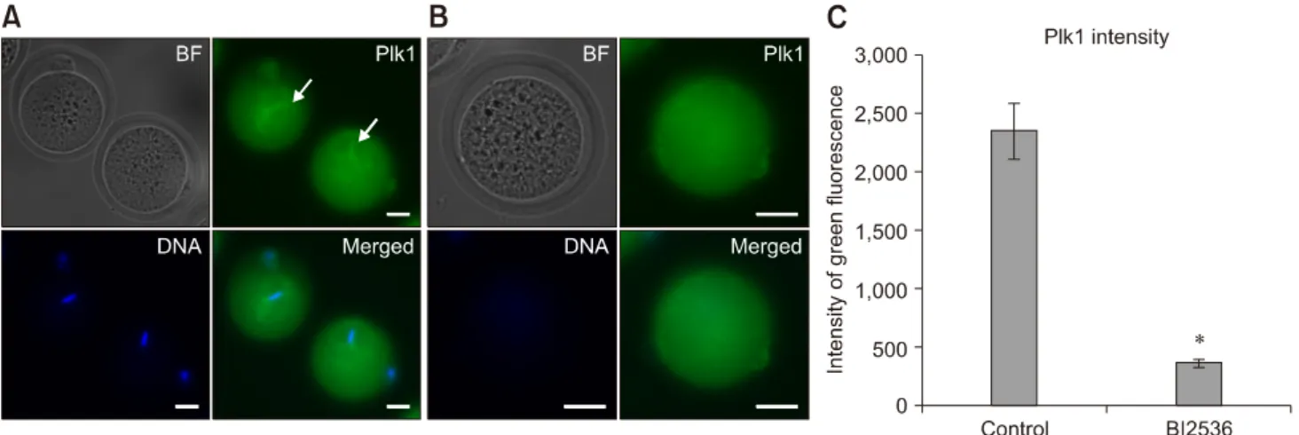
(3) 4 Jeonghyeon Moon, Sangho Roh o. KSOM medium until analysis. Treatment of a Plk1 inhibitor, BI2536 As Plk1 is essential for mitotic division in in vivo-derived embryos, embryos were treated with 2, 4, 10, 20, 100, or 500 nM of BI2536 to investigate the optimal concentration required for Plk1 inhibition. The embryos used in the control group were cultured in KSOM medium with 1% dimethyl sulfoxide. The embryos used in the experimental groups were placed in KSOM medium with the indicated concentrations of BI2536. All embryos were cultured in a 37oC humidified incubator for 30 min. Immunofluorescence analysis was performed at the times in the boxes with dotted lines as described in panel B in Fig. 1. Immunofluorescence, live imaging and image analysis The reconstructed oocytes were fixed with 4% paraformaldehyde in phosphate-buffered saline (PBS) for 30 min, then the oocytes were washed three times in PBS containing 0.5% PVP and 0.1% Triton X-100 washing buffer. The oocytes were placed in PBS containing 0.25% Triton X-100 for 4 h to make the membrane permeable and, then, incubated in PBS with 0.1% Triton X-100 and 1% BSA for. Table 1. First cleavage rates of in vivo-fertilized and somatic cell nuclear transfer (SCNT) murine embryos Group In vivo-fertilized (n = 449) SCNT (n= 612). No. of embryos cleaved (%) 409 (90.9 ± 5.9) 325 (53.1 ± 2.3)*. Data are presented as mean ± SD. *p < 0.05.. blocking for 2 h in a 37 C incubator. Plk1 was detected by rabbit polyclonal IgG antibodies (1:100; Santa Cruz Biotechnology, USA) and goat-anti-rabbit polyclonal IgG antibodies (1:500; Millipore, USA). DAPI staining was used to assess nuclear morphology. Confocal microscopy images were acquired by using an LSM 700 microscope (Zeiss, Germany). A JuLI Stage recorder (NanoEntek, Korea) was used to obtain live cell images. The confocal microscopy images were analyzed using Zen2 BLUE edition software (Zeiss). Statistical analysis All experiments were iterated three times at least. All percentage data obtained in this study are presented as mean ± SD values. The embryonic development and first mitotic division data were analyzed by applying Student’s t-test. A probability of p < 0.05 was considered significant.. Results Comparison of first mitotic division efficiencies of SCNT murine embryos and in vivo-fertilized embryos The success rate for the first mitotic division of SCNT murine embryos (53.1 ± 2.3%) was significantly lower than that of in vivo-fertilized embryos (90.9 ± 5.9%; p < 0.05) (Table 1). The spindle and chromatin together with adjacent cytoplasm were removed by an enucleation pipette during the SCNT process, resulting in the removal of the greatest proportion of spindle-binding proteins including Plk1. The loss of Plk1 may have caused the low mitotic division rate of the SCNT murine embryos. Consequently, experiments were designed to analyze the expressions of Plk1 before and after mitosis in both SCNT and in vivo-fertilized murine embryos.. Fig. 2. Immunofluorescence expression of polo-like kinase 1 (Plk1) in mouse oocytes. (A) Plk1 (green) localized in the company of chromosomes (blue) in metaphase II oocytes (arrows). (B) Low intensity Plk1 in oocytes after enucleation. No chromosomes were detected in enucleated oocytes. (C) Quantization data for the fluorescence intensity of Plk1 in normal (control) and BI2536-treated oocytes. BI2536-treated oocytes show significantly higher fluorescence intensity. BF, bright field. *p < 0.05. Scale bars = 20 m (A and B). Journal of Veterinary Science.
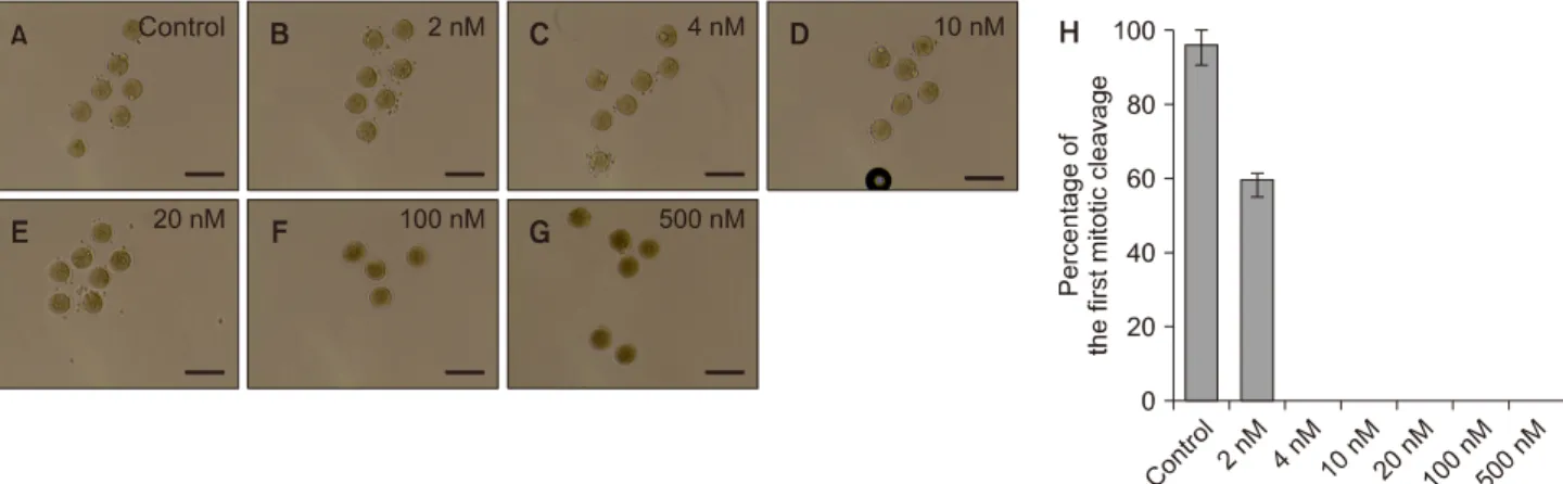
(4) Role of polo-like kinase 1 in nuclear transfer murine embryos. Intensity of Plk1 was significantly lower in enucleated oocytes than in MII oocytes The fluorescence intensity of Plk1 expression was measured by performing immunofluorescence analysis. In MII oocytes, marked fluorescence intensity of Plk1 was observed around the chromosomes and the spindle apparatus (panel A in Fig. 2; green). However, enucleated oocytes had low Plk1 fluorescence intensity as Plk1 was removed with the chromosomes during the enucleation process (panel B in Fig. 2). Quantization data obtained by confocal microscope analysis showed that the fluorescence intensity of Plk1 in MII oocytes was over five times higher than the intensity of Plk1 in enucleated oocytes (panel C in Fig. 2). Mitotic division of embryos was blocked by BI2536, a Plk1 inhibitor The images in panels A–G in Fig. 3 show the morphology of embryos that were treated with different concentrations of BI2536. About 60% of the 2 nM BI2536-treated embryos and more than 90% of the untreated in vivo-fertilized embryos developed into 2-cell embryos, whereas all embryos treated with more than 4 nM of BI2536 failed to attain 2-cell division (panel H in Fig. 3). Further, the groups of embryos that were treated with highs concentrations of BI2536, such as 100 nM and 500 nM, were dead within 30 min. The dead embryos showed membrane collapse and shrinkage (panels F and G in Fig. 3). Based on these results, 4 nM of BI2536 was applied to block Plk1 in the next series of experiments. In addition, the effect of BI2536 was tested in parthenogenetic murine embryos. All parthenogenetic embryos that were treated with 4 nM of BI2536 did not develop to 2-cell embryos (0.0 ± 0.0%), while most untreated embryos developed to the 2-cell stage (96.7 ± 4.3%) (Table 2, Fig. 4). Taken together, the results above show that BI2536, a Plk1 inhibitor, causes blocking of mitotic. 5. Table 2. First cleavage rates of BI2536-treated, and control parthenogenetic murine embryos Group Control (n = 30) BI2536-treated (n = 31). No. of embryos cleaved (%) 29 (96.7 ± 4.3) 0 (0.0 ± 0.0)*. Data are presented as mean ± SD. *p < 0.05.. Fig. 4. Effect of BI2536 on the first mitotic division in parthenogenetic murine embryos. Embryos were cultured in KSOM medium with 1% of dimethyl sulfoxide (A) or with 4 nM BI2536 (B). Scale bars = 100 m (A and B).. Fig. 3. Effect of BI2536 treatment in 1-cell embryos. (A) In vivo-fertilized embryos cultured in KSOM medium including 1% of dimethyl sulfoxide. (B–G) Embryos treated with BI2536 concentration from 2 to 500 nM. (H) Cleavage rate of embryos after BI2536 treatments at different concentrations. Scale bars = 100 m (A–G). www.vetsci.org.
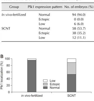
(5) 6 Jeonghyeon Moon, Sangho Roh division in both in vivo-fertilized and parthenogenetic murine embryos. The experimental results confirm that Plk1 has a critical role in mitosis in early-stage mouse embryos. Therefore, in the following experiments, the focus was placed on the localization of Plk1 within the mouse embryo. Abnormal expression pattern of Plk1 was shown in SCNT murine embryos with developmental failure From fertilization to the 2-cell stage, the dual immunofluorescence labeling images showed that Plk1 was located around the nuclei in embryos that developed normally (panel A in Fig. 5). These results show that Plk1 gathers around the nuclear membrane from fertilization to the 2-cell stage under normal conditions. In addition, Plk1 expression was present on the nuclear membrane in 2-cell stage embryos. Interestingly, Plk1 exhibited a bridge-like morphology by being present between the two nuclei in 2-cell stage embryos with normal development. However, the SCNT murine embryos, which failed to reach the 2-cell developmental stage, presented two notable Plk1 results: ectopic Plk1 localization and low Plk1 expression. Among the in vivo embryos, 94% showed normal Plk1 expression patterns with only 6% of those embryos showing a low Plk1 expression pattern. However, among the SCNT murine embryos, the low Plk1 expression pattern was twice that in the in vivo-fertilized group. In addition, the ectopic pattern, in which nuclei and Plk1 proteins were not co-located, was observed in the 35.2% of the SCNT murine embryos (panel B in Fig. 5, Table 3). Next, to describe Plk1 expression in more detail, Plk1 and DNA were double-stained and monitored by using confocal. microscopy in both in vivo-fertilized and SCNT murine embryos. Images of 2-cell stage in vivo-fertilized embryos showed a bridge-like Plk1 expression morphology by Plk1 expression (panel A in Fig. 6). The Plk1 intensity levels in embryos with developmental failure were less than 1.0e04, while those in normally developed embryos were 2.0e04 and above, which is twice higher, than in developmental failure group. Based on those results, embryos with a Plk1 intensity value under 1.0e04 were classified as low intensity. The SCNT 2-cell embryos that developed normally also exhibited a bridge-like Plk1 expression morphology (panel B in Fig. 6). However, compared to the normally developed embryos, the SCNT murine embryos that failed to develop to the 2-cell stage exhibited the two Plk1 expression pattern types: ectopic Plk1 expression (panel C in Fig. 6) and low Plk1 expression (panel D in Fig. 6). The Plk1 expression intensity was higher in the nuclear membrane boundary region than on the other side of the cytoplasm (panels. Table 3. Comparison of Plk1 expression patterns in somatic cell nuclear transfer (SCNT) and in vivo-fertilized embryos Group In vivo-fertilized. SCNT. Plk1 expression pattern No. of embryos (%) Normal Ectopic Low Normal Ectopic Low. 94 (94.0) 0 (0.0) 6 (6.0) 58 (53.7) 38 (35.2) 12 (11.1). Fig. 5. Localization of polo-like kinase 1 (Plk1) in early-stage embryos. (A) Immunofluorescence images of Plk1 (green) and DNA (blue). Plk1 is located around the nucleus in normally developed in vivo-fertilized (a) and SCNT (b) murine embryos (b). The somatic cell nuclear transfer (SCNT) murine embryos which failed to undergo 2-cell division showed abnormal Plk1 expression patterns: ectopic expression (c) and low expression (d). (B) Percentages of Plk1 localization patterns in SCNT and in vivo-fertilized embryos. Scale bars = 20 m (A).. Journal of Veterinary Science.
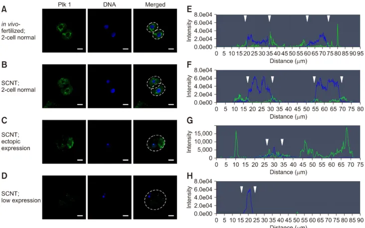
(6) Role of polo-like kinase 1 in nuclear transfer murine embryos. 7. Fig. 6. Quantitative analysis of polo-like kinase 1 (Plk1) in somatic cell nuclear transfer (SCNT) and in vivo-fertilized embryos. (A–D) Immunofluorescence images of Plk1 and DNA stained with Plk1 antibodies (green) and DAPI (blue), respectively. (E–H) Graphs of immunofluorescence intensity values measured in the area of the dotted circles in Fig. 6 A–D. Plk1 (green) was located around the nuclear membrane (blue) in in vivo-fertilized (A and E) and SCNT (B and F) 2-cell embryos that developed normally. Plk1 expression concentrated on the nuclear membranes in in vivo-fertilized and SCNT murine embryo of normal development (arrowheads). (C and G) Ectopic expression of Plk1 in SCNT murine embryos that failed to develop. (D and H) The low Plk1 expression pattern in SCNT murine embryo that failed to develop. Scale bars = 20 m (A).. E and F in Fig. 6; arrowheads). In the group showing Plk1 ectopic expression, the expression of Plk1 was significantly lower in the nuclear membrane boundary (arrowheads) than on the other side of the cytoplasm (panel G in Fig. 6). Twelve percent of the SCNT murine embryos had a low-intensity Plk1 expression pattern (panel H in Fig. 6). Taken together, the Plk1 expression location in the embryos that developed to the 2-cell stage normally, including SCNT embryos, was around the nuclear membrane boundary in both 1-cell and 2-cell stage embryos. However, the Plk1 expression of the SCNT murine embryos that failed to develop showed both a low level of Plk1 expression and ectopic Plk1 expression.. Discussion In previous studies, Plk1 has been shown to be an essential element in mitotic cell division [14]. However, cell division of embryos during development is different from that of somatic cells [26]. Recently, some studies have demonstrated that Plk1. is a critical protein in meiosis and at the embryo pre-implantation stage [2,40]. Mouse and primate SCNT procedures include an enucleation process that removes the MII spindle. This method results in the loss of chromosomes and spindle-binding proteins. However, oocytes and embryos before the 2-cell stage do not have an intact zygotic genome activation ability [34]. Accordingly, protein rescue of early-stage embryos depends on maternal mRNA. Therefore, early-stage SCNT murine embryos have an inadequate protein rescue ability for chromosomes and spindle-binding proteins. Hence, we focused on the effect of Plk1 loss during the development of SCNT murine embryos in this study. First, our results confirmed that Plk1 was located together with the chromosomes in MII stage oocytes. Consequently, after enucleation, there was a low Plk1 expression in the enucleated oocytes. This result shows that Plk1 is removed with the chromosomes during the enucleation process. Next, the embryos were treated with BI2536, a Plk1 inhibitor, to determine whether BI2536 affects early mitotic division in www.vetsci.org.
(7) 8 Jeonghyeon Moon, Sangho Roh mouse embryos. Although the 50% inhibitory concentration value of BI2536 is reported to be 0.83 nM in human cancer cells [32], some research groups have used higher concentrations of BI2536 such as 100 to 500 nM to inhibit mitosis [1,19]. In this study, BI2536 treatments over 100 nM produced rapid apoptosis of embryos similar to results reported by another research group studying the toxicity of BI2536 [28]. In this study, 4 nM of BI2536 was sufficient to block in vivo-fertilized and parthenogenetic embryos from reaching the 2-cell stage. Taken together, these results confirm that Plk1 is a crucial cytokinesis protein in mouse pre-implantation embryos. For verification of the location and expression of Plk1, assessment of immunofluorescence assay results for Plk1 and DNA followed. It was observed that Plk1 was expressed around the nuclei in all of the normally developed embryos. However, the embryos that failed to reach the 2-cell stage exhibited two difference in Plk1 expression patterns; first, there was a low level of Plk1 expression in the embryo’s cytoplasm and second, there was a pattern of ectopic Plk1 expression. In those embryos, the Plk1 was not co-located with the nucleus. Ectopic expression of Plk1 was only observed in the SCNT murine embryos that did not develop past the 2-cell stage. These results suggest that ectopic localization of Plk1 might be the result of removing Plk1 recruiting elements, such as Aurora family kinase, inner centromere protein, and survivin [33]. In conclusion, even though the accurate mechanism involved in SCNT murine embryo development remains incompletely described, the results of this study demonstrate that Plk1 has a critical role in the first mitotic division in mouse SCNT murine embryos. In particular, this study suggests the nuclear membrane boundary localization of Plk1 is crucial for successful development in SCNT murine embryos. These results provide a new insight to elucidating the molecular mechanisms in SCNT murine embryos.. Acknowledgments This study was supported by a grant from the National Research Foundation of Korea (NRF-2016R1D1A1B03931864) and by the Technology Development Program (S2519744) funded by the Ministry of SMEs and Startups (Republic of Korea). The authors also thank Eura Roh for assisting in correcting the report’s grammatical errors.. Conflict of Interest The authors declare no conflicts of interest.. References 1. Baran V, Solc P, Kovarikova V, Rehak P, Sutovsky P. Polo-like kinase 1 is essential for the first mitotic division in Journal of Veterinary Science. the mouse embryo. Mol Reprod Dev 2013, 80, 522-534. 2. Bennabi I, Terret ME, Verlhac MH. Meiotic spindle assembly and chromosome segregation in oocytes. J Cell Biol 2016, 215, 611-619. 3. Betthauser J, Forsberg E, Augenstein M, Childs L, Eilertsen K, Enos J, Forsythe T, Golueke P, Jurgella G, Koppang R, Lesmeister T, Mallon K, Mell G, Misica P, Pace M, Pfister-Genskow M, Strelchenko N, Voelker G, Watt S, Thompson S, Bishop M. Production of cloned pigs from in vitro systems. Nat Biotechnol 2000, 18, 1055-1059. 4. Bucur O, Stancu AL, Muraru MS, Melet A, Petrescu SM, Khosravi-Far R. PLK1 is a binding partner and a negative regulator of FOXO3 tumor suppressor. Discoveries (Craiova) 2014, 2, e16. 5. Bui HT, Seo HJ, Park MR, Park JY, Thuan NV, Wakayama T, Kim JH. Histone deacetylase inhibition improves activation of ribosomal RNA genes and embryonic nucleolar reprogramming in cloned mouse embryos. Biol Reprod 2011, 85, 1048-1056. 6. Byrne JA, Pedersen DA, Clepper LL, Nelson M, Sanger WG, Gokhale S, Wolf DP, Mitalipov SM. Producing primate embryonic stem cells by somatic cell nuclear transfer. Nature 2007, 450, 497-502. 7. Campbell KH, McWhir J, Ritchie WA, Wilmut I. Sheep cloned by nuclear transfer from a cultured cell line. Nature 1996, 380, 64-66. 8. Chung YG, Eum JH, Lee JE, Shim SH, Sepilian V, Hong SW, Lee Y, Treff NR, Choi YH, Kimbrel EA, Dittman RE, Lanza R, Lee DR. Human somatic cell nuclear transfer using adult cells. Cell Stem Cell 2014, 14, 777-780. 9. Combes G, Alharbi I, Braga LG, Elowe S. Playing polo during mitosis: PLK1 takes the lead. Oncogene 2017, 36, 4819-4827. 10. Deuse T, Wang D, Stubbendorff M, Itagaki R, Grabosch A, Greaves LC, Alawi M, Grünewald A, Hu X, Hua X, Velden J, Reichenspurner H, Robbins RC, Jaenisch R, Weissman IL, Schrepfer S. SCNT-derived ESCs with mismatched mitochondria trigger an immune response in allogeneic hosts. Cell Stem Cell 2015, 16, 33-38. 11. Hai T, Hao J, Wang L, Jouneau A, Zhou Q. Pluripotency maintenance in mouse somatic cell nuclear transfer embryos and its improvement by treatment with the histone deacetylase inhibitor TSA. Cell Reprogram 2011, 13, 47-56. 12. Holtrich U, Wolf G, Bräuninger A, Karn T, Böhme B, Rübsamen-Waigmann H, Strebhardt K. Induction and down-regulation of PLK, a human serine/threonine kinase expressed in proliferating cells and tumors. Proc Natl Acad Sci U S A 1994, 91, 1736-1740. 13. Huang J, Zhang H, Yao J, Qin G, Wang F, Wang X, Luo A, Zheng Q, Cao C, Zhao J. BIX-01294 increases pig cloning efficiency by improving epigenetic reprogramming of somatic cell nuclei. Reproduction 2016, 151, 39-49. 14. Kang YH, Park JE, Yu LR, Soung NK, Yun SM, Bang JK, Seong YS, Yu H, Garfield S, Veenstra TD, Lee KS. Self-regulated Plk1 recruitment to kinetochores by the Plk1-PBIP1 interaction is critical for proper chromosome segregation. Mol Cell 2006, 24, 409-422. 15. Kato Y, Tani T, Sotomaru Y, Kurokawa K, Kato J, Doguchi H, Yasue H, Tsunoda Y. Eight calves cloned from somatic.
(8) Role of polo-like kinase 1 in nuclear transfer murine embryos. cells of a single adult. Science 1998, 282, 2095-2098. 16. Kim J, Ishiguro K, Nambu A, Akiyoshi B, Yokobayashi S, Kagami A, Ishiguro T, Pendas AM, Takeda N, Sakakibara Y, Kitajima TS, Tanno Y, Sakuno T, Watanabe Y. Meikin is a conserved regulator of meiosis-I-specific kinetochore function. Nature 2015, 517, 466-471. 17. Kishigami S, Wakayama S, Thuan NV, Ohta H, Mizutani E, Hikichi T, Bui HT, Balbach S, Ogura A, Boiani M, Wakayama T. Production of cloned mice by somatic cell nuclear transfer. Nat Protoc 2006, 1, 125-138. 18. Lee KS, Oh DY, Kang YH, Park JE. Self-regulated mechanism of Plk1 localization to kinetochores: lessons from the Plk1-PBIP1 interaction. Cell Div 2008, 3, 4. 19. Lénárt P, Petronczki M, Steegmaier M, Di Fiore B, Lipp JJ, Hoffmann M, Rettig WJ, Kraut N, Peters JM. The small-molecule inhibitor BI 2536 reveals novel insights into mitotic roles of polo-like kinase 1. Curr Biol 2007, 17, 304-315. 20. Li Z, Liu J, Li J, Kong Y, Sandusky G, Rao X, Liu Y, Wan J, Liu X. Polo-like kinase 1 (Plk1) overexpression enhances ionizing radiation-induced cancer formation in mice. J Biol Chem 2017, 292, 17461-17472. 21. Li Z, Sun X, Chen J, Liu X, Wisely SM, Zhou Q, Renard JP, Leno GH, Engelhardt JF. Cloned ferrets produced by somatic cell nuclear transfer. Dev Biol 2006, 293, 439-448. 22. Mallol A, Santaló J, Ibáñez E. Improved development of somatic cell cloned mouse embryos by vitamin C and latrunculin A. PLoS One 2015, 10, e0120033. 23. Matoba S, Liu Y, Lu F, Iwabuchi KA, Shen L, Inoue A, Zhang Y. Embryonic development following somatic cell nuclear transfer impeded by persisting histone methylation. Cell 2014, 159, 884-895. 24. Miyamoto T, Akutsu SN, Fukumitsu A, Morino H, Masatsuna Y, Hosoba K, Kawakami H, Yamamoto T, Shimizu K, Ohashi H, Matsuura S. PLK1-mediated phosphorylation of WDR62/MCPH2 ensures proper mitotic spindle orientation. Hum Mol Genet 2017, 26, 4429-4440. 25. Mizutani E, Yamagata K, Ono T, Akagi S, Geshi M, Wakayama T. Abnormal chromosome segregation at early cleavage is a major cause of the full-term developmental failure of mouse clones. Dev Biol 2012, 364, 56-65. 26. Niakan KK, Han J, Pedersen RA, Simon C, Pera RA. Human pre-implantation embryo development. Development 2012, 139, 829-841. 27. Otsuki J, Nagai Y, Chiba K. Association of spindle midzone particles with polo-like kinase 1 during meiosis in mouse and human oocytes. Reprod Biomed Online 2009, 18, 522-528. 28. Raab M, Kappel S, Krämer A, Sanhaji M, Matthess Y, Kurunci-Csacsko E, Calzada-Wack J, Rathkolb B, Rozman. 29. 30.. 31.. 32.. 33. 34. 35.. 36.. 37.. 38.. 39. 40.. 9. J, Adler T, Busch DH, Esposito I, Fuchs H, Gailus-Durner V, Klingenspor M, Wolf E, Sänger N, Prinz F, Angelis MH, Seibler J, Yuan J, Bergmann M, Knecht R, Kreft B, Strebhardt K. Toxicity modelling of Plk1-targeted therapies in genetically engineered mice and cultured primary mammalian cells. Nat Commun 2011, 2, 395. Schmucker S, Sumara I. Molecular dynamics of PLK1 during mitosis. Mol Cell Oncol 2014, 29, e954507. Solc P, Kitajima TS, Yoshida S, Brzakova A, Kaido M, Baran V, Mayer A, Samalova P, Motlik J, Ellenberg J. Multiple requirements of PLK1 during mouse oocyte maturation. PLoS One 2015, 10, e0116783. Soung NK, Park JE, Yu LR, Lee KH, Lee JM, Bang JK, Veenstra TD, Rhee K, Lee KS. Plk1-dependent and independent roles of an ODF2 splice variant, hCenexin1, at the centrosome of somatic cells. Dev Cell 2009, 16, 539-550. Steegmaier M, Hoffmann M, Baum A, Lénárt P, Petronczki M, Krssák M, Gürtler U, Garin-Chesa P, Lieb S, Quant J, Grauert M, Adolf GR, Kraut N, Peters JM, Rettig WJ. BI 2536, a potent and selective inhibitor of polo-like kinase 1, inhibits tumor growth in vivo. Curr Biol 2007, 17, 316-322. Sun SC, Liu HL, Sun QY. Survivin regulates Plk1 localization to kinetochore in mouse oocyte meiosis. Biochem Biophys Res Commun 2012, 421, 797-800. Svoboda P. Mammalian zygotic genome activation. Semin Cell Dev Biol 2018, 84, 118-126. Tao J, Zhang Y, Zuo X, Hong R, Li H, Liu X, Huang W, Cao Z, Zhang Y. DOT1L inhibitor improves early development of porcine somatic cell nuclear transfer embryos. PLoS One 2017, 12, e0179436. Vazquez-Martin A, Oliveras-Ferraros C, Cufí S, Menendez JA. Polo-like kinase 1 regulates activation of AMPactivated protein kinase (AMPK) at the mitotic apparatus. Cell Cycle 2011, 10, 1295-1302. Wakayama T, Perry AC, Zuccotti M, Johnson KR, Yanagimachi R. Full-term development of mice from enucleated oocytes injected with cumulus cell nuclei. Nature 1998, 394, 369-374. Wang YS, He X, Du Y, Su J, Gao M, Ma Y, Hua S, Quan F, Liu J, Zhang Y. Transgenic cattle produced by nuclear transfer of fetal fibroblasts carrying Ipr1 gene at a specific locus. Theriogenology 2015, 84, 608-616. Weng Ng WT, Shin JS, Roberts TL, Wang B, Lee CS. Molecular interactions of polo-like kinase 1 in human cancers. J Clin Pathol 2016, 69, 557-562. Zhang Z, Chen C, Cui P, Liao Y, Yao L, Zhang Y, Rui R, Ju S. Plk1 inhibition leads to a failure of mitotic division during the first mitotic division in pig embryos. J Assist Reprod Genet 2017, 34, 399-407.. www.vetsci.org.
(9)
수치
+2
관련 문서
잔여접근법 (residual approach) 또는 차감법 : 거래가격에서 판매가격이 알 려진 이행의무의 판매가격을 차감한 나머지 금액을 판매가격이 알려지지 않 은
진행기준에 의한 수익인식은 다음과 같은 이유에서 특정 회계기간 의 의무이행활동과 성과의 정도에 대한 유용한 정보를 제공.. ① 거래가 발생하는 기간에 거래의 영향을 보고함으로써
개별판매가격 (stand-alone selling price): 해당 제품 또는 용역을 별도로 판매하였을 때 받게 될 금액.. 가장 쉽고 객관적인 방법.. 그러나 게임사용권은
약국은 당초 수집 목적과 합리적으로 관련된 범위에서 정보주체에게 불이익이 발생하는지 여부, 암호화 등 안전성 확보에 필요한 조치를 하였는지 여부 등을
(Taekwondo, Weight Lifting Players) (90 min × 6 days/week) Warming
개인적인 것에서 사회적인 것으로 문제를 확장하여 공동체 사회에서 나의 역할에 대해 고민하고, 문제해결과정을 창의적으로 표현하며 디지털
과수농민이 한국농산물품질관리원의 농산물 품질규격에 따라 과일을 출하할 때 의무적 으로 표시해야 하는 등급은 크기와 색택(빛깔), 신선도, 결함 여부로
[r]
