서 론
아토피 피부염(atopic dermatitis)은 주된 증상으로 피부발진 과 가려움증을 호소하며, 대부분 유아기에 발생하는 염증성 피부질 환이다. 전 세계적으로 유병률이 매년 증가하고 있으며, 국내에서 도 최근 지속적으로 증가하고 있다1-3). 아토피 피부염은 유전적 취 약성, 면역계의 불균형, 소양증, 피부장벽 기능의 이상 등이 상호 복합적으로 작용하여 발병하는 것으로 알려져 있다4).
아토피 피부염을 치료하기 위한 다양한 의학적 접근이 시도되 어 왔으며, 이중 약물치료법으로 항히스타민제, 면역억제제, 항생 제, 스테로이드제 등이 주로 사용되었다. 그러나 장기간 치료 시 부작용과 내성이 문제가 되어 최근에는 천연물 유래 치료제 개발에 대한 높은 관심이 대두되고 있다5,6).
영유아에게서 다발하는 아토피 피부염은 태열(胎熱), 혹은 태독 (胎毒)의 범주에 해당되며, 주로 산모가 맵거나 기름진 음식을 과식 하여 그 열독(熱毒)이 태아에게 전해져서 생기거나, 산모가 우울증
두시를 이용한 하태독법의 NC/Nga 생쥐에서 DNFB로 유발된 피부손상 완화 효과
송지훈1·안상현2·천진홍1,4·박선영3·김호현3·김기봉1,4*
1 : 부산대학교 한의학전문대학원 소아과학교실, 2 : 부산대학교 한의과학연구소, 3 : 세명대학교 한의과대학 생리학교실, 4 : 부산대학교한방병원 한방소아과
Effects of Hataedock with Douchi on 2,4-dinitrofluorobenzene-induced Atopic Dermatitis-like Skin Lesion in NC/Nga Mice
Ji-hoon Song
1, Sang-Hyun Ahn
2, Jin-Hong Cheon
1,4, Sun-young Park
3, Ho-Hyun Kim
3, Ki-Bong Kim
1,4*
1 : Department of Pediatrics, School of Korean Medicine, Pusan National University, 2 : Research Institute for Korean Medicine, Pusan National University, 3 : Department of Physiology, College of Korean Medicine, Semyung University, 4 : Department of Pediatrics, Korean Medicine Hospital, Pusan National University
Hataedock is a Korean medical treatment that administers herbal extracts orally to newborn infants. This method is used for alleviating harmful heat and excreting fetal wastes by meconium. The purpose of this study was to evaluate anti-inflammatory effect of Hataedock method with Douchi on 2,4-dinitrofluorobenzene (DNFB)-induced atopic dermatitis (AD). The 3-week-old NC/Nga mice were divided into 3 groups: the control group (Ctrl), the AD-induced group (AE), and the Hataedock-treated group (GT). Only the GT group was treated with Hataedock at the 3rd week.
After 28 days from Hataedock treatment, we induced AD-like dermatitis to the AE and GT group by DNFB. The effects of Hataedock were evaluated by immunohistochemical method. In the epithelium, PKC-positive reaction of the GT group was decreased by 57%. In the dermal papillae, IL-4-positive reaction was decreased by 34%. In the dermis, the distribution of degranulated mast cells was decreased and substance P-positive reaction was decreased by 49%. In the skin tissue, edema was decreased and MMP-9-positive reaction was decreased by 71%. Tissue damage such as epithelial cell hyperplasia, infiltration of granulocyte and lymphocyte, and capillary distribution were also decreased.
The Hataedock method with Douchi maintained skin barrier and inhibited skin-damaging factors via regulating Th2 differentiation. In conclusion, Hataedock has a potential for preventative treatment of AD. Further studies are necessary to investigate the immune-regulating mechanism and verify the safety and efficacy of the Hataedock method.
keywords : Hataedock(下胎毒), Douchi(豆豉), Atopic dermatitis, PKC, IL-4, NC/Nga Mice
* Corresponding author
Ki-bong Kim, Department of Pediatrics, Korean Medicine Hospital, Pusan National University, 20, Geumo-ro, Mulgeum-eup, Yangsan-si, Gyeongsangnam-do, 50612, Republic of Korea
·E-mail : [email protected] ·Tel : +82-55-360-5952
·Received : 2016/03/28 ·Revised : 2016/04/19 ·Accepted : 2016/04/19
ⓒ The Society of Pathology in Korean Medicine, The Physiological Society of Korean Medicine pISSN 1738-7698 eISSN 2288-2529 http://dx.doi.org/10.15188/kjopp.2016.04.30.2.109 Available online at http://www.hantopic.com/kjopp/KJOPP.htm
이 있거나 근심이 많고, 혹은 자주 화가 나는 등 감정의 기복이 심 하여 오지(五志)가 화(火)로 변하여 태아에게 영향을 주어 발생한다 고 하였다. 환경적으로는 산모가 지나치게 몸을 덥게 하거나 더운 곳에 기거하여 화기(火氣)가 태아에게 영향을 끼치면 발생한다고 하였다7).
한의학에서는 태독을 영유아에서 발병하는 다양한 질환들의 원 인으로 인식하고, 이를 제거하기 위해 출생 직후 하태독법(下胎毒 法)을 시행하였다8).
하태독법은 두시법(豆豉法), 감초법(甘草法), 황련법(黃連法), 주밀법(朱蜜法) 등이 있으며, 부드러운 비단을 한약재를 달인 약물 에 담갔다가 입속의 더러운 것들을 닦아주면서 소량 먹이는 방법이 다9). 이 중 두시는 신량해표약(辛凉解表藥)으로 체표(體表)의 열을 발산시키는 효능이 있으며, 최근 연구에서 콩과 식물들이 피부의 열로 인해 발생하는 각종 염증 증상들을 완화시킨다는 보고들이 있 다10,11).
이러한 내용을 근거로 두시를 이용한 하태독법이 아토피 피부 염 발진 억제에 효과가 있을 것으로 생각되나, 하태독법의 면역조 절 작용에 의한 효능을 검증한 연구가 전무한 실정이다. 본 연구에 서는 두시를 이용한 하태독법이 아토피 피부염 유발 조절에 활용될 수 있는 근거를 마련하기 위해 2,4-dinitrofluorobenzene(DNFB) 으로 아토피 유사 피부염을 유발한 NC/Nga mice를 이용하여 면 역조직학적 지표를 측정 검사하였다. 또한 비만세포의 침윤과 분 포, 염증세포의 이주조절 여부를 검사하였으며 피부조직을 분리하 여 피부에 대한 하태독법의 반응을 관찰하였다. 이를 통해 유의한 결과를 얻었기에 보고하는 바이다.
재료 및 방법
1. 아토피 유사 피부염 동물모델
동물 모델로는 중앙실험동물(Seoul, Republic of Korea)에서 분양받은 태령 3주된 13~15 g의 NC/Nga 수컷 생쥐를 선별하여 사용하였다. 대조군(Ctrl군), 아토피 유사 피부염 유발군(AE군), 하 태독 처리 후 아토피 유사 피부염 유발군(GT군)으로 나누었으며, 각 군에 각 10마리씩 배정하였다. 하태독법 실시 4주 후에 아토피 유사 피부염을 유발하였다. 우선 생쥐의 등 쪽 부위 피부를 면도한 다음 5% sodium dodecyl sulfate(SDS: Sigma-Aldrich, USA) 1
㎖를 면봉으로 20회 문질러서 각질층의 lipid lamella를 제거하고 acetone/olive(4:1)에 희석된 1% DNFB 100 ㎕로 감작시켰다. 감 작 후 7일과 14일째 2% DNFB 100 ㎕를 도포하여 아토피 유사 피부염을 유발하였다. 피부염 유발 72시간 후 sodium pentobarbital 용액으로 마취 후 처치하였다. 얻어진 등 쪽 피부를 10% neutral buffered formalin(NBF)으로 실온에서 24시간 동안 고정한 후 통상적인 방법으로 paraffin에 포매하고 5 ㎛ 두께로 연 속절편을 만들었다. 만들어진 연속절편은 phloxine-tartrazine 염 색하여 표본을 제작하였다(Fig. 1).
본 연구과정은 부산대학교 동물실험윤리위원회 승인을 받아 시 행되었으며(IACUC number: PNU-2014-0732), 실험실 동물의 관 리와 사용에 대해서는 NIH 가이드라인에 따라 시행되었다.
Fig. 1. Protocol of Hataedock for DNFB-induced dermatitis.
Mice were sensitized on day 28 by exposing to 1% DNFB 100 ㎕. After 7 and 14 days from initial sensitization, the mice were challenged with 2% DNFB 100 ㎕. Hataedock treatment was orally administered on day 1, 2 and 3.
Abbreviations: PBS, phosphate-buffered saline; DNFB, 2,4-dinitrofluorobenzene.
2. 하태독법 약물의 제조와 처리
두시(豆豉)는 남영제약영농조합(Muju, Republic of Korea)에 서 구입하여 사용하였다. 두시는 청호(靑蒿)와 소엽(蘇葉)의 전탕액 에 흑대두를 담가 흡수시킨 후, 남은 전탕액으로 흑대두를 삶고, 흑대두를 37~38 ℃에서 3~5일간 발효시켜 제조되었다. 두시 100 g을 증류수 1000 ㎖에 넣고 3시간동안 전탕한 후 여과하였다. 그 여액을 rotary evaporator를 이용하여 50 ㎖로 감압, 농축한 후 동결 건조하여 추출물 20.2 g(수득률 20.2%)을 얻어 연구에 사용 하였다. 3주령 NC/Nga 생쥐 GT군에 3일 동안 두시 추출물 20 ㎎ /㎏을 경구 투여하는 하태독법을 실시하였다.
3. Fingerprinting analysis
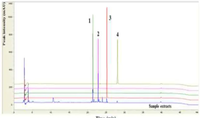
시료의 분석은 HPLC를 이용하였으며, 두시의 성분 분석에 사 용한 표준성분은 isoflavone 유도체인 daidzin 및 daidzein을 사 용하였다. 분석 결과, daidzin은 약 21.4분에 검출되었고 daidzein 은 약 25.1분에 검출되었다(Fig. 2).
Fig. 2. Comparative chromatogram of isoflavones in standard solution and Douchi extract.
Peak number 1: daidzin; 2: genistin; 3:
daidzein; and 4: genistein in standard solution. Separation was performed by HPLC-DAD.
4. 조직화학
신경 펩티드를 분비하는 비만세포의 분포와 형태 변화를 조사
하기 위해 Luna's method를 실시하였다. 우선 진피 내 비만세포 과립을 aldehyde fuchsin에 30분간 염색한 후, Weigert's iron hematoxylin과 methyl orange 용액에 각각 1분, 5분 동안 대조 염색한 후 관찰하였다.
부종 완화는 Masson trichrome 염색을 통해 조사하였다. 우 선 50~60 ℃ Bouin 용액에서 1시간동안 매염 처리한 다음 70%
에탄올에서 picric acid를 제거하였다. Weigert iron hematoxylin 에서 10분 동안 반응시켜 핵을 염색하고, Biebrich scarlet-acid fuchsin과 phosphomolybdic-phosphotungstic acid에서 각각 15분간, aniline blue에서 5분간 처리한 후 관찰하였다.
5. 면역조직화학
피부 절편을 proteinase K(20 ㎍/㎖)에 5분 동안 proteolysis 과정을 거친 후, blocking serum인 10% normal goat serum에 4시간 동안 실온에서 반응시켰다. 그리고 1차 항체인 goat anti-PKC 1:100, Santa Cruz Biotec, USA), goat anti-IL-4 1:100, Santa Cruz Biotec, USA), goat anti-substance P 1:100, Santa Cruz Biotec, USA) 그리고 goat anti-MMP-9 1:200, Santa Cruz Biotec, USA)에 4 ℃ humidified chamber에 서 72시간 동안 반응시켰다. 그런 다음 2차 항체인 biotinylated rabbit anti-goat IgG(1:100, Santa Cruz Biotec, USA)에 실온에 서 24시간 반응시켰고, avidin-biotin complex kit(Vector Lab, USA)에 1시간 동안 실온에서 반응시켰다. 0.05%
3,3'-diaminobenzidine과 0.01% HCl이 포함된 0.05 M tris-HCl 완충용액(pH 7.4)에서 발색시킨 후, hematoxylin으로 대조염색하 였다.
6. 영상분석
면역조직화학의 결과는 Image Pro Plus(Media Cybernetics, USA)를 이용한 영상분석을 통해 수치화(mean ± standard error)했다. 영상분석은 각 군의 표본에서 임의로 선정된 상피와 진피유두를 400배 확대하여 촬영한 다음 positive pixels/전체 pixel(10,000,000 pixels)로 나타내었다.
7. 통계
통계는 SPSS software(SPSS 23, SPSS Inc., USA)로 이루어 졌으며, one-way ANOVA 시행을 통해 유의성(P<0.001)을 검증하 고 Duncan’s multiple range test로 사후 검증하였다.
결 과
1. PKC 활성 억제에 따른 Th2 분화 조절
상피에서 관찰되는 PKC 양성반응은 세포질에서 강하게 나타났 으며. AE군에 비해 GT군에서 PKC 양성반응 세포가 감소한 것으로 관찰되었다(Fig. 3). 영상분석 결과, GT군이 AE군에 비해 57% 감 소한 것으로 관찰되었다(Fig. 6).
진피유두에서 관찰되는 IL-4 양성반응은 세포질에서 강하게 나타났으며. AE군에 비해 GT군에서 IL-4 양성반응 세포가 감소된
것으로 관찰되었다(Fig. 3). 영상분석 결과, GT군이 AE군에 비해 34% 감소한 것으로 관찰되었다(Fig. 6).
Fig. 3. The decrease in Th2 differentiation by Douchi-HTD.
The PKC-positive cells (arrow) in GT were remarkably decreased (PKC immunohistochemistry; bar size, 50 ㎛). The IL-4-positive cells (arrow) in GT were remarkably decreased (IL-4 immunohistochemistry; bar size, 50
㎛) Abbreviations: Douchi-HTD, Glycine Semen Preparatum Hataedock;
EPI, epithelium; DER, dermis.
2. 비만세포 활성 조절
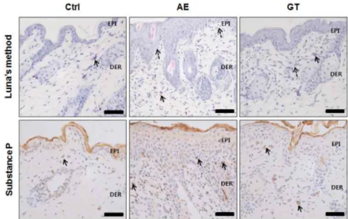
AE군의 진피유두에서 피하 인접부까지 많은 수의 비만세포가 분포하였으며, 주로 탈과립형(degranulated type)으로 관찰되었다.
이에 비해 GT군에서는 AE군에 비해 적은 수가 관찰되었으며, 주로 과립형이었다(Fig. 4).
또한, 진피유두에서 관찰되는 substance P 양성반응은 세포질 에서 강하게 나타났으며. AE군에 비해 GT군에서 substance P 양 성반응세포가 감소된 것으로 관찰되었다(Fig. 4). 영상분석 결과 GT군이 AE군에 비해 49 % 감소한 것으로 관찰되었다(Fig. 6).
Fig. 4. The decrease in itching by Douchi-HTD.
The degranulated mast cells (dotted line arrow) in dermal papillae were increased in AE but decreased in GT (Luna's method; full line arrow, granulated mast cell; bar size, 50 ㎛). The substance P-positive cells (arrow) in GT were remarkably decreased (substance P immunohistochemistry; bar size, 50 ㎛).
Abbreviations: Douchi-HTD, Glycine Semen Preparatum Hataedock; EPI, epithelium; DER, dermis.
3. 부종 조절
AE군에서는 진피유두에서 피하 인접부까지 아교섬유의 분포가 감소하였지만, GT군에서는 AE군에 비해 많은 수가 관찰되었다
(Fig. 5). 즉, GT군에서는 AE군에 비해 부종 양상이 완화되었다.
한편 진피유두에서 관찰되는 MMP-9 양성반응은 세포질에서 강하게 나타났으며, AE군에 비해 GT군에서 MMP-9 양성반응 세포 가 감소한 것으로 관찰되었다(Fig. 5). 영상분석 결과, GT군이 AE 군에 비해 71% 감소한 것으로 관찰되었다(Fig. 6).
Fig. 5. The decrease in edema by Douchi-HTD at the dermatitis-induced skin.
The distribution of collagen fiber were decreased in AE but increased in GT (Masson trichrome method; bar size, 50 ㎛). The matrix metalloproteinases (MMP)-9-positive cell (arrow) in GT were remarkably decreased (MMP-9 immunohistochemistry; bar size, 50 ㎛). Abbreviations:
↔, edema-evoked region; Douchi-HTD, Glycine Semen Preparatum Hataedock; EPI, epithelium; DER, dermis.
Fig. 6. Image analysis of immunohistochemistry. Abbreviation: #, p<0.001 compared with AE.
4. 피부 손상 완화
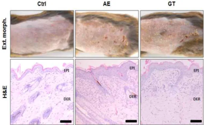
외부 형태 변화를 관찰한 결과, AE군의 피부 대부분의 지역에 서 각질층이 손상된 습진(eczema)이 나타났으며, 일부 가장자리 지역에서는 탈락과정에 있는 혈병(blood clot)과 표피 잔재도 관찰 되었다. GT군은 피부 손상이 적은 것으로 관찰되었다(Fig. 7).
AE군의 피부 조직에서 나타난 변화로는 표피에서 각질형성세 포(keratinocyte)의 과형성(hyperplasia)으로 인한 피부 상피 층수 의 급격한 증가, 기저층의 원주상피세포와 기저판의 결합이 파괴된 공간으로 침윤(infiltration)하는 림프구의 증가가 확인되었다. 또한 가시층과 기저층 사이에서 핵의 수축으로 인한 핵막과 세포질 사이 공간의 확장된 상피세포의 증가와 세포 사이 공간(intercellular space)의 확장이 관찰되었다. 표피 쪽 진피에서는 염증 관여 세포
의 이주(migration) 증가와 혈관 증가가 관찰되었다. GT군에서의 형태 변화도 AE군과 유사한 경향성을 보였으나, 전반적으로 피부 염이 약화된 양상으로 나타났다. 특히 이러한 피부 조직 손상의 완 화 증거로는 AE군에 비해 표피세포의 과형성 감소, 침윤하는 림프 구 수의 감소, 표피 원주상피세포 손상 감소, 그리고 세포 사이 공 간의 회복 등이 관찰되었다. 또한, 진피에서 염증 관여 세포의 침 윤 감소와 혈관 수의 감소가 확인되었다(Fig. 7).
Fig. 7. The mitigative effect of Douchi-HTD for dermatitis-induced skin lesion.
In external morphology, the skin damages as eczema were mitigated in GT. In histological morphology, the decrease in skin damages such as hyperplasia, edema, infiltration of lymphocytes, and increase of capillary were alleviated in GT compared with AE (hematoxylin and eosin;
bar size, 100 ㎛). Abbreviations: Douchi-HTD, Glycine Semen Preparatum Hataedock; EPI, epithelium; DER, dermis.
고 찰
아토피 피부염의 원인은 유전, 음식 알레르기, 면역학적 이상, 피부 장벽 이상, 환경적 요인, 심인성 요인 등이 서로 연관되어 있 으며, IgE 증가, mast cell, T세포, 호산구, CD4+ T세포, CD8+ T 세포, Th2 매개 cytokine 등의 불균형이 작용하여 발병하는 것으 로 알려져 있다12,13). 주된 증상으로 피부 수분 손실, ceramide, filaggrin, antimicrobial peptides 등의 감소 등이 나타난다14). 이 러한 피부 장벽 손상은 더 많은 항원이 피부를 투과하게 하여 침윤 하는 단핵구(monocyte) 수가 늘어나고, 만성적인 습진과 염증이 발생하게 된다15). 또한 이러한 습진과 염증이 반복되면서 피부 표 피층이 과증식하여 태선화(lichenification)를 유발하게 된다14). 최 근에는 아토피 피부염이 IgE와 관련되어 있어 IgE 생성을 억제하려 는 연구들이 많이 진행되고 있다16-18). 이런 연구들과 아울러 치료 제로 보편적으로 사용되고 있는 스테로이드제나 항히스타민제로는 부작용과 역치값의 상승 등으로 근본적인 치료가 이루어지지 못함 에 따라, 대체요법이나 한약을 이용한 치료제 개발에 관한 연구가 증가하고 있다19-22).
본 연구에서는 아토피 피부염에 대한 하태독법 치료의 객관적 효능을 평가하기 위하여 면역학적 지표를 측정 검사하였다. 기존 연구에서는 아토피 피부염을 유발한 후 약물 투약을 통한 아토피 피부염의 호전 정도를 파악하였다면, 본 연구는 하태독법을 시행하 는 목적에 부합하고자 두시를 이용하여 하태독법을 먼저 시행하고 일정 기간 경과 후 아토피 피부염을 유발함으로써 약물의 직접적인
약리적 효과로 인한 증상 경감보다는 하태독법에 의한 면역조절 작 용에 의한 효능을 확인하고자 하였다.
아토피 피부염에서는 각질층 내 ceramide 감소로 인하여 sphingosine이 감소하고 protein kinase C(PKC) 활성을 유도하여 상피 기저층에서 세포분열이 증가하게 된다23). 이는 TNF-α, IL-1β 등의 proinflammatory cytokine 분비 증가에 의한 NF-κB/Rel의 활성을 유도하며, 염증 반응에 관여하는 효소인 iNOS가 생성되어 과도한 산화적 스트레스로 의한 조직 손상이 일어난다24). 특히 IL-4와 PKC는 Th2 cell의 분화에 중요한 역할을 하는 것으로 알 려져 있다25,26). 본 연구에서는 하태독법에 의한 Th2 분화 조절 효 능에 대해 알아보기 위해 PKC와 IL-4의 발현 억제 여부를 확인하 였다. 그 결과, AE군에 비해 GT군에서 PKC와 IL-4 양상반응 세포 가 유의하게 감소한 것을 관찰하였다. 이는 하태독법이 Th2 분화 를 제어함으로써 아토피 피부염 유발 조절에 관여할 수 있음을 의 미한다.
알러지 반응을 일으키는 과정에서 중요한 역할을 하는 비만세 포는 세포 표면에 FcεRI 수용체를 발현하며, 항원들이 이 수용체에 결합된 IgE와 교차 결합하면 비만세포의 활성화와 탈과립화가 진행 된다. 탈과립화는 항원 결합 후 몇 분 내에 발생하여 histamine, substance P, matrix metalloprotinases, serotonin 등 다양한 화학적 매개물질들을 세포 밖으로 방출하게 된다27). 이 중 substance P는 신경 펩티드(neuropeptide)로 신경 생성에 주로 관여하며 피부, 근육, 관절의 감각신경 말단에서 분비된다28,29). 또 한 미세혈관을 확장하여 투과성을 증가시키고, mast cell의 탈과립 과 염증세포 이주 및 증식을 유도하는 등 신경성 염증 및 피부 소 양증과 연관되어 있다30,31). 본 연구에서는 하태독법의 알러지 반응 에 대한 조절 효과를 확인하기 위해 피하에 위치한 비만세포의 탈 과립 현상과 substance P의 발현 정도를 관찰하였다. 그 결과, GT군에서 AE군에 비해 탈과립형 비만세포의 분포가 적게 나타났 으며, substance P의 발현도 GT군에서 감소함을 확인하였다. 이 는 하태독법이 비만세포의 탈과립을 억제하여 화학매개물질과 염증 성 cytokine의 분비로 인한 비만세포 매개 알레르기 반응을 억제 하는 것으로 생각되며, substance P의 발현 억제를 통한 소양증 완화에 효과가 있음을 시사한다.
MMP-9은 cytokines, growth factor와 같은 여러 자극에 의 해 합성, 분비되어 세포 구조에 영향을 주며32), laminin과 type IV collagen을 선택적으로 분해하고 기저막을 파괴하여 염증관련 세포 의 이주를 용이하게 한다33,34). 본 연구에서는 하태독법의 진피 내 부종 변화 및 염증세포들의 이주 조절 효능을 확인하기 위해 MMP-9의 발현 정도와 Masson trichrome 염색에 의한 진피 내 부종 변화 양상을 관찰하였다. 관찰 결과, GT군에서는 AE군에 비 해 부종이 완화되었으며 MMP-9의 활성이 감소하였다. 이러한 결 과는 하태독법이 MMP-9를 억제하고 염증 관련 세포의 이주를 조 절하여 진피 내 부종도 경감시켰음을 의미한다.
본 연구에서는 하태독법에 의한 피부 손상 완화 효과를 확인하 기 위해 하태독법을 시행하고 일정기간 후 등 쪽 피부에 아토피 피 부염을 유발하였다. AE군에서는 대부분 각질층이 손상된 습진이 나타났으며, 일부 가장자리 지역에서는 탈락과정에 있는 혈병과 표
피 잔재도 관찰되었다. 이에 비해 GT군은 피부 손상이 적은 것으 로 관찰되었다. 피부조직의 손상 정도에서도 AE군에 비해 GT군이 상피세포 과형성 및 기저막 주변으로의 과립백혈구와 림프구 침윤 이 적었고, 진피에서 염증 관여 세포의 이주 증가와 모세혈관 분포 증가 정도가 적었다. 이를 통해 하태독법이 피부염으로 유발된 부 종의 감소뿐만 아니라, 피부 손상을 감소시키는 효과가 있음을 확 인할 수 있었다.
본 실험은 DNFB 유발 아토피 피부염 동물모델을 이용하여 유 전적 요인을 고려하지 못하였다는 단점이 있다. 아울러 향후 후속 연구를 통해 하태독법의 면역 조절에 관련된 기전과 안전성 및 유 효성의 검증이 필요할 것으로 생각된다.
결 론
하태독법의 아토피 피부염 유발 조절효과를 조사하기 위해 행 해진 본 연구는 태령 3주에 하태독 처리한 NC/Nga 생쥐에 DNFB 를 이용해 인위적 피부염을 유발한 후 Th2 분화 조절, 피부소양증 감소 효과, 염증세포 이주 조절 작용, 피부 손상의 면역조직학적 변화를 관찰한 결과 다음과 같은 결과를 얻었다.
하태독법 처리 후 상피 내 PKC 양성반응이 57%, 진피유두 내 IL-4 양성반응이 34% 감소하였다.
하태독법 처리 후 피하에 위치한 탈과립형 비만세포의 분포가 감소하였으며, substance P 발현이 49% 감소하였다.
하태독법 처리 후 조직 내 부종이 감소하였고, MMP-9의 활성 이 71% 감소하였다.
조직 내 형태학적 변화를 관찰한 결과, 상피세포 과형성, 과립 백혈구와 림프구의 침윤, 모세혈관 분포가 완화되었다.
이상의 결과로 하태독은 Th2 분화를 제어하여 피부 상피장벽 의 유지 및 피부 손상을 선제적으로 제어한다고 생각된다.
감사의 글
이 연구는 2014년도 정부(교육부)의 재원으로 한국연구재단의 지원을 받아 수행된 기초연구사업임(No. NRF-2014R1A1A2055061).
References
1. Korean Dermatological Association Textbook Compilation Committee. Dermatology. Seoul, RyoMoonGak, pp 170-171, 2008.
2. Blume-Peytavi, U., Metz, M. Atopic dermatitis in children: management of pruritus. J. Eur. Acad.
Dermatol. Venereol. 26: 2-8, 2012.
3. Addor, F.A., Takaoka, R., Rivitti, E.A., Aoki, V. Atopic dermatitis: correlation between non-damaged skin barrier function and disease activity. Int. J. Dermatol.
51: 672-676, 2012.
4. Kabashima, K. New concept of the pathogenesis of
atopic dermatitis: Interplay among the barrier, allergy, and pruritus as a trinity. J. Invest. Dermatol. 70: 3-11, 2013.
5. Leung, D.Y. Atopy dermatitis: new insight and opportunities for therapeutic intervention. J. Allergy Clin. Immunol. 105: 860-867, 2000.
6. Beltrani, V.S. The role of house dust mites and other aeroallergens in atopic dermatitis. Clin. Dermatol.
21(3):177-182, 2003.
7. Jiangyu, R. Practical TCM Pediatrics. Shanghai, Shanghai Science and Technology Press, 2005.
8. Kang, M.Y., Jang, G.T., Kim, J.H. A study on fetal toxicosis removal therapy. J. Pediatr. Korean Med.
17(1):29-51, 2003.
9. Im, G.M., Jeong, H.W., Kim, H.S., Jeong, W.Y. Oriental Medical Approach on the Allergic Disease. Kor. J.
Oriental Physiology & Pathology. 16(5):831-839, 2002.
10. Miller, A.K., Benson, J.M., Muanza, D.N., Smith, J.R., Shepherd, D.M. Anti-inflammatory effects of natural product formulations on murine dendritic cells. J. Diet Suppl. 8(1):19-33, 2011.
11. Yeh, C.Y., Jung, C.J., Huang, C.N., Huang, Y.C., Lien, H.T., Wang, W.B., Wang L.F., Chia J.S. A legume product fermented by Saccharomyces cerevisiae modulates cutaneous atopic dermatitis-like inflammation in mice. BMC Complement. Altern. Med. 14: 194, 2014.
12. Sandstrom, M.H., Faergemann, J. Prognosis and prognostic factors in adult patient with atopic dermatitis : a long-term follow-up qeustionnaire study.
Br. J. Dermatol. 150: 103-110, 2004.
13. Dupuy, P. Does atopic dermatitis result from cytokine dysregulation? J. invest. Dermatol. 92: 44-47, 1994.
14. Cork, M.J., Danby, S.G., Vasilopoulos, Y., Hadgraft, J., Lane, M.E., Moustafa, M., Guy, R.H., Macgowan, A.L., Tazi-Ahnini, R., Ward, S.J. Epidermal barrier dysfunction in atopic dermatitis. J. Invest. Dermatol.
129(8):1892-1908, 2009.
15. Ye, Y.M., Kim, B.E., Shin, Y.S., Park, H.S., Leung, D.Y.M. Overexpression of Epidermal Filaggrin in Patients with Chronic Idiopathic Urticaria Correlates with Urticaria Severity. J. Allergy Clin. Immunol.
131(2):AB56, 2013.
16. Kim, J.W. Allergy and immunological perspective in atopic dermatitis. J. of Korean Dermatological Association. 41(6):687-689, 2003.
17. Ahn, P.S., Bang, H.D., Jung, J.H., Kim, G.H., Park, K.C., Yoon, J.I. Effect of recombinant interferon gamma on the treatment of atopic dermatitis. J. Immunol.
16(3):291-298, 1996.
18. Sung, H.C., Lee, W.J., Lee, S.J., Kim, D.W. A Study on the Relationship of Allergy-related Labaratory Findings with the Clinical Severity of Atopic Dermatitis. J. of Korean Dermatological Association. 44(9):1051-1057, 2006.
19. Sidbury, R., Hanifin, J.M. Old, New, And Emerging Therapies for Atopic Dermatitis. Dermatologic Clinics.
18(1):1-11, 2000.
20. Schreiber, S.L., Crabtree, G.R. The mechanism of action of cyclosporin A and FK506. Immunol. Today.
13(4):136-142, 1992.
21. Kim, S.H., Kim, S., Lee, S.H., Park, H.W., Chang, Y.S., Min, K.U., Cho, S.H. The effects of PG102, a water-soluble extract from actinidia arguta, on serum total IgE levels:a double-blind, randomized, placebo-controlled exploratory clinical study. Eur. J.
Nutr. 50(7):523-529, 2011.
22. Kim, D., Kim, S.H., Park, E.J., Kang, C.Y., Cho, S.H., Kim, S. Anti-allergic effects of PG102, a water-soluble extract prepared from actinidia arguta, in a murine ovalbumin-induced asthma model. Clin. Exp. Allergy.
39(2):280-289, 2009.
23. Lee, H.H. Atopic Dermatitis and Epidermal Barrier.
Allergy Asthma & Respiratory Disease. 19(2):95-105, 2009.
24. Connelly, L., Palacios-Callender, M., Ameixa, C., Moncada, S., Hobbs, A.J. Biphasic regulation of NF-kappa B activity underlies the pro- and anti-inflammatory actions of nitric oxide. J. Immunol.
166(6):3873-3881, 2001.
25. Chen, L., Grabowski, K.A., Xin, J.P., Coleman, J., Huang, Z., Espiritu, B., Alkan, S., Xie, H.B., Zhu, Y., White, F.A., Clancy, J. Jr., Huang, H. IL-4 Induces Differentiation and Expansion of Th2 Cytokine-Producing Eosinophils. J. Immunol.
172(4):2059-2066, 2004.
26. Marsland, B.J., Soos, T.J., Späth, G., Littman, D.R., Kopf, M. Protein kinase C theta is critical for the development of in vivo T helper (Th)2 cell but not Th1 cell responses. J. Exp. Med. 200(2):181-189, 2004.
27. Amin, K. The role of mast cells in allergic inflammation. Respir. Med. 106(1):9-14, 2012.
28. Park, S.W., Yan, Y.P., Satriotomo, I., Vemuganti, R., Dempsey, R.J. Substance P is a promoter of adult neural progenitor cell proliferation under normal and ischemic conditions. J. Neurosurg. 107(3):593-599, 2007.
29. Donkin, J.J., Turner, R.J., Hassan, I., Vink, R. Substance P in traumatic brain injury. Prog. Brain Res. 161:
97-109, 2007.
30. Ebner, K., Singewald, N. The role of substance P in stress and anxiety responses. Amino Acids.
31(3):251-272, 2006.
31. Yamaoka, J., Kawana, S. Rapid changes in substance P signaling and neutral endopeptidase induced by skin-scratching stimulation in mice. J. Dermatol. Sci.
48(2):123-132, 2007.
32. Woessner, J.F.Jr. MMPs and TIMPs-an historical perspective.
Mol. Biotechnol. 22(1):33-49, 2002.
33. Burrage, P.S., Mix, K.S., Brinckerhoff, C.E. Matrix metalloproteinases ; role in arthritis. Front Biosci. 11:
529-543, 2006.
34. Onodera, S., Kaneda, K., Mizue, Y., Koyama, Y., Fujinaga, M., Nishihira, J. Macrophage migration inhibitory factor up-regulates expression of matrix metalloproteinases in synovial fibroblast of rheumatoid arthritis. J. Biol. Chem. 275(1):444-450, 2000.