Antioxidant and Antifungal Activity of Endophytic Fungi Associated with Agarwood Trees 1
Asep Hidayat
2,†⋅Maman Turjaman
2⋅Sarah Asih Faulina
2⋅Fadel Ridwan
3⋅ Aryanto
2⋅ Najmulah
2⋅ Tun Tedja Irawadi
3⋅ Apri Heri Iswanto
4ABSTRACT
1)Several species of Aquilaria and Gyrinops are native to Indonesia and well known as agarwood-producing trees with a high economic value. Their bioactive compounds have a wide spectrum of uses, such as in medicine and cosmetics.
These genera have undergone extensive search for novel bioactive compounds. The purpose of this study was to isolate, identify, and characterize the endophytic fungi community associated with Aquilaria malaccensis, A. microcarpa, Gyrinops versteegii, and A. crassna trees and investigate their bioactive properties as antioxidant agents and antagonists. A total of 50 fungi were successfully isolated from different tissues of the four species of agarwood-producing trees. Two isolates exhibited strong antioxidant activity, namely, Apodus oryzae (R2MC3A, IC
5060.92 mg/mL) and Diaporthe sp. (P1DS1[C], IC
5076.65 mg/mL). Two isolates, Pestalotiopsis theae (P3BS3[B]) and Curvularia sp. (P2CD3A), showed
>75% antifungal activity against pathogenic Fusarium solani. The results revealed that endophytic fungi associated with the studied agarwood-producing trees had potential antioxidant and antifungal activities for further applications in biotechnology.
Keywords: Aquilaria, Gyrinops, endophytic fungi, antioxidant, antagonists
1. INTRODUCTION
Thymelaeceae is the major plant family producing agarwood, also called Gaharu in Indonesian. Agarwood have been used as one of the essential ingredients in fragrance, aromatherapy, pharmaceutical and herbal medicines for centuries. It also hold social, cultural, and economic values in local communities (Beek and Philips, 1999; Barden et al., 2000; Donovan and Puri,
2004). Indonesian agarwood has been known worldwide and retained long historical story. It was first recorded as the main commodity bartered between the empires of China and the kingdoms of Indonesia since the Silk Road era (Turjaman et al., 2016). Wider spectrums of agarwood uses have been known for sedative in oriental medicine, antimicrobial, antitumor and antioxidant activity (Takemoto et al., 2008; Wetwitayaklung et al., 2009; Chen et al., 2011). Agarwood is the resinous
1
Date Received March 16, 2019, Date Accepted July 15, 2019
2
Forest Microbiology Laboratory, Forest research and Development Centre, Research, Development and Innovation Agency, Ministry of Environmental and Forestry. Jl. Gunung Batu No. 5., Bogor 16001, West Java, Indonesia
3
Department of Chemistry, Faculty of Mathematics and Natural Sciences, Bogor Agricultural University. Jl. Tanjung Kampus IPB, Dramaga, Bogor 16680, West Java, Indonesia
4
Department of Forest Product, Faculty of Forestry, Universitas Sumatera Utara, Padang Bulan, Medan 20155, Indonesia
†
Corresponding author: Asep Hidayat (e-mail: [email protected], ORCID: 0000-0003-3755-072X)
Original Article
and fragrant heartwood that may not be formed in normal wood tissue but formed in wounded trees.
Among them, Aquilaria and Gyrinops are the most common agarwood-producing genus in Indonesia (Gong and Guo, 2009). Different parts of various plant have been known to have various benefit (Jung et al., 2017.
Ham and Kim, 2018; Li et al., 2018), healthy wood of Aquilaria and Gyrinops have been acknowledge to contain bioactive compound; leaves demonstrated inhibition activity against polymorphonuclear neutro- phils (PMNs) respiratory burst stimulated by PMA (Qi et al., 2009), anti-diabetic, anti-HIV, anti-cancer, immuno-modulatory anti-inflammatory and laxative effects (Yoshimi et al., 2001); while fresh steam ex- hibited cytotoxicity against SGC-7901 and SMMC- 7721 cell lines (Wang et al., 2010).
Parallel with the huge loss of agarwood producing- tree in the wild, the losses of endophytic fungi associated with the trees automatically determine the loss of Indonesia biodiversity as well. Agarwood-producing tress are host for many endophytic fungi which produce similar or even higher amount of bioactive substrates compare to their host plant (Stierle et al., 1993; Strobel et al., 1996; Li et al., 1998). Previous studies have recorded that 1) Fusarium spp. isolated from leaves, steam and root of A. sinensis showed the most potent antimicrobial activity, 2) several endophytes fungi (Gong and Guo, 2009) isolated form A. crassna showed capability to produce indole-3-acetic acid (IAA) and siderophores (Nimnoi et al., 2010), 3) F. verticillioides SHTr3 and Colletotrichum truncatum SHTrHc7 isolated from A. crassna showed comparable scavenging abilities on DPPH-free radicals (Chi et al., 2016), 4) Xylaria mali, Lasiodiplodia theobromae and Phaeoacremonium rubrigenum isolated form A. sinensis exhibited cyto- toxicity against 293-T, 293-T, and SKVO3 cells lines (Cui et al., 2011), and 5) fungus AL-2 strain isolated from A. malaccensis had antibacterial activities against gram positive and negative bacteria as well as general
toxicity based on brine shrimp lethality (Shoeb et al., 2010). A. sinensis have been used as traditional Chinese medicinal, a tropical evergreen tree distributed in Hainan, Guangdong, Guangxi, Yunnan and Taiwan (Chen et al., 2016); A. crassna used also as traditional medical plant and widely distributed in Indochina and Thailand (Turjaman and Hidayat, 2017); and so that for A. malaccensis which is the most popular species for its agarwood production and distributed from different countries, Bangladesh, Bhutan, Cambodia, India, Indonesia, Iran, Lao PRD, Malaysia, Myanmar, Philippines, Singapore, Thailand, Vietnam and Papua New Guinea (Oldfield et al., 1998; Premalatha and Kalra, 2013).
The genus Aquilaria has 27 species spread world- wide, which 6 are found in Indonesia (Soehartono and Newton, 2000; Saikia and Khan, 2013). The genus Gyrinops is distributed in at least 12 countries, and 7 species were found naturally in Indonesia (Barden et al., 2000). Several studies have been previously conducted to those species, particularly on exploration and investigation of their phytochemical constituents and their respective endophytic fungi bioactivities.
There was vast variations of endophytic fungal commu- nities related to living conditions, host plant species, as well as host tissue types (Lamit et al., 2014); indi- cating that further investigations are important. For this reason, this study aimed to isolate the understudied endophytic fungi associated with three Indonesia-native species of agarwood (A. malaccensis and A. microcarpa, G. versteegii) and one exotic species (A. crassna), as well as their antioxidant and antifungal activities.
2. MATERIALS and METHODS
2.1. Chemicals
Alcohol, NaClO 4%, Dextrose, KOH 10%, HCl,
Glycerol 20%, acetic acid anhydrite, magnesium (Mg),
H
2SO
4, Folin–Ciocalteu (FC), ethyl acetate and meth- anol were purchased from Merck chemical (Germany).
1,1-diphenyl-2-picrylhydrazyl (DPPH) was purchased from Sigma-Aldrich chemical (USA) and other che- micals were provided by Himedia chemicals (India).
2.2. Samples and cultures medium for isolation
Different parts of plant materials (healthy segment;
bark, leaves, shoot and root) from Aquilaria and Gyrinops trees originated from West Java and Bangka were collected (diameter breast high /dbh > 15 cm, either grown naturally or planted). The potato dextrose agar (PDA), which consist of potato extract 20%, dextrose 2%, and agar 2% was used for isolation. Pure fungal strains were sub-cultured and maintained on PDA prior to use. Malt Yeast Glucose Agar (MYGA, containing yeast extract 0.4%, malt extract 1%, sucrose 0.4%, and agar 2%), and Malt Yeast Glucose Pepton Agar (MYGPA, which consist of yeast extract 0.3%, malt extract 0.3%, peptone 0.5%, sucrose 1.05%, and agar 2%) were also used to obtain optimum fungal growth (Atlas, 2004).
2.3. Endophytic fungal isolation
Fungal strains were isolated from bark, leaves, shoot, and root of A. malaccensis, A. microcarpa, A. crasna and G. verstigii trees. The tree parts were collected at least from three different trees. In order to minimize the growth of epiphytic fungi during internal tissue sampling, tree parts were surface-sterilized by washing, followed by soaking in alcohol 70% for 2 min, and lastly in sodium hypochlorite for 2 min (4% Cl active).
The outer tissues were removed, and the inner tissues of 0.5 x 0.5 cm size were carefully dissected and placed in PDA media plates. Chloramphenicol (100 mg/L) was added to media to suppress bacterial growth. The plates were then incubated at room temperature for up to 14 days, during which the development of different colonies
was periodically observed and verified. Each colonies of different fungi appeared from tissue fragment were cut out by dissecting microscope and purified on PDA plates. All pure cultures obtained were used for further investigation.
To select the most suitable medium for each isolate, the pure cultures were tested on PDA, MYGA and MYGPA media. A disk (5 mm, diameter) of actively growing fungal colony on agar media was obtained with a cork-borer and was sub-cultured into petri dish containing PDA, MYGA and MYGPA media. The plate was incubated for 7 d, and the fungal growth was observed daily. All the experiments were conducted in triplicate.
2.4. Screening of endophytic fungi against F. solani in dual culture
F. solani was obtained from Laboratory of Forest Microbiology (INTROF-CC 00509, Forest Research and Development Centre, Bogor, Indonesia) All pure strains of fungal endophytes were screened against F.
solani in dual cultures on PDA. An agar disk (5 mm diameter) of endophytic and F. solani was co-inoculated 4 cm apart on PDA in petri dish. The plates were in- cubated at room temperature up to 7 days. All experi- ments were carried out in triplicates. Growth inhibition percentage was determined by the following formula (Dennis and Webster, 1971).
Percentage of inhibition = [(J1-J2)/J1] x 100%
Where J1 is radial covered by pathogen toward petri dish wall (outward), and J2 is radial covered by pathogen (F. solani) toward endophytic fungi.
2.5. Screening of fungal endophytes for antioxidant activity
2.5.1. Preparation of fungal extraction
Each of endophytic fungal isolates was grown in 250
mL Erlenmeyer flasks containing 50 ml of selected liquid medium (previously described) and incubated for 14 days in stationary condition. After incubation, mycelia biomass was harvested by filtering through Whatman No. 1 filter paper. The filter papers containing mycelia mat were oven dried until constant weight was reached, then extracted by ethyl acetate. The filtrate was also extracted by ethyl acetate using funnel separation. Each organic phase was mixed, and the solvent was then removed under reduced pressure using rotary vacuum evaporator to obtain a concentrate. The yield of ethyl acetate soluble was determined by calculation with dried weight of fungal mycelium (%, w/w) and volume of culture liquid medium (%, w/v).
2.5.2. Antioxidant assay
Antioxidant activity of each extract was determined according to published methods with slight modifi- cations (Kamonwannasit et al., 2013; Marson Ascêncio et al., 2014; Sadeghi et al., 2015). In a series of test tubes, extract was diluted with ethyl acetate to obtain in the range of concentration into which 0.25 mL of a DPPH (1 M) and added methanol to obtain 2.5 mL of final reaction volume. After 30 min incubation at room temperature, the absorbance was monitored at 517 nm. The ethyl acetatesolution without extracts was selected as control. All experiments were conducted in triplicated test. The inhibitory activity was calculated from [(A
0– A
1)/A
0] × 100, where A
0is the absorbance of the control, and A
1is the absorbance of the extract or standard. The antioxidant activity was expressed as IC
50, in which the amount of sample necessary to reduce the initial DPPH concentration by half.
2.5.3. Phytochemical assay
Phytochemical assay was carried out according to Tapwal et al. (2016) with slight modification, including detection of some important constituents (phenolic, flavonoid, tannin, steroid, terpenoid, and alkaloid). All ethyl acetate soluble obtained from extraction (pre-
viously described) with concentration of 1000 µg/L was subjected for phytochemical assay. In general, phenolic and tannin were reacted by ferric chloride test, flavonoid was determined by addition of magnesium and hydro- chloric acid, steroid was monitored by addition of acetate anhydrate and sulfuric acid, terpenoid was determined by addition of sulfuric acid, and alkaloid was monitored by using Mayer’s and Wagner’s reagents.
2.6. Identification of fungi
Fungal identification was done with molecular meth- ods, focusing on fungi with antioxidant and antifungal properties. DNA was obtained by extraction of mycelia cultured in 7 d grown on potato dextrose broth (PBD) using DNA Wizard Kit (Promega, USA) according to the published method (Kumar and Shukla, 2005). The internal transcribed spacer ITS region (Jellison and Jasalavich, 2000) was amplified by polymerase chain reaction (PCR) using ITS1 (5’-CTTGGTCATTTAG AGGAAGTAA-3’) and ITS4-B (5’-CAGGAGACTT GTACAGGTCCAG-3’). The PCR was performed in 20 µl of solution containing 10 ng of genomic DNA, 5 pmol each of the forward and reverse primers, and 10 µl of Go Taq® Hot Start Colourless Master Mix (Promega, Wisconsin USA) according to the manu- facturer’s instructions. Prior to sequencing, the PCR products were purified using rAPid Alkaline Phosphatase (Roche, Germany) and exo-nuclease I (New England Biolabs, Massachusetts, USA). The purified PCR prod- ucts were Sanger sequenced (First Base Sequencing Service, Singapore). Homology searches were con- ducted by using the BLASTn program in the National Center for Biotechnology Information (NCBI) GenBank database (http://www.ncbi.nlm.nih.gov/).
2.7. Statistical analysis
All results were presented as the mean ± the standard
deviation. When necessary, results were also analysed
using SPSS (version 15) for Windows.
3. RESULTS and DISCUSSION
3.1. Endophytic fungi isolation
Total of 50 endophytic fungal isolate was successfully purified from 232 different part of fresh and healthy plant material belonging to A. malaccensis, A.
microcarpa, A. crassna and G. Versteegii collected from West Java and Bangka Belitung Provinces. There were 24, 16, 7 and 3 pure endophytic fungal isolates from bark, leaves, shoot, and root, respectively. All fungal isolates were able to grow in all three media, with most of them showed highest colony growth in PDA (80%), MYGA (14%) and MYGPA (6%) (data not shown here).
Hence, PDA and its equivalent liquid medium, potato dextrose broth (PDB), were selected for further investigation on antioxidant and antifungal properties.
Endophytic microorganisms, mostly fungi in general, are localized internally in their host plants and become an important issue in the past few decades (Faeth, 2002; Staniek et al., 2008). Endophytic fungi thrive asymptomatically in the tissues of plants including stems, leaves, barks and/or roots. They may help clarify the role and biological change in stressful environments.
Pure endophytic fungal isolates from G. versteegii were most abundant (21 strains) among other host plant species in this study; A. crassna (14 strain), A. microcarpa (7 strains) and A. malaccensis (3 strains). The isolation of endophytic fungi by traditional method in this study have revealed a new finding, particularly originated from G. verstegii and A. microcarpa, which has never been conducted before and could serve as a reference for the next studies. Whereas the diversity of endophytic fungi and their various potential bioactive metabolites from A. crassna and A malaccensis in this study has contributed to wider knowledge of the potential use of endophytic fungi for agarwood-producing trees as
has been previously reports (Nimnoi et al., 2010; Shoeb et al., 2010; Chi et al., 2016).
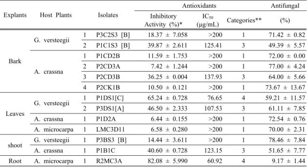
3.2. Antioxidant activity
In this study, 50 endophytic fungal isolates were evaluated for antioxidant activity. The results revealed that among 50 isolates, six had ability to reduce the absorbance of DDPH free radical with the range was 36% - 82% (Table 1); P1C1S3 (39.87 ± 2.61), P2CD3B (36.25 ± 0.01), P1DS1[C] (65.25 ± 0.73), P3DS1[A]
(46.5 ± 2.33), P1B1C (40.67 ± 0.73), and R2MC3A (82.08 ± 5.99). Further analysis (Table 1) indicated that the ethyl acetate extracts from two isolates, R2MC3A and P1DS1[C] had IC
50of 60.92 µg/mL and 76.65 µg/mL, fall into the range of strong category (50-100 µg/mL). Whereas other isolates those had IC
50fall under the range of medium category (100-150 µg/mL). The ethyl acetate extract of R2MC3A resulted in higher yield, 0.06% (w/v) or 11.97% w/w of dried mycelia, than of P1DS1[C] (Table 2). These results indicated that the highest potential antioxidant was generated from endophytes isolated from fresh leaves (P1DS1[C]) and roots (R2MC3A) of G. verstegii and A. microcarpa trees.
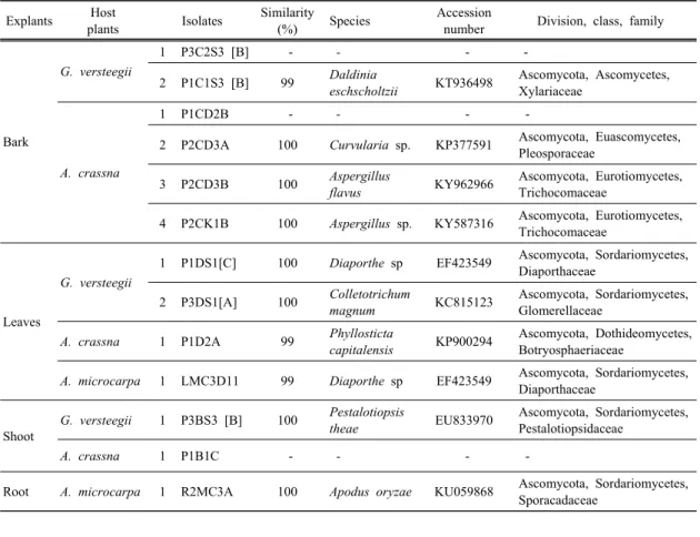
Molecular identification clearly identified Diaporthe sp. (P1DS1[C]) and Apodus oryzae. (R2MC3A), both have IC
50less than 80 µg/mL (Table 3). Interestingly, Diaporthe sp. has been known as a genus of plant pathogenic, saprobic and endophytic fungi (Gomes et al., 2013); while Apodus oryzae (R2MC3A) has been known as aminor endophytic and rhizospheric fungi associated with several plants (Cai et al., 2006; Zhou et al., 2015). In contrast, Apodus oryzae isolate (R2MC3A) showed greater scavenging activity (IC
5060.92 µg/mL) than Diaporthe sp. (P1DS1[C]) isolate.
This study is the first report to discover that Apodus
oryzae (R2MC3A) and Diaporthe sp. played as
endophytic fungi which resided in the leaves tissue of
Explants Host Plants Isolates
Antioxidants Antifungal
Inhibitory Activity (%)*
IC
50(μg/mL) Categories** (%)
Bark
G. versteegii 1 P3C2S3 [B] 18.37 ± 7.058 >200 1 71.42 ± 0.82
2 P1C1S3 [B] 39.87 ± 2.611 125.41 3 49.39 ± 5.57
A. crassna
1 P1CD2B 11.59 ± 1.753 >200 1 72.00 ± 0.00
2 P2CD3A 7.42 ± 1.244 >200 1 77.00 ± 4.24
3 P2CD3B 36.25 ± 0.004 137.93 3 64.00 ± 5.66
4 P2CK1B 10.50 ± 0.121 >200 1 73.67 ± 13.67
Leaves
G. versteegii 1 P1DS1[C] 65.24 ± 0.728 76.65 4 59.21 ± 11.57
2 P3DS1[A] 46.50 ± 2.333 107.53 3 61.11 ± 7.85
A. crassna 1 P1D2A 6.44 ± 0.155 >200 1 72.54 ± 0.76
A. microcarpa 1 LMC3D11 6.58 ± 0.280 >200 1 70.00 ± 2.31
shoot G. versteegii 1 P3BS3 [B] 14.44 ± 3.611 >200 1 78.46 ± 7.84
A. crassna 1 P1B1C 40.60 ± 0.728 123.15 3 51.65 ± 7.77
Root A. microcarpa 1 R2MC3A 82.08 ± 5.990 60.92 4 9.17 ± 1.44
Note: * concentration of fungal crude extract (100 mg/L);
** 1 = very weak, 2 = weak, 3 = medium, 4 = strong, 5 = very strong
Table 1. Antioxidant and antifungal activity of thirteen potential endophytic fungi
G. verstegii and the roots of A. microcarpa. Previous studies reported that only Fusarium, Colletotrichum, Xylaria, Lasiodiplodia, Phaeoacremonium, have been isolated from A. sinensis, A. crassna andA malaccensis (Gong and Guo, 2009; Nimnoi et al., 2010; Shoeb et al., 2010; Cui et al., 2011; Chi et al., 2016). Diaporthe sp. has been previously described as endophyte and produce bioactive secondary metabolites which are antioxidant potential. However, antioxidant activity between isolates varies greatly. For example, Diaporthe sp isolated from Costus spiralis have shown antioxidant activity (IC
50) between 500-1750 µg/mL (Marson Ascêncio et al., 2014), whereas IC
50of 76.65 µg/mL were obtained from Diaporthe sp (P1DS1[C]) isolated from leaves tissue of G. verstegii. Diaporthe sp. was reported to produce bioactiverelated to phenolic com- pounds, such as benzene acetaldehyde, benzyl benzoate, salicylaldehyde, benzoin and benzyl cinnamate (Tanapichatsakul et al., 2018).
Other interesting finding is the antioxidant capacity of Apodus sp. Apodus sp. was previously reported to be found in Hedyotis diffusa and Pseudotsuga menziesii (Zhou et al., 2015; Daniels, 2017). However, the information on its antioxidant activity was not yet investigated. The antioxidant activity of Apodus oryzae (R2MC3A), among other isolates in this study, was the highest (60.92 µg/mL) and confirmed to contain a phenolic compound (Table 2); achieving the total phenolic content (TPC) 96.10 mg gallic acid equivalent per g sample (data not shown here). The detection of TPC content clearly supported other findings on antioxidant capacity closely related with TPC (Sadeghi et al., 2015; Jeong et al., 2017; Hidayat et al., 2018;
Kim et al., 2018). Although, the specific phenolic
compound was not yet identified, the phenolic content
itself is important due to its unique structure for redox
property which acts as high reducing agents, hydrogen
donators, and singlet oxygen quenchers which may lead
Explants Host
Plants Isolates
Mycelium Weight (mg/L)
Yield of Ethyl Acetate Soluble
Extracts
Phytochemical Properties
% (w/v)*
% (w/w)**
Alkaloid
Flavonoid* Fenol* Terpenoid Steroid Mayer Wagner
Bark
G. versteegii
1 P3C2S3 [B] 6,354 0.02 3.02 - - + + + -
2 P1C1S3 [B] 59,813 0,07 1.14 - - + + - +
A. crassna
1 P1CD2B 9,957 0,05 4.86 - - - + + -
2 P2CD3A 9,083 0,04 4.36 - - + - + +
3 P2CD3B 5,850 0,03 4.99 - - - + + -
4 P2CK1B 5,570 0,03 6.13 - - + + + -
Leaves
G. versteegii
1 P1DS1[C] 7,461 0.03 4.66 - - + + + -
2 P3DS1[A] 6,082 0.06 10.06 - - + + + -
A. crassna
1 P1D2A 4,315 0,06 13.52 - - + - + -
A. microcarpa 1 LMC3D11
10,395 0.02 2.20 - - - + - +
Shoot
G. versteegii1 P3BS3 [B] 6,936 0.03 3.81 - - + - + -
A. crassna
1 P1B1C 3,069 0,04 12.49 - - - + + -
Root
A. microcarpa 1 R2MC3A4,691 0.06 11.97 - - - + - +
Noted: * calculated with volume of liquid medium, ** calculated with mycelium weight, + = indicates presence, - = indicates absence
Table 2. Biomass, yield and phytochemical analyisis of crude extract etyl acetate soluble of 13 isolates endophytic fungi after fermentation at 14 days
to antioxidant activity (Khoddami et al., 2013; Kim et al., 2017).
3.3. Antifungal against F. solani activity
Antifungal activity tested by means of dual culture/confrontation against F. solani. Fusarium was recorded as a significant plant pathogen and an inducer of agarwood formulation (Sitepu et al., 2011; Turjaman et al., 2016). This pathogenic fungi was commonly used to induce agarwood formation and one of the most effective agent for agarwood formation in Indonesia (Turjaman et al., 2016). The effectiveness of agarwood formation was affected by many factors such as host tree, type of fungal pathogen, and environment (Santoso, 2013), as well as the presence of endophytic fungi inside the host tree (Nimnoi et al., 2011; Zhang et al., 2014).
In this study, 50 endophytic fungi showed variation
in the capability to inhibit the growth of F. solani (9-78%). Seven isolates inhibited the growth of F. solani over than 70%, (Table 1), those were P2CK1B (73%), P1D2A (72%), P1CD2B (72%), P2CD3A (77%), P3BS3[B] (78%), P3C2S3[B] (71%), and LMC3D11 (70%). Most above-ground tree isolates had strong antifungal activity against F. solani, while R2MC3A, which was originated from the root, had a weak antifungal activity. It revealed that R2MC3A was not compatible to work against F. solani, which produced symptoms on the infected root (Sitepu et al., 2011;
Turjaman et al., 2016). More detailed observation on
tissue origin and host plant species revealed that the
strongest inhibitions (> 75%) were originated from shoot
(P3BS3[B]) and bark (P2CD3A) of G. verstegii and
A. crassna trees. According to molecular identification
(Table 3), isolate P3BS3[B] and P2CD3A were clearly
identified as P. theae and Curvularia sp.
Explants Host
plants Isolates Similarity
(%) Species Accession
number Division, class, family
Bark
G. versteegii
1 P3C2S3 [B] -
-- -
2 P1C1S3 [B] 99
Daldiniaeschscholtzii
KT936498 Ascomycota, Ascomycetes, Xylariaceae
A. crassna
1 P1CD2B -
-- -
2 P2CD3A 100
Curvularia sp.KP377591 Ascomycota, Euascomycetes, Pleosporaceae
3 P2CD3B 100
Aspergillusflavus
KY962966 Ascomycota, Eurotiomycetes, Trichocomaceae
4 P2CK1B 100
Aspergillus sp.KY587316 Ascomycota, Eurotiomycetes, Trichocomaceae
Leaves
G. versteegii
1 P1DS1[C] 100
Diaporthe spEF423549 Ascomycota, Sordariomycetes, Diaporthaceae
2 P3DS1[A] 100
Colletotrichummagnum
KC815123 Ascomycota, Sordariomycetes, Glomerellaceae
A. crassna
1 P1D2A 99
Phyllostictacapitalensis
KP900294 Ascomycota, Dothideomycetes, Botryosphaeriaceae
A. microcarpa
1 LMC3D11 99
Diaporthe spEF423549 Ascomycota, Sordariomycetes, Diaporthaceae
Shoot
G. versteegii
1 P3BS3 [B] 100
Pestalotiopsistheae
EU833970 Ascomycota, Sordariomycetes, Pestalotiopsidaceae
A. crassna