Original Article
원고 접수일 2011년 9월 7일, 원고 수정일 2011년 9월 27일, 게재 확정일 2011년 10월 3일
책임저자 손동석
(705-718) 대구시 남구 대명4동 3056-6, 대구가톨릭대학교병원 치과 구강악안면 외과
Tel: 053-650-4291, Fax: 053-622-7067, E-mail: [email protected]
RECEIVED September 7, 2011, REVISED September 27, 2011, ACCEPTED October 3, 2011
Correspondence to Dong-Seok Sohn
Department of Dentistry and Oral and Maxillofacial Surgery, Department of Dentistry, Daegu Catholic University Medical Center 3056-6, Daemyung-4-dong, Nam-gu, Daegu 705-718, Korea Tel: 82-53-650-4291, Fax: 82-53-622-7067, E-mail: [email protected]
CC This is an open access article distributed under the terms of the Creative Commons Attribution Non-Commercial License (http://creativecommons.org/licenses/
by-nc/3.0) which permits unrestricted non-commercial use, distribution, and reproduction in any medium, provided the original work is properly cited.
하악 제3대구치와 하악 우각부 골절과의 상관관계
유석현ㆍ이형주ㆍ문지원ㆍ손동석
대구가톨릭대학교병원 치과 구강악안면외과
Abstract
A Correlation between Mandibular Angle Fracture and the Mandibular Third Molar
Seok-Hyun Yu, Hyung-Ju Lee, Jee-Won Moon, Dong-Seok Sohn Department of Dentistry and Oral and Maxillofacial Surgery, Department of Dentistry, Daegu Catholic University Medical Center
Purpose: This study evaluated correlation and risk factors between position of the mandibular third molars and mandibular angle fractures using clinical and radiographic findings.
Methods: Medical records and panoramic radiographs of 188 patients with mandibular fractures were retrospectively reviewed.
The presence and position of the third molars were assessed for each patient and were related to the occurrence of mandibular angle fractures.
Results: The incidence of mandibular angle fracture was found to be greater when a lower third molar was present, particularly at the occlusal plane positioned on the 2nd molar occlusal surface (by Archer system) and the third molar is impacted in mandibular ramus (by Pell & Gregory system). Of the 192 sites with a lower third molar, 32 (16%) had an angle fracture.
Of the 184 site without lower third molars, 16 (8%) had an angle fracture.
Conclusion: This study confirmed an increased risk of angle fractures in the presence of a lower third molar as well as variable risk for angle fracture, depending on positioning of the third molar.
Key words: Mandibular angle fracture, Mandibular third molar
서 론
하악골은 해부학적 구조상 다른 안면골에 비해 상대적으로 돌출되어 있기 때문에, 골절이 빈번하게 발생한다[1]. Lee 등[2]은
악안면부 골절의 발생부위별 진도를 조사하여 하악골이 349예로 가장 많았고 관골 및 관골중, 상악골, 비골 순으로 나타났다고 보고하였다. Ugboko 등[3]은 안면 골절 치료를 받은 환자의 64%
에서 하악골 골절이 나타난다고 하였으며 특히 Azevedo 등[4]은
Fig. 1. Pell - Gregory classification; Depth of impacted mandibular 3
rd molar.치아의 존재 여부 등 여러 가지 요인에 영향을 받을 수 있으며[7], Neal 등[8]은 하악골 골절의 50%가 치아와 연관되어 나타난다고 보고하기도 하였다. 하악 우각부의 경우 하악골의 이부나 과두 하부에 힘이 가해져도 쉽게 응력이 집중되므로 골절 가능성이 높다[9]. 세부적인 부위별 발생빈도를 보면 Kruger와 Schilli[10]
은 이부, 과두부, 우각부의 순으로 발생빈도가 높다고 하였고, Kelly와 Harrigan[11]은 우각부위가 최대 호발부위라고 보고하였 다. Haug 등[12]은 하악 우각부 골절이 전체 하악골 골절의 약 40%를 차지한다고 보고하였다.
하악 골절에 영향을 미치는 요소 중 하나인 하악 제3대구치는 흔히 매복되어 있고 존재 유무와 존재 양상에 따라 하악 우각부 골절에 영향을 미칠 수 있다. Tevepaugh와 Dodson[13]은 제3대 구치가 존재하는 환자에서 그렇지 않은 환자에 비해 우각부 골절 이 2.8배 호발한다고 보고하였다. Safdar과 Meechan[14] 역시 제3대구치의 위치와 하악골 우각부 골절 위험성이 상관관계가 있다고 주장하였다. 또한, 하악 제3대구치를 포함한 하악 우각부 골절에서 골절 부위의 정복 시, 하악 제3대구치의 발치 여부는 술 후 감염에도 영향을 준다. 우각부 골절 시 제3대구치가 매복되 어 있으면 치주인대를 통해 구내, 구외가 서로 교통하게 되고 치수는 괴사될 수 있으며 이것이 복합적으로 작용해 감염과 같은 합병증을 유발할 수 있다[15]. 이 같은 합병증을 고려해 최근에는 하악 우각부 골절과 관련된 하악 제3대구치가 심하게 변위된 경 우, 치근단이 노출된 경우, 치주염이나 지치주위염에 이환된 경우 등에서 선택적으로 치아를 발거하는 것이 추천되고 있다[16].
이와 관련해 본 연구에서는 본원에 내원한 우각부 골절 환자를 대상으로 하악 제3대구치의 위치와 맹출 양상에 따른 하악 우각부 골절의 발생 빈도와의 관계를 분석하였으며 골절편 정복 시 하악 제3대구치의 발치 여부에 따른 술 후 감염에 대해 조사하여 골절
1. 연구대상
이 연구는 2007년 1월부터 2009년 12월까지 대구가톨릭대학 교병원 치과 구강악안면외과에 내원한 하악골 골절 환자 188명의 진료 기록지 및 파노라마 방사선 사진을 토대로 조사하였다.
2. 연구방법
1) 성별, 연령 및 골절 원인별 분류
진료 기록지를 토대로 연구 대상자를 성별, 연령 및 골절 원인별 로 분류하였다. 상해의 원인은 교통사고, 폭행, 낙상, 넘어짐, 운동에 의한 외상 및 기타로 구분하였다.
2) 하악 제3대구치의 교합평면과의 관계
하악 제3대구치의 교합 평면과의 관계는 Archer의 분류법을 따랐다[17]. 교합 평면은 하악 제1, 2대구치의 교합면을 이은 선으 로 정하였다. 제3대구치의 최상방점이 하악 제2대구치의 교합면 수준인 경우를 class A, 하악 제3대구치의 교합면이 제2대구치 교합면과 백악법랑경계 사이에 존재하는 경우를 class B, 하악 제3대구치의 교합면이 제2대구치의 백악법랑경계 하방에 있을 때를 class C라고 분류하였다(Fig. 1).
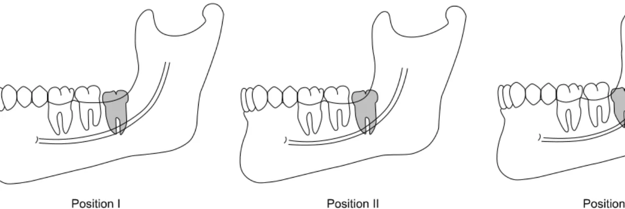
3) 하악 제3대구치의 하악지 전방부와의 관계
하악 제3대구치의 하악지 전방부와의 관계는 Pell과 Gregory 의 분류법을 따른다[18]. Level I은 하악지의 전방부와 하악 제2대 구치 사이에 하악 제3대구치가 맹출할 수 있는 충분한 공간이 있는 경우이다. Level II는 공간이 불충분한 경우이다. Level III은 하악 제3대구치가 대부분 하악지에 덮혀 있는 경우이다(Fig.
2).
Fig. 3. Angulation of mandibular 3
rd molar. Clockwise from top left:Mesioangulated, Horizontal, Ver- tial, and Distoangulated.
Fig. 2. Pell - Gregory classification; Amount of bone covering mandibular 3
rd.4) 하악 제3대구치의 경사도
하악 제3대구치의 경사도는 Shiller의 연구 및 Winter의 분류 에 따라 근심경사, 원심경사, 수직경사, 수평경사로 나눈다 [19,20]. 교합 평면과 제3대구치의 교합면이 이루는 각으로 ±10 도 이내를 수직 경사, ±11∼70도를 근심 혹은 원심 경사, 70도 이상을 수평경사로 분류하였다(Fig. 3).
5) 하악 제3대구치의 매복정도
임상적으로 하악 제3대구치의 매복 정도는 3개의 그룹으로 분류하였다. 구내 점막을 천공시키지 못한 경우를 미맹출, 구내 점막을 천공시키고 부분적으로 교합면 등을 노출시킨 경우를 부분 맹출, 인접치와 흡사하게 교합면이 맹출한 경우를 맹출로 분류하
였다.
우각부 골절이 있는 환자 중 제3대구치가 있는 경우는 골절과 관련된 쪽의 제3대구치 상태를 분석하였고 우각부 골절이 있으나 골절부 반대측에만 제3대구치가 있는 경우는 제3대구치가 없는 것으로 간주하였다. 진료 기록지상 외력이 가해진 부위가 명확하 게 기록된 경우는 거의 없었으므로 이 부분에 대한 분류 및 검토는 생략하였다.
통계적 분석은 SPSS statistics, version 17.0 program을 사용
하였다. chi-square로 통계적 유의성을 검증하였으며, P value
값이 0.05 미만인 경우 통계적 유의성을 가지는 것으로 판단하였
다.
Table 1. Mandibular angle fracture associated with presence and
absence of mandubular third molarThird molar Mandibular angle fracture
Total
Present Absent
Present 32 160 192
Absent 16 168 184
Total 48 328 376
x2=5.361, P <0.05.
Table 2. Mandibular angle fracture associated with occlusal plane
of mandibular third molar positionOcclusal position
Mandibular angle fracture
Total Relative Present Absent risk
Class A 10 (11%) 83 (89%) 93 1
Class B 15 (20%) 60 (80%) 75 1.8
Class C 7 (30%) 17 (70%) 24 2.7
Total 32 (17%) 160 (83%) 192
x2=3.086, P >0.05.
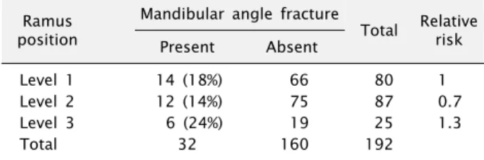
Table 3. Mandibular angle fracture associated with ramus space
of mandibular third molar positionRamus position
Mandibular angle fracture
Total Relative Present Absent risk
Level 1 14 (18%) 66 80 1
Level 2 12 (14%) 75 87 0.7
Level 3 6 (24%) 19 25 1.3
Total 32 160 192
x2=5.486, P >0.05.
Table 4. Mandibular angle fracture associated with mandibular
third molar angulationAngulation Mandibular angle fracture
Total Relative Present Absent risk
Mesial 13 (18%) 60 (82%) 73 1
Vertical 17 (15%) 99 (85%) 116 0.8
Distal 2 (67%) 1 (33%) 3 3.7
Horizontal 0 (0%) 0 (0%) 0 0
Total 32 160 192
x2=5.486, P >0.05.
총 인원은 188명(376부위)이었으며 이 중에서 남자가 152명 (80%), 여자가 36명(20%)이었다. 전체 환자 중 47명(94부위)이 하악 우각부 골절을 보였고 이 중에서 남자가 41명(87%), 여자가 6명(13%)이었다.
연령별 분포를 살펴보면 전체 골절 환자는 20∼29세(24%)에 서 가장 호발하는 것으로 나타났고, 하악 우각부 골절 환자 역시 20∼29세(37%)에서 호발하는 것으로 나타났다. 전체 환자에 비 해서 우각부 골절 환자에서 20대가 차지하는 비율이 더 많은 것으로 나타났다.
하악골 골절의 원인별 분포는 교통사고 88명(47%), 폭행 32명 (17%), 낙상 20명(11%), 넘어짐 26명(14%), 스포츠 상해 6명 (3%), 기타 16명(8%)의 순으로 나타났다. 하악 우각부 골절을 보인 환자 중에서는 교통사고 12명(25%), 폭행 17명(37%), 낙상 1명(2%), 넘어짐 9명(19%), 스포츠 상해 2명(4%), 기타 6명 (13%)이었다.
2. 하악 제3대구치의 유무에 따른 하악 우각부 골절의 유무 전체 환자 188명 중 골절이 있는 부위는 총 376부위이며, 그 중 제3대구치가 있는 192부위(51%)에서 우각부 골절이 있는 경 우는 32부위(67%)이며, 제3대구치가 없는 184부위(49%) 중 우 각부 골절이 있는 경우는 16부위(33%)로 하악 제3대구치가 존재 하는 경우 우각부 골절의 발생 빈도가 높았으며 통계적으로 유의 성을 나타내었다( P <0.05)(Table 1).
골절의 유무를 살펴보면 32부위의 하악 제3대구치를 가진 하악 우각부 골절부 중 class B 군이 15부위로 가장 많은 수를 차지하였 다. 발현 빈도로 살펴보면 Class C의 제3대구치를 가진 24부위 중 7부위(30%)에서 우각부 골절이 발생하여 상대적인 위험도가 높게 나타났으나 통계적으로 유의성을 나타내지는 않았다(Table 2).
4. 하악 제3대구치의 하악지 전방부와의 관계에 따른 하악 우각부 골절의 유무
하악지 전방부와의 관계에서는 총 32부위의 하악 제3대구치를 가진 하악 우각부 골절 부위 중 Level I군이 14개로 가장 많았으나 발현 빈도로 분석해보면 상대적인 위험도는 Level III군에서 가장 높게 나타났으나 통계적으로 유의성을 나타내지는 않았다(Table 3).
5. 하악 제3대구치의 매복 경사도에 따른 하악 우각부 골절 의 유무
하악 제3대구치의 매복 경사도에 따른 하악 우각부 골절에서는
총 32부위의 하악 제3대구치를 가진 하악 우각부 골절 부위 중
수직 경사가 17개로 가장 많이 나타났고 그 다음으로 근심 경사,
원심 경사의 순으로 나타났다. 상대적인 위험도가 원심경사에서
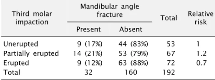
Table 5. Mandibular angle fracture associated with mandibular
third molar impactionThird molar impaction
Mandibular angle
fracture Total Relative Present Absent risk
Unerupted 9 (17%) 44 (83%) 53 1
Partially erupted 14 (21%) 53 (79%) 67 1.2
Erupted 9 (12%) 63 (88%) 72 0.7
Total 32 160 192
x2=1.325, P >0.05.
Table 6. Post-operative infection associated with extraction and
non-extraction case of mandibular third molarPost-operative infection
Total Present Absent
Extraction 6 6 12
Non-extraction 0 20 20
Total 6 26 32
x2=12.308, P <0.05.
높게 나타났으나 통계적으로 유의성을 나타내지는 않았다(Table 4).
6. 하악 제3대구치의 매복 정도에 따른 하악 우각부 골절의 유무
하악 제3대구치의 매복 정도에 따른 하악 우각부 골절에서는 총 32부위의 하악 제3대구치를 가진 하악 우각부 골절 부위 중 부분 맹출이 14개로 가장 많이 나타났고 그 다음으로 미맹출, 완전 맹출이 동일한 수로 나타났다. 상대적인 발생 빈도에서도 부분 맹출이 가장 높게 나타났으나 통계적으로 유의성을 나타내지 는 않았다(Table 5).
7. 하악 제3대구치의 발치 여부에 따른 술 후 감염의 유무 32부위의 하악 제3대구치를 가진 골절된 하악 우각부 중 발치 를 시행한 12부위 중 6부위에서 감염이 발생하였고, 발치를 시행 하지 않은 20부위 중에서는 감염이 발생하지 않았다(Table 6).
고 찰
본 연구에서는 하악 제3대구치의 위치와 맹출 양상을 알아보고 하악 우각부 골절의 발생 빈도와의 관계를 분석하였다. 먼저 성별 에 따른 분포를 살펴보면 전체 골절 환자에서는 남성이 여성에 비해 4배 가량 호발하였고, 우각부 골절 환자에서는 남성이 6.7배 가량 호발하는 것으로 나타났다. 이러한 남성 호발 경향은 Park 등[21], Han과 Yoon[22]의 성별 연구에서도 비슷하게 나타났다.
연령에 따른 분포를 살펴보면 전체 골절 환자와 우각부 골절 환자 모두 20대(20∼29세)에서 가장 호발하는 것으로 나타났다.
일반적으로 20대의 남성은 사회 활동의 빈도나 신체적 활동이 왕성한 시기로 외상에 대해 노출될 확률이 상대적으로 높은 것이 이 같은 결과의 원인이라 생각된다. 또한 20대에는 제3대구치가 존재할 가능성이 많은 연령이고 매복되어 있을 가능성도 높으므로 우각부 골절 빈도가 높게 나타난다고 할 수 있다.
하악 우각부 골절빈도와 하악 제3대구치의 존재 유무와의 상관 관계에 대해서는 대부분의 연구에서 어느 정도 의견의 일치가
있다[23-27]. 본 연구의 결과에서도 제3대구치의 존재시 우각부 골절 발생률이 높은 경향을 보였고 존재하지 않을 시 발생률이 낮은 경향을 보였다. 이것은 통계적으로도 유의성이 있는 것으로 나타났으므로 이런 결과를 통해 하악 제3대구치가 하악 우각부 골절에 있어서 위험인자가 될 수 있다고 생각된다.
하악 제3대구치의 교합 평면과의 관계에 따른 하악 우각부 골절은 class C>class B>class A 순으로 나타났다. Halmos 등[28], Lee와 Dodson[29]의 연구에서는 하악 제3대구치의 위치 는 우각부 골절의 중요한 위험 인자일 뿐 매복 깊이에 따라 골절 발생률이 좌우되지는 않는다고 하였다. 그러나 본 연구에서는 매복 깊이가 깊을수록 우각부 골절 발생률이 높게 나타났으며 이것은 Safdar와 Meechan[14]의 연구에서도 비슷하게 나타났다.
하악지 전방부와 제3대구치와의 관계에서는 Level 3에서 우각부 골절 발생 확률이 가장 높게 나타났는데 이 두 범주의 공통점은 하악 제3대구치가 골내에서 차지하는 부피가 크다는 것이다. 이러 한 결과로 볼 때 우각부 골 면적이 감소하게 되면 골절 발생 가능성이 높아진다고 생각된다.
제3대구치 장축이 이루는 각도별로 보면 수직경사인 경우 빈도 가 가장 많이 나타났고 원심경사된 경우에서 상대적인 위험도가 높게 나타났다. 그러나 우각부 골절 유무와 관련 없이 전체 대상범 위 중 제3대구치의 원심경사를 보인 경우는 3부위에 불과하며 통계적으로 유의성을 나타내지 않아 의미 있는 결과라고 볼 수는 없었다.
악골 골절선에 치아가 포함된 경우, 골절편 정복 시 치아의 맹출 상태 및 발치 여부는 술 전, 술 후 감염에 영향을 미칠 수 있다. 해당 치아의 치주인대를 통해 구강과 개통이 될 수 있으므로 치수, 치주, 치근단 병변과 복합적으로 작용하여 골절 치유에 있어 합병증이 발생할 수 있다[15]. 이에 대해 Thoma[30]
는 골수염, 비유합, 유합지연 등의 합병증을 예방하기 위해서 즉시 발치를 시행해야 한다고 하였으며 이와 반대로 Wagner 등[31]은 해당 치아가 골절편을 안정화시키는 역할을 담당할 수 있으므로 골 치유과정이 끝난 후 발치하는 것을 추천하였다.
Schonberger[32]는 이와 관련된 연구에서 치아를 보존한 환자가
18%의 감염을 보이고 치아를 발치한 환자에서 6%의 감염을 보여
골절선을 포함하는 치아는 반드시 발치해야 한다고 하였다. 이와
는 감염이 발생하지 않았다. 6개의 감염 증례를 살펴보면, 관혈적 정복술 시행과 동시에 발치한 경우가 3개, 환자 내원 직후 발치하 고 지연 수술을 시행한 경우가 2개, 환자 초진 시 치주염으로 인해 해당 제3대구치의 sulcus에서 pus가 나온 경우가 1개였다.
Pus가 발생한 경우 역시 즉시 발치를 시행하였다. 발치를 한 경우와 하지 않은 경우에서 통계적으로 유의성이 있다고 판단되어 발치를 한 경우 감염의 위험성이 높다고 생각된다. 발치 증례에서 는 발치로 인해 발생된 공간을 완전히 닫는 것에 한계가 있으므로 고정시킨 plate와 screw 주변이 감염에 노출될 가능성이 높아진 다. 또한 관혈적 정복술 후에도 악간 고정기간이 있으므로 양호간 감염 관리가 이뤄지기 어렵다는 것이 감염을 발생시킨 원인이라 생각된다. 따라서 James 등[33]과 Ellis[34]의 기준에 따라 치아에 심한 치아 동요가 있거나 치근이 파절된 경우, 치근단 병소가 존재하거나 골절선 상의 치아가 골절편 정복을 방해하는 경우에 선택적으로 제3대구치를 발치하는 것이 좋겠다.
결 론
1. 하악 제3대구치가 존재하고 맹출 공간이 부족할 때 하악 우각부 골절이 일어날 확률은 높아진다.
2. 외력에 많이 노출되는 20대 남성에서 하악 우각부 골절이 호발하며 골절 예방 차원에서도 매복된 하악 제3대구치를 발치하 는 것이 추천된다.
3. 하악 우각부의 정복 시에는 하악 제3대구치가 동요도, 파절, 감염, 위치 이상 등을 나타낼 때 선택적으로 발치하는 것이 좋다.
4. 하악 제3대구치의 발치가 필요한 우각부 골절환자의 경우에 는 관혈적 정복술을 시행할 때 발치를 시행하고, 관혈적 정복술을 시행할 수 없는 경우에는 발치만을 시행하여 술 후 감염을 예방하 는 것이 추천된다.
References
![Fig. 1. Pell - Gregory classification; Depth of impacted mandibular 3 rd molar.치아의 존재 여부 등 여러 가지 요인에 영향을 받을 수 있으며[7], Neal 등[8]은 하악골 골절의 50%가 치아와 연관되어 나타난다고 보고하기도 하였다](https://thumb-ap.123doks.com/thumbv2/123dokinfo/5114528.331043/2.892.83.591.913.1119/classification-mandibular-치아의-요인에-영향을-연관되어-나타난다고-보고하기도.webp)