하악 제 3대구치의 맹출 양상과 제 2대구치의 후방 치아우식과의 상관관계
640
Ⅰ. 서 론
제3대구치의 외과적 발거는 구강악안면영역에서 이루어지 는 가장 많은 수술 중 하나이다.1)제3대구치는 지치주위염, 감 염, 악골골절, 낭성종양, 제2대구치우식 등을 일으키는 원인이 될 수 있다.2-6)제3대구치로 인한 지치주위염의 경우 제3대구치 의 발거로 치료가능하고, 제3대구치로 인한 농양의 경우는 절 개 및 배농과 항생제의 사용으로 치료가능 하다. 그러나 이 중 제2대구치의 우식의 경우는 그 치료가 제3대구치 발치에 그치 는 것이 아니라 제2대구치 우식 치료, 심하게는 제2대구치의 발치까지 이를 수 있게 되어 환자에게 큰 저작계의 손실을 일 으킬 수 있다. 조기에 제2대구치의 원심우식의 위험인자를 알 아내어 제2대구치가 제3대구치에 의해 위험을 받게되는 경우 라면 예방적 발치를 권유하는 것도 좋은 치료방법이 될 수 있 다.
하악 제2대구치 원심 우식과 제3대구치의 관계에 대한 연구 는 2005년에 Louis W. McArdle7)이 발표한 연구가 유일하다. 그 는 하악 제2대구치 원심 우식을 주소로 제3대구치를 발거한
100명의 122치를 대상으로 조사하였으며, 조사 치아 중 82%가 원심으로 40-80의 기울기를 보였다고 보고하였다. 이 연구는 하악 제2대구치의 원심 우식과 제3대구치의 위치와의 관계를 설명을 했다는데 그 의의가 있겠다. 하지만 비우식군과의 관 계를 설명하지 않았을 뿐 아니라, 그가 제시한 40-80도 근심경 사 제3대구치의 발거의 기준은 참고하기에는 그 범위가 너무 넓고, 다양한 제3대구치의 매복양상과 접목시키기 어려운 한 계가 있다. 따라서 다양한 제3대구치 매복양상과 제2대구치와 제3대구치와의 관계를 포함한 정확하고 적용하기 용이한 제3 대구치의 발거의 기준이 필요하다.
본 연구의 목적은 파노라마방사선 사진을 이용하여 하악 제 2대구치와 제3대구치의 위치관계가 하악 제2대구치 원심치경 부 우식과의 상관관계를 파악하고, 제2대구치 원심 비우식군 과 비교하여, 제2대구치 원심면 치아우식의 위험군의 파악을 통해 추후 하악 제2대구치의 우식예방을 위한 제3대구치의 예 방적발거의 이론적 배경을 제시함에 있다.
Ⅱ. 연구 대상 및 방법 1. 연구 대상
2002년 3월부터 2007년 12월까지 삼성서울병원 구강악안면 외과에서 파노라마 방사선 사진 촬영 후 하악 제3대구치를 발 거한 786명의 883개의 제3대구치를 연구대상으로 의무기록을 통해 후향적 코호트 연구를 시행하였다. 152개는 제2대구치의
하악 제 3대구치의 맹출 양상과 제 2대구치의 후방 치아우식과의 상관관계
이명환∙설정은∙장인걸∙홍종락∙김창수 성균관대학교 의과대학 삼성서울병원 구강악안면외과
Abstract (J. Kor. Oral Maxillofac. Surg. 2008;34:640-643)
CORRELATION OF DISTAL CARIES IN THE MANDIBULAR SECOND MOLAR AND ERUPTION STATE OF THE MANDIBULAR THIRD MOLAR.
Myeong-Hwan Lee, Jung-Eun Seol, In-Gul Jang, Jongrak Hong, Chang-Soo Kim Department of Oral & Maxillofacial Surgery, Samsung Medical Center
Sungkyunkwan University, School of Medicine, Seoul, Rep. of Korea
Distal caries of the second molar is common indication for the mandibular third molar surgery and there are no universally acceptable predictive cri- teria for distal caries of the second molar. To analyze the correlation of the distal caries of the second molar and the eruption state of the mandibular third molar using panoramic radiographs statistically and propose the acceptable guideline for preventive extraction of the mandibular third molar. 786 patients who were extracted the mandibular third molar from 2002 to 2006 at Samsung medical center were examined. The presence and absence of distal caries of mandibular second molar, age, gender, angulation, impaction degree, distance between distal cementoenamel junction of the second molar and mesial cementoenamel junction of the mandibular third molar were assessed. 79.6% of third molars had a mesial angulation of between 40�
and 80�. The mean age of third molar removal for distal caries of second molar was 33.86±9.81.
The prophylactic removal of a mesio-angular third molar about 40�and 80�could prevent distal cervical caries forming in the mandibular second molar.
홍 종 락
135-710 서울 강남구 일원동50
성균관의대 삼성서울병원 구강악안면외과 Jongrak Hong
Dept. of OMFS, Samsung Medical Center, Sungkyunkwan University School of Medicine, #50, Irwon-dong, Kangnam-gu, Seoul, 135-710, Rep. of Korea Tel: 82-2-3410-2420 Fax: 82-2-3410-0038,
E-mail: [email protected]
대구외지 2008;34:640-643
641
원심치경부 우식을 제3대구치 발거 후 치과용 거울을 이용하 여 확인하였고, 731개는 제2대구치의 원심치경부 우식이 없는 것을 제3대구치 발거 후 치과용 거울을 이용하여 확인하였다.
786명 중 309명은 남성이었고, 477명은 여성이었다. 연령은 14 세에서 75세까지 분포하였고, 평균 연령은 28.3세였다.
2.방법
파노라마방사선 사진과 진료기록지를 토대로 하여 제3대구 치 발거 후 제2대구치 원심우식의 유무를 확인하였고, 성별, 연 령(10대, 20대, 30대, 40대, 50대 이상), 제3대구치의 원심경사정 도(0�에서 20�, 21�에서 40�, 41�에서 60�, 61�에서 80�, 81�이 상), 제3대구치의 매복정도, 제2대구치 원심치경부와 제3대구 치의 근심치경부 사이의 거리를 측정하였다. 제3대구치의 원 심경사정도는 Shiller’s classification (1979)을 이용하여 제2대구 치의 교합면에서 수직인 선과 제3대구치의 교합면에서의 수 직선 사이의 각을 측정하였다(Fig.1). 제3대구치의 매복정도는 Pell-Gregory’s classification (1942)을 이용하였으며(Fig.2), 제2대 구치의 교합면과 치경부를 기준으로 제3대구치의 최상방점의 위치가 교합면 상방의 경우 Level A, 교합면과 치경부 사이의 경우 Level B, 치경부 하방인 경우 Level C로 분류하였다. 제2대 구치 원심면 치경부와 제3대구치 근심면 치경부 사이(1mm에 서 3mm, 4mm에서 6mm, 7mm에서 9mm, 10mm에서 12mm, 13mm 이상)으로 는 Leone’s classification(1986)을 이용하였으며 (Fig.3), 두 사이의 거리를 측정하였다. Pearson chi-square test로 각 변수의 유의성을 판단하였고 유의 수준(α)는 0.05로 설정하 였다.
Ⅲ. 결 과 1. 연령
제2대구치 원심치경부 우식의 호발 연령대로는 30대(57명, 37.5%), 20대(48명, 31.6%), 40대(32명, 21.1%), 50대 이상(10명 6.6%), 10대(5명, 3.3%) 순이었으며, 비우식군의 경우 20대(447 명, 61.1%), 30대(133명, 18.2%), 10대(84명, 11.5%), 40대(43명,
5.9%), 50대 이상(24명, 3.3%) 순이다. 우식군의 평균연령은 33.86±9.81세 이었으며, 비우식군의 연령은 27.11±8.64세 이 였다. 연령은 제2대구치 원심 우식과 유의한 상관관계를 보였 다 (p<0.001) (Table. 1).
2. 제3대구치의 근심경사각도
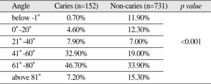
제2대구치 원심치경부 우식의 호발각도 순으로는 61�에서 80�(71치, 46.7%), 41�에서 60�(50치, 32.9%), 21�에서 40�(12치, 7.9%), 81�이상(11치, 7.2%), 0�에서 20�(7치, 4.6%), -1�이하(1 치, 0.7%) 순이었다. 비우식군의 분포로는 61�에서 80�(248치, 33.9%), 41�에서 60�(143치, 19.0%), 81�이상(112치, 15.3%), 0�에 서 20�(90치, 12.3%), -1�이하(87치, 11.9%), 21�에서 40�(51치, 7.0%) 순이었다. 제3대구치의 근심경사각도는 제2대구치 우식 과 유의한 상관관계를 나타내었다 (p<0.001) (Table. 2).
3. 제3대구치의 매복정도
제3대구치의 매복정도의 경우 제2대구치 원심치경부 우식 의 호발 순으로는 Level A(125치, 82.2%)로 그 대부분을 차지하 였고, Level B(27치, 17.8%), Level C(0치, 0%) 순이었다. 비우식 군의 경우, Level A(452치, 61.8%), Level B(252치, 34.5%), Level C(27치, 3.7%) 순이었다. 제3대구치의 매복정도는 제2대구치의 원심 우식과 유의한 상관관계를 나타내었다(p<0.001) (Table. 3).
4. 제2대구치의 원심치경부와 제3대구치의 근심치경부 사이의 거리
제2대구치 원심치경부 우식의 호발 순으로는 7mm에서 9mm(87치, 57.2%), 10mm에서 12mm(30치, 19.7%), 4mm에서 6mm(25치, 16.4%), 1mm에서 3mm(9치, 5.9%), 13mm 이상(1치,
0.7%) 순이었다. 비우식군의 경우 7mm에서 9mm(320치,
43.8%), 4mm에서 6mm(195치, 26.7%), 1mm에서 3mm(158치, 21.6%), 10mm에서 12mm(56치, 7.7%), 13mm 이상(2치, 0.3%) 순 이었다. 제2대구치의 원심치경부와 제3대구치의 근심치경부 사 이의거리는유의한상관관계를나타내었다(p<0.001) (Table. 4).
Fig. 1.Shiller’s classification Fig. 2.Pell-Gregory’s classification Fig. 3.Leone’s classification
하악 제 3대구치의 맹출 양상과 제 2대구치의 후방 치아우식과의 상관관계
642
Ⅳ. 고 찰
하악 제3대구치의 발거 후 합병증으로는 출혈, 피하기종, 악 관절증, 치조골염, 감염, 종창, 하치조신경손상, 인접치손상, 악 골골절 등이 있다8). 이에 제3대구치의 발거는 적응증의 경우를 명확히 진단하고 발치에 임하여, 발치 후 합병증의 발생을 줄 여야 할 것이다. Libersa P9)등의 연구에 의하면 750,000건의 발 치중에서 37건의 하악골 골절이 발생했다고 보고하였다. 이는 발생률이 0.0049%로 발생 가능성이 상당히 낮다고 하겠다.
Krimmel M10)등은 하악 제3대구치 발거 후 골절이 발생한 6명을 대상으로 조사한 결과, 42세에서 50세의 유치악 환자였다. 이 연구에 의하면 하악 제3대구치 발거 후 하악골 골절은 연령과 관련이 있다고 하였다. 치조골염의 경우, 문헌에 따라 발생률 이 1%에서 30%까지11,12)다양하다. 하치조신경의 손상의 경우 발생률은 문헌에 따라 0.4%에서 8.4%로 보고하고 있고13-16), 대 부분의 경우 정상적으로 회복되지만 0.1%에서 1.2%에서는 영 구적인 신경손상증상으로 지속적인 불편감을 나타낼 수 있다
13-16). 증상이 없는 하악 제3대구치의 예방적 발거 시 위의 합병
증이 발생할 경우 환자에게 저작계의 손실과 일상생활의 불편 감을 초래하게 된다. 이는 환자의 불편감에만 그치는 것이 아
니라 의료소송으로 이어질 수 있다. Hupp JR17)의 연구에 의하 면 치과의료소송 중 발치에 관한 건수가 증가하고 있다고 보 고하고 있다. 발치관련 소송 중 신경손상이 14%로 제일 많고 그 뒤로 잘못된 치아 발치가 14%로 같은 비율을 보였다. 이러 한 의료소송의 증가로 인한 의료인의 부담 또한 커질 수 있다.
그래서 여러 문헌들5,18,19)은 적절한 발치 guide-line들을 제시하고 발치에 대한 risk & benefit를 고려하여 발치에 임하기를 권유하 고 있으며, 또한 술 전에 환자에게 이러한 합병증들을 충분히 설명 후에 발치를 권하고 있다. 본 연구의 목적 중의 하나가 임 상에서 무증상의 하악 제3대구치 발치를 할 경우, 발치에 관한 적절한 근거를 제시하고자 함에 있다. 그리하여 환자의 발치 에 대한 동의는 물론 술자의 적절한 판단에 대한 기준을 제시 하여 환자의 합병증, 부작용을 줄이고 저작계의 효율 향상에 그 목적을 두는 것이다.
제2대구치의 원심면 우식이 발생할 가능성은 우식군에서 연 령이 높았는데, 이는 제3대구치의 발거가 지연되어 비우식군 에 비해 원심면 우식이 발생할 가능성이 더욱 높았기 때문이 며, 비우식군인 경우 증상없이 치과검진 후 예방적 발거를 권 고 받았으리라 추정된다. 또한 결과적으로 우식군에서 제3대 구치와 제2대구치의 사이가 7-9mm 정도의 치경부 거리에 40- 80�정도 경사로 치조골정에 가깝게 매복이 될 때, 가장 우식 확 률이 높았다는 것은 이경우에 치태나 음식물 잔사가 저류가 되었다는 의미로 해석될 수 있다.
인접면 우식의 경우 각 치아별 발생율의 차이가 있다. Meja`re I20)의 연구에 의하면, 21세를 기준으로 상악의 경우 경우 제1대 구치 원심면(18%), 제2소구치 원심면(17%) 제1대구치 근심면 (15%), 제1소구치 원심면(14%) 순이며, 평균 14.5% 이었고, 하 악의 경우 제1대구치 원심면(26%), 제1대구치 근심면(16%), 제 2대구치 원심면(14%), 제2소구치 원심면(13%) 순으로 평균 13%으로 보고하였다. 본 연구에서는 총 883개의 하악 제2대구 치를 관찰하였고 그 중 152개가 우식에 이환되어 있었다. 제2 대구치 원심면 우식 이환률은 17.21%로 Meja`re I20)의 연구결과 와 비교하여 볼 때 하악 제1대구치 원심면(26%) 다음으로 높은 이환률을 나타내고 있다. 이는 단순 우식증으로만 보더라도 높은 확률이며 제2대구치의 원심우식은 제3대구치와의 연관 성으로 볼 때, 그 영향은 제1대구치 원심면 우식보다 더 위험하 다고 볼 수 있다. 원심우식의 경우 class II 와동으로 수복을 하 여야 한다. 이는 수복 시 기술적 어려움이 있고 수복 후에도 수 복물의 변연부 누출, 수복물의 부피증가로 인한 중합 시 수축 증가, 변연 능선의 부재로 인한 교합 시 수복물에 응력의 증가 로 수복제의 파절 가능성이 높아진다. 이렇듯 그 치료와 유지 가 어려운 제2대구치의 인접면 우식을 예방하기 위해서 제3대 구치의 예방적 발거가 필요하다.
본 연구의 주제와 유사한 Louis W. McArdle7)의 연구 결과와 비교해 보면, 그의 연구에서는 하악 제2대구치 원심치경부우 식 환자의 평균 연령이 30세였으며, 그 범위는 16세에서 64세 였다. 본 연구의 평균 나이는 33.86±9.81세이고, 14세에서 75세 까지 분포하였다. 평균 연령의 경우 Louis W. McArdle7)의 연구 Table 1. Age comparison between Caries group and
Non-caries group
Caries (n=152) Non-caries (n=731) p value (Mean±SD) (Mean±SD)
Age 33.86±9.81세 27.11±8.64세 <0.001
Table 2.Shiller’s classification distribution
Angle Caries (n=152) Non-caries (n=731) p value
below -1� 0.70% 11.90%
0�-20� 4.60% 12.30%
21�-40� 7.90% 7.00% <0.001
41�-60� 32.90% 19.00%
61�-80� 46.70% 33.90%
above 81� 7.20% 15.30%
Table 3.Pell-Gregory’s classification
Level Caries (n=152) Non-caries (n=731) p value
Level A 82.20% 61.80%
Level B 17.80% 34.50% <0.001
Level C 0% 3.70%
Table 4.Leone’s classification
Distance Caries (n=152) Non-caries (n=731) p value
1-3mm 5.90% 21.60%
4-6mm 16.40% 26.70%
7-9mm 57.20% 43.80% <0.001
10-12mm 19.70% 7.70%
above 13mm 0.70% 0.30%
대구외지 2008;34:640-643
643
결과와 약 3년의 차이가 있었다. 비우식의 경우는 27.11±8.64 세로 약 5년 정도의 차이가 있었다. 하악 제2대구치의 근심경 사 각도의 경우 Louis W. McArdle7)의 연구 결과는 82%가 40�에 서 80�, 10%가 40�이하, 8%가 80�이상으로 보고 하였고, 본 연 구결과는 46.7%가 61�에서 80�, 32.9%가 41�에서 60�, 7.9%가 21�에서 40�, 7.2%가 81�, 4.6%가 0�에서 20�, 0.7%-1�이하 순이 였다. 본 연구결과의 40�에서 80�를 합산한 경우 79.6%로 Louis W. McArdle7)의 연구 결과인 82%와 유사한 결과를 나타내었다.
이는 매우 의의있는 결과로 사료되며, 제2대구치 원심면우식 과 제3대구치의 우식과의 관계를 지역과 인종의 차이 없이 유 사하게 나타내주고 있다.
본 연구에서 제3대구치의 매복정도, 근심경사각도, 제2대구 치와의 거리 등의 수치를 이용하여 제2대구치의 원심우식의 가능성을 평가해본 결과 일정한 관계를 파악할 수 있었다. 그 관계를 임상적으로 유용하게 적용할 수 있을 것이라고 사료된 다.
Ⅴ. 결 론
외과적 하악 제3대구치 발치를 시행받은 786명의 환자의 883 개의 치아를 대상으로 파노라마방사선사진을 이용하여 제2대 구치 원심치경부 우식과 제3대구치의 위치와의 관계를 조사 한 결과 다음과 같은 결과를 얻었다.
1. 제3대구치의 근심경사각도 : 40�에서 80�
2. 제3대구치의 매복정도 : Level A
3. 제2대구치의 원심치경부와 제3대구치의 근심치경부 사 이의 거리 : 7mm에서 9mm
제1항, 2항, 3항에 모두 속해있는 경우 제2대구치 원심치경부 우식을 예방하기 위한 제3대구치의 발거를 추천한다.
References
1. Almendros-Marques N, Alaejos-Algarra E, Quinteros-Borgarello M, Berini-Aytes L, Gay-Escoda C: Factors influencing the pro- phylactic removal of asymptomatic impacted lower third molars.
Int J Oral Maxillofac Surg 2008;37:29-35.
2. Al-Khateeb TH, Bataineh AB: Pathology associated with impact- ed mandibular third molars in a group of Jordanians. J Oral Maxillofac Surg 2006;64:1598-1602.
3. Elter JR, Cuomo CJ, Offenbacher S, White RP, Jr.: Third molars associated with periodontal pathology in the Third National Health and Nutrition Examination Survey. J Oral Maxillofac Surg
2004;62:440-445.
4. Halmos DR, Ellis E, 3rd, Dodson TB: Mandibular third molars and angle fractures. J Oral Maxillofac Surg 2004;62:1076-1081.
5. Jamileh Y, Pedlar J: Effect of clinical guidelines on practice for extraction of lower third molars: study of referrals in 1997 and 2000. Br J Oral Maxillofac Surg 2003;41:371-375.
6. van der Linden W, Cleaton-Jones P, Lownie M: Diseases and lesions associated with third molars. Review of 1001 cases. Oral Surg Oral Med Oral Pathol Oral Radiol Endod 1995;79:142-145.
7. McArdle LW, Renton TF: Distal cervical caries in the mandibular second molar: an indication for the prophylactic removal of the third molar? Br J Oral Maxillofac Surg 2006;44:42-45.
8. Susarla SM, Blaeser BF, Magalnick D: Third molar surgery and associated complications. Oral Maxillofac Surg Clin North Am 2003;15:177-186.
9. Libersa P, Roze D, Cachart T, Libersa JC: Immediate and late mandibular fractures after third molar removal. J Oral Maxillofac Surg 2002;60:163-165; discussion 165-166.
10. Krimmel M, Reinert S: Mandibular fracture after third molar re- moval. J Oral Maxillofac Surg 2000;58:1110-1112.
11. Catellani JE, Harvey S, Erickson SH, Cherkin D: Effect of oral contraceptive cycle on dry socket (localized alveolar osteitis). J Am Dent Assoc 1980;101:777-780.
12. Larsen PE: Alveolar osteitis after surgical removal of impacted mandibular third molars. Identification of the patient at risk. Oral Surg Oral Med Oral Pathol 1992;73:393-397.
13. Robert RC, Bacchetti P, Pogrel MA: Frequency of trigeminal nerve injuries following third molar removal. J Oral Maxillofac Surg 2005;63:732-735; discussion 736.
14. Schultze-Mosgau S, Reich RH: Assessment of inferior alveolar and lingual nerve disturbances after dentoalveolar surgery, and of recovery of sensitivity. Int J Oral Maxillofac Surg 1993;22:214- 217.
15. Tay AB, Go WS: Effect of exposed inferior alveolar neurovascu- lar bundle during surgical removal of impacted lower third mo- lars. J Oral Maxillofac Surg 2004;62:592-600.
16. Wofford DT, Miller RI: Prospective study of dysesthesia follow- ing odontectomy of impacted mandibular third molars. J Oral Maxillofac Surg 1987;45:15-19.
17. Hupp JR: Legal implications of third molar removal. Oral Maxillofac Surg Clin North Am 2007;19:129-136.
18. Bagheri SC, Khan HA: Extraction versus nonextraction manage- ment of third molars. Oral Maxillofac Surg Clin North Am 2007;19:15-21.
19. Marciani RD: Third molar removal: an overview of indications, imaging, evaluation, and assessment of risk. Oral Maxillofac Surg Clin North Am 2007;19:1-13.
20. Mejare I, Kallestal C, Stenlund H, Johansson H: Caries develop- ment from 11 to 22 years of age: a prospective radiographic study. Prevalence and distribution. Caries Res 1998;32:10-16.