Immediate changes in the mandibular dentition after maxillary molar distalization using headgear
The purpose of this study was to investigate immediate changes in the mandibular dentition after maxillary molar distalization using headgear in non- growing patients. Sixteen patients (mean age, 18.9 ± 2.0 years) with Class II molar relationship and crowding were included in the present study. To correct the molar relationship, headgear was used for maxillary molar distalization.
Cone-beam computed tomography-generated half-cephalograms (CG Cephs) and dental casts were used to evaluate dental changes for each subject before and immediately after molar distalization using headgear. The mean duration that subjects wore the headgear was 6.3 months. CG Cephs showed that the first maxillary molars were distalized 4.2 ± 1.6 mm with 9.7
o± 6.1
oof distal angulation. The intercanine, interpremolar, and intermolar widths of the mandible increased after maxillary molar distalization. The present study’s results suggest that maxillary molar distalization using headgear induces a spontaneous response in the untreated mandibular dentition of non-growing patients.
[Korean J Orthod 2017;47(2):142-147]
Key words: Molar distalization, Mandibular dentition, Cone-beam computed tomography-generated half cephalograms
Sung-Ja Kang Hyun-Hee Kim Hyeon-Shik Hwang Kyung-Min Lee
Department of Orthodontics, School of Dentistry, Chonnam National University, Gwangju, Korea
Received May 11, 2016; Revised July 17, 2016; Accepted August 8, 2016.
Corresponding author: Kyung-Min Lee.
Associate Professor, Department of Orthodontics, School of Dentistry, Chonnam National University, 33 Yongbong-ro, Buk-gu, Gwangju 61186, Korea
Tel +82-62-530-5864 e-mail [email protected]
*This work was supported by a grant (CRI 14018-1) of Chonnam National University Hospital Biomedical Research Institute.
© 2017 The Korean Association of Orthodontists.
The authors report no commercial, proprietary, or financial interest in the products or companies described in this article.
This is an Open Access article distributed under the terms of the Creative Commons Attribution Non-Commercial License (http://creativecommons.org/licenses/by-nc/4.0) which permits unrestricted non-commercial use, distribution, and reproduction in any medium, provided the original work is properly cited.
pISSN 2234-7518 • eISSN 2005-372X
https://doi.org/10.4041/kjod.2017.47.2.142
INTRODUCTION
Maxillary molar distalization with skeletal anchorage is an efficient and noncompliance-dependent treatment modality for patients with a Class II molar relationship.
1-3However, orthodontists are occasionally in challenging situation dealing with skeletal anchorage devices. Root damage caused by miniscrews is considered a reason for failure when the screws are placed in interdental areas.
4,5Some patients have reported discomfort during the placement and removal of midpalatal miniscrew implants or onplants; thus, they require extensive local anesthesia and often postsurgical analgesia.
6,7In cases of screw loosening or failure, the use of headgear is a potential alternative approach to implants.
Cervical headgear is a commonly used appliance for treating growing patients with Class II correction.
8-10Although cervical headgear is used to modify sagittal growth, headgear traction can also be used to move the maxillary molars distally. During non-extraction treatment, the space required to relieve maxillary crowding or to correct the molar relationship can be achieved through distalization of the maxillary molars, whereas mandibular crowding is relieved by labial flaring of the incisors upon spontaneous alignment.
11,12Interproximal reduction of the mandibular incisors can improve alignment and leveling; however, this is limited to no more than a quarter to a half of a millimeter of enamel thickness.
13Proclination of the mandibular incisors after treatment in patients with Class II non- extraction may be challenging for clinicians.
In the present study, we found the immediate change of untreated mandibular dentition after the use of headgear for maxillary molar distalization. Based on this finding, the authors proposal was to investigate the immediate changes in the mandibular dentition after the use of headgear.
MATERIALS AND METHODS
This study was approved by the institutional review board of the Chonnam National University Dental Hospital, and informed consent was obtained from all subjects (CNUDH-2015-005). Subjects included 16 post- pubertal, female patients with a mean age of 18.9 ± 2.0 years who were treated by one of the authors as part of routine clinical orthodontic care from 2007 through 2014 (Table 1). All patients had a bilateral or unilateral Angle Class II molar relationship at the beginning of treatment. Exclusion criteria were as follows: (1) growing patients, (2) missing tooth or prosthetic restoration, and (3) orthodontic treatment plan that included premolar extraction.
Before starting orthodontic treatment, the patients
were unwilling to undergo the standard surgical procedure, which involved the placement of miniscrews.
Therefore, after being given an alternative treatment option that required the use of headgear, they agreed to wear the headgear for 6 to 9 months to achieve molar distalization. Patients were treated using a two-step treatment protocol, i.e., maxillary molar distalization was followed by fixed appliance treatment with 0.018- inch slot brackets. The fixed appliance treatment for alignment and leveling started after sufficient molar distalization had been achieved. To prevent unwanted tooth movement, especially flaring of the maxillary incisors, the teeth were moved to the spaces created through molar distalization.
Subjects were instructed to wear the cervical headgear for 12 to 14 hours per day, mainly at night. The inner bow of the headgear was expanded 2 mm wider than the distance between the first two maxillary molar tubes, and it was positioned parallel to the occlusal plane. The inner bow was engaged 3 mm labial to the incisors to avoid contact with these teeth. To achieve maxillary molar distalization, a traction force of 14 to 16 oz (400 to 450 g) per side was applied through the headgear.
14The amount of traction force was
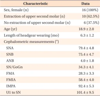
Table 1. Patients’ demographic characteristics
Characteristic Data
Sex, female (n) 16 (100%)
Extraction of upper second molar (n) 10 (62.5%) No extraction of upper second molar (n) 6 (37.5%)
Age (yr) 18.9 ± 2.0
Length of headgear wearing (mo) 6.3 ± 1.2 Cephalometric measurements (
o)
SNA 79.4 ± 4.8
SNB 75.4 ± 4.7
ANB 4.0 ± 1.8
SN/GoGn 34.3 ± 4.1
FMA 28.3 ± 3.3
FMIA 58.4 ± 4.0
IMPA 92.4 ± 5.3
U1 to SN 101.4 ± 9.5
Values are presented as number or mean ± standard devia- tion.
SNA, Sella-nasion-A point angle; SNB, sella-nasion-B
point angle; ANB, A point-nasion-B point angle; SN/GoGn,
mandibular plane angle to the anterior cranial base; FMA,
mandibular plane angle to the Frankfort plane; FMIA, lower
incisor angle to the Frankfort plane; IMPA, lower incisor
angle to the mandibular plane; U1 to SN, upper incisor angle
to the anterior cranial base.
adjusted during appointments scheduled every 4 weeks.
Molar distalization was performed until a super Class I relationship was achieved.
Cone-beam computed tomography (CBCT) scans and dental casts were obtained from the patients before (T0) and immediately after (T1) maxillary molar distalization.
CBCT scans were obtained using an Alphard Vega system (Asahi Roentgen Ind. Co., Ltd., Kyoto, Japan).
CBCT scans were obtained with a reference ear plug (REP) and head posture aligner (HPA) to ensure standardized volume data.
15CBCT scan data were exported to InVivo 5 software (version 5.2; Anatomage, San Jose, CA, USA).
The head image was reoriented to the standard position using two ball markers and a wire indicator.
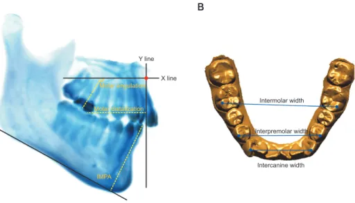
16To ensure that the head position was identical in the images taken at T0 and T1, the CBCT volume data were superimposed 3-dimensionally on the anterior cranial base using InVivo software. Next, CBCT-generated half-cephalograms (CG Cephs) were generated from volume data from the right side of the CBCT scan at T0 and T1. Using the import orientation function of the program, we oriented two volume images in the same position. The original coordinates (0, 0, 0) were set at the center point of the line passing through the two ball markers of the REP. Cephalograms before and after molar distalization were precisely placed in the same orientation in the coordinate system. On the CG Ceph, a horizontal reference line (X line) was established parallel to the HPA. The vertical reference line (Y line) was perpendicular to the X line passing through point A
(Figure 1A).
To evaluate treatment changes after molar distaliza- tion, we obtained the following cephalometric measure- ments with landmarks and reference planes: the distance (mm) between the Y line to the first maxillary molar mesial cusp tip, angle (
o) between the X line and the long axis of the first maxillary molar, and mandibular incisor inclination (IMPA) (Figure 1A). Maxillary molar movement was evaluated relative to an xy coordinate system set at point A.
17Dental casts at T0 and T1 were scanned using a TRIOS
®scanner (3Shape, Copenhagen, Denmark). Then the casts were evaluated using a software program (OrthoAnalyzer
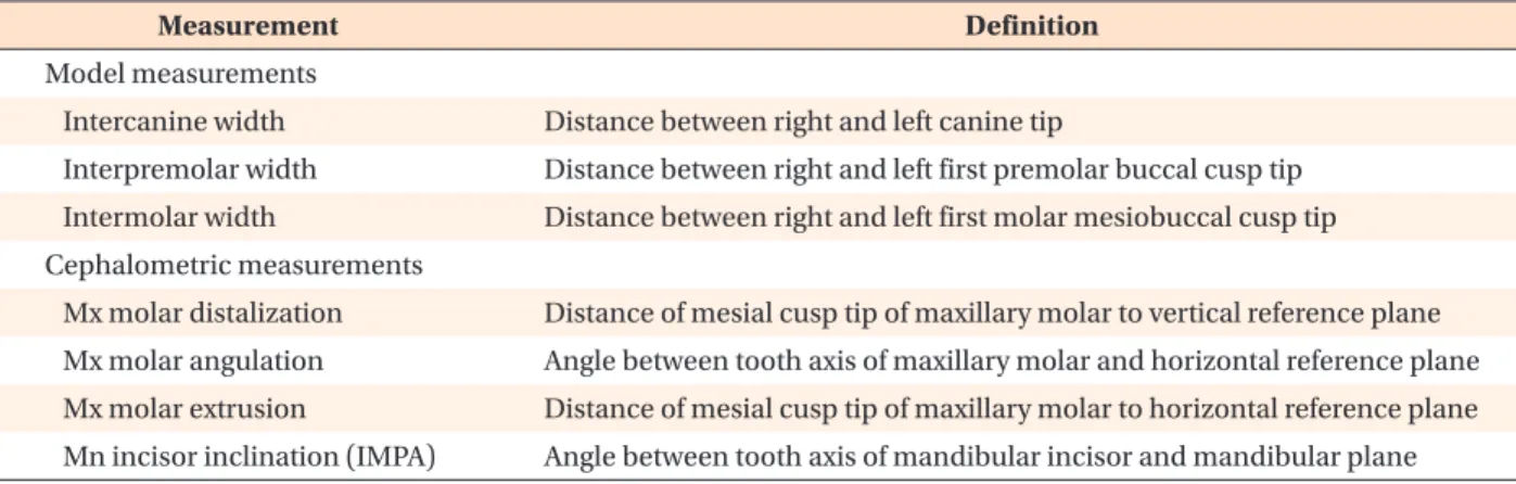
TM, 3Shape). To assess changes in the mandibular dentition between T0 and T1, we obtained the following measurements: the intercanine width (ICW), interpremolar width (IPW), and intermolar width (IMW) of the mandibular dentition. In addition, the IMW of the maxilla was evaluated (Figure 1B). Definitions of these measurements are presented in Table 2.
The sample size of this study was not calculated a priori; however, the post hoc power analysis according to the G*power program (version 3.1.9.2; Heinrich- Heine-University, Düsseldorf, Germany) showed more than 93% power for all the measurements.
18To assess the method errors, we repeated the measure- ments at an interval of 2 weeks. The method errors were calculated using the Dahlberg formula.
19Measurement errors ranged from 0.1 to 0.4 mm for the linear measurements and from 0.2
oto 0.7
ofor the angular
Y line
A B
X line Molar angulation
Molar distalization
IMPA
Intermolar width
Interpremolar width
Intercanine width