Introduction
The third molar (M3) varies more than the other molars in terms of shape, size, timing of eruption, and even tend- ency toward impaction. In modern societies, M3s get im- pacted far more than any other teeth, and impaction of mandibular M3 is more common than its maxillary coun-
terpart.1According to several studies, there is no sex pre- dilection in the impaction of M3.1-3 However, Hugoson and Kugelberg showed a higher frequency in females than males.4
It has been noted that M3 crypt formation starts at the age of 3 to 4, and calcification begins at 7 to 10 years of age. However, the time of eruption varies from 14 to 24 years in different populations.1,5Several factors are involv- ed in impaction of mandibular M3. First, the shortage of space between the anterior border of the ascending ramus and the distal area of the mandibular second molar (M2) has been identified as a major factor in M3 impaction.1
Position of impacted mandibular third molar in different skeletal facial types:
First radiographic evaluation in a group of Iranian patients
Abbas Shokri1, Majid Mahmoudzadeh2, Maryam Baharvand3, Hamed Mortazavi3,*, Javad Faradmal4, Samira Khajeh1, Faezeh Yousefi1, Maruf Noruzi-Gangachin1
1Department of Oral and Maxillofacial Radiology, Dental School, Hamadan University of Medical Sciences, Hamadan, Iran
2Department of Orthodontics, Dental School, Hamadan University of Medical Sciences, Hamadan, Iran
3Department of Oral Medicine, Dental School, Shahid Beheshti University of Medical Sciences, Tehran, Iran
4Department of Epidemiology and Statistics, Medical School, Hamadan University of Medical Sciences, Hamadan, Iran
ABSTRACT
Purpose: This study was performed to evaluate the position of impacted mandibular third molars in different skeletal facial types among a group of Iranian patients.
Materials and Methods: A total of 400 mandibular third molars in 200 subjects with different types of facial growth were radiographically investigated for their positions according to their types of facial growth on the basis of the β angle. The subjects were divided into three groups (class I, II, and III) according to ANB angle, representing the anteroposterior relationship of the maxilla to the mandible. Meanwhile, the subjects were also divided into three groups (long, normal, and short face) according to the angle between the stella-nasion and mandibular plane (SNGoGn angle). ANOVA was used for statistical analysis.
Results: The mean β angle showed no significant difference among class I, II, and III malocclusions (df==2, F==0.669, p==0.513). The same results were also found in short, normal, and long faces (df==1.842, F==2, p==0.160). The mesio- angular position was the most frequent one in almost all of the facial growth patterns. Distoangular and horizontal positions of impaction were not found in the subjects with class III and normal faces. In the long facial growth pat- tern, the frequency of vertical and distoangular positions were not different.
Conclusion: In almost all of the skeletal facial types, the mesioangular impaction of the mandibular third molar was the most prevalent position, followed by the horizontal position. In addition, β angle showed no significant difference in different types of facial growth. (Imaging Sci Dent 2014; 44 : 61-5)
KEY WORDS: Mandible; Molar, Third; Tooth, Impacted; Malocclusion
Received July 25, 2013; Revised August 16, 2013; Accepted September 1, 2013
*Correspondence to : Prof. Hamed Mortazavi
Department of Oral Medicine, Dental School, Shahid Beheshti University of Medical Sciences, Daneshjoo Blvd, Tabnak St, Chamran Highway, Tehran Zip code: 198396 3113, Iran
Tel) 98-21-29902311, Fax) 98-21-22403194, E-mail) [email protected]
Copyright ⓒ 2014 by Korean Academy of Oral and Maxillofacial Radiology
This is an Open Access article distributed under the terms of the Creative Commons Attribution Non-Commercial License (http://creativecommons.org/licenses/by-nc/3.0) which permits unrestricted non-commercial use, distribution, and reproduction in any medium, provided the original work is properly cited.
Imaging Science in Dentistry∙pISSN 2233-7822 eISSN 2233-7830
Björk demonstrated reduction of the alveolar arch space behind the mandibular M2 in 90% of patients with M3 impaction.6Second, a short mandibular length is thought to be another etiologic factor in M3 impaction.2However, Kaplan did not find any significant difference in mandi- bular length between subjects with erupted and impacted molars.7Third, the size of mandibular M3 has a possible role in this regard as well.1 Hattab and Alhaija showed that impacted M3s were larger in size than erupted ones.8
Finally, impaction of mandibular M3 has also been asso- ciated with the pattern of facial growth.9In contrast, Lego- vi´c et al showed no significant difference between the position of mandibular M3 and the type of facial growth.10 Due to these controversies, further studies should be done to clarify the factors associated with M3 impaction.
Therefore, the aim of this study was to evaluate the im- pacted mandibular M3 positions in different skeletal facial types. To best of our knowledge, this is the first study on the topic in a group of Iranian patients.
Materials and Methods
A list of patients referred to the Orthodontics Depart- ment of Hamadan Dental School, Iran in 2012 and 2013 was identified (1,650 cases: 921 women and 629 men).
The inclusion criteria for this study were as follows: 1) adequate data records and complete history of surgical and orthodontic treatments, 2) panoramic radiographs showing complete dentition dating back to pre-orthodon- tic treatment, 3) lateral cephalometric image pertaining to pre-orthodontic treatment, 4) presence of mandibular M3s with at least two-thirds of root formation being complete, 5) no missing or extracted permanent teeth or any previous orthodontic procedures, 6) presence of bilateral impaction in both left and right sides of the mandible for comparison, and 7) no history of medical problems with a potential effect on facial growth. Patients with pathological condi- tions related to mandibular M2 and M3 such as extensive caries or cystic lesions were excluded.
In this study, 265 out of 1,650 cases had one or two im- pacted mandibular M3. However, only 200 cases (66 males and 134 females) met the inclusion criteria. The age of the patients included in this study ranged between 19 and 32 years, and the average age was 22.5±2.03.
The ANB angle representing the relative position of the maxilla and mandible anteroposteriorly, and the SNGoGn angle representing the angle between the stella-nasion and mandibular plane, were used to detect different skeletal facial types. The patients were divided into three subgroups
on the basis of their ANB angle as follows: skeletal class I (ANB: 1-5 degrees), skeletal class II (ANB more than 5 degrees), and skeletal class III (ANB less than 1 degree).
At the same time, the patients were divided into three subgroups according to their SNGoGn angle as follows:
short face (low angle: SNGoGn less than 27 degrees), nor- mal (SNGoGn between 27 and 37 degrees), and long face (long angle: SNGoGn more than 37 degrees).11
The type of mandibular M3 impaction was determined according to Winter’s classification by using β angle as follows: distoangular position (angle from -11 to -79 degrees), vertical position (angle from -10 to 10 degrees), mesioangular position (angle from 11 to 79 degrees), and horizontal position (angle from 80 to 100 degrees). The β angle was formed between the intersecting long axes of the M2 and M3 and drawn through the midpoint of the occlu- sal surface and midpoint of the root bifurcation.12We did not consider the buccal/lingual obliquity (transverse) because we needed an occlusal radiograph to confirm this position.
Lateral cephalometric radiographs were taken for each patient in centric occlusion with the lips in repose and the Frankfort plane horizontal, according to the natural head position, using a Cranex D X-ray unit (Sordex, Helsinki, Finland) at 66 to 70 kVp, 10 mA, and 14.2 s exposure.
Lateral cephalometric radiographs were used to allocate the subjects to their groups according to the ANB angle and SNGoGn angle to identify the type of facial growth.
Panoramic radiographs were also taken for each patient with the upper and lower incisors in an edge-to-edge rela- tionship using the Cranex D X-ray unit (Soredex, Helsinki, Finland) at 66 to 70 kVp, 10 mA, and 17.6 s.
All of the images were displayed on a 17-inch Samsung monitor (Syncmaster 740N, Seoul, Korea) with the screen resolution set at 1,280×1,024 pixels and color set to 32- bit depth. Finally, tracing was done using Scanora soft- ware (Soredex, Helsinki, Finland) by two investigators for measurement of the ANB, SNGoGn, and β angles.
The reliability and degree of agreement between investi- gators were also determined by the mean of Cohen’s kappa analysis so that intra- and inter-examiner reliability was above 0.70 and 0.88, respectively. The data were statisti- cally analyzed by the analysis of variance (ANOVA) and t-test using SPSS software (ver. 15.0, SPSS Inc, Chicago, USA). A p value less than 0.05 was considered to indicate statistical significance.
Results
The incidence of mandibular M3 impaction was 16.06%
(265 out of 1650 cases). The incidence of bilateral impac- tion of the mandibular M3 was 12.12% (200 out of 1650 cases).
The means of the β angle in class I, II, and III were 29.63
±16.29, 30.15±17.69, and 33.27±12.17, respectively.
Meanwhile, it was found to be 31.21±16.53, 30.69±
14.66, and 27.57±16.53 in different skeletal facial types of short face, normal face, and long face, respectively.
The mean of the β angle in females (30.66±17.24) and males (29.04±15.12) did not show a significant difference (p==0.657).
According to ANOVA, the mean of the β angle did not differ significantly among class I, II, and III patients (df== 2, F==0.669, p==0.513). The same results were also found among the short, normal, and long face groups (df==1.842, F==2, p==0.160).
In addition, according to ANOVA, the mean of the β angle in both sides (the left and right sides of the mandible) was higher in class III than class I and II. However, the β angle showed no significant difference between the left and
right sides in class I, II, and III (right side: F==0.239, df== 2.197, p==0.788; left side: F==0.491, df==2.197, p==0.613) (Table 1). Although the β angle showed no significant dif- ference between the left and right sides in the short, nor- mal, and long faces (right side: F==0.128, df==2.197, p== 0.880; left side: F==0.491, df==2.197, p==0.613), in short faces, the mean of the β angle in the right side was almost equal to the left side.
In both genders, mesioangular and distoangular positions were found to be the most and the least prevalent types of mandibular M3 impaction, respectively. In addition, the frequency of vertical and horizontal types of mandibular M3 impaction was equal in the men in our study (Table 2).
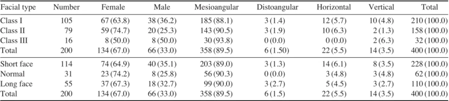
In all of facial growth patterns, the mesioangular posi- tion was found to be the most common type of mandibular M3 impaction (Table 3). In almost all facial growth pat- terns, except for class III, a horizontal position of the im- pacted mandibular M3 was the second most common one after the mesioangular position, followed by the vertical and distoangular positions (Table 3).
Discussion
The results of this study demonstrated an overall rate of mandibular M3 impaction of 16.06%. According to Andrea- sen et al, impaction of mandibular M3 varies from 18% to 32% in different populations.13This rate was also estimat- ed by Dachi and Howell as 17.5% and 21.9% for mandibu- lar and maxillary M3s, respectively.14 In addition, in a recent study, Breik and Grubor showed a rate of 58.76%
for mandibular M3 impaction in Melbourne, Australia.2
Table 1.Distribution of β angle (mean±SD) in the right and left sides of the mandible according to different skeletal facial types Facial types β angle in the right side β angle in the left side
Class I 26.11±16.89 26.72±15.16
Class II 27.10±16.26 24.96±18.01
Class III* 28.89±10.37 32.66±13.85
Short face 27.06±16.84 27.34±16.49
Normal 27.15±13.47 26.45±15.26
Long face 25.78±16.36 24.68±16.62
*: significantly different from Class I and II
Table 2.Distribution (number and percentage) of different positions of mandibular M3 impaction according to the genders
Genders Number Mesioangular Distoangular Horizontal Vertical Total
Female 134 (67.0) 239 (89.2) 3 (1.1) 17 (6.4) 9 (3.4) 268 (100.0)
Male 66 (33.0) 119 (90.2) 3 (2.3) 5 (3.8) 5 (3.8) 132 (100.0)
Total 200 (100.0) 358 (89.5) 6 (1.5) 22 (5.5) 14 (3.5) 400 (100.0)
Table 3.Distribution (Number and percentage) of different positions of mandibular M3 impaction according to different skeletal facial types
Facial type Number Female Male Mesioangular Distoangular Horizontal Vertical Total
Class I 105 67 (63.8) 38 (36.2) 185 (88.1) 3 (1.4) 12 (5.7) 10 (4.8) 210 (100.0)
Class II 79 59 (74.7) 20 (25.3) 143 (90.5) 3 (1.9) 10 (6.3) 2 (1.3) 158 (100.0)
Class III 16 8 (50.0) 8 (50.0) 30 (93.8) 0 (0.0) 0 (0.0) 2 (6.3) 32 (100.0)
Total 200 134 (67.0) 66 (33.0) 358 (89.5) 6 (1.50) 22 (5.5) 14 (3.5) 400 (100.0)
Short face 114 74 (64.9) 40 (35.1) 203 (89.0) 3 (1.3) 14 (6.1) 8 (3.5) 228 (100.0)
Normal 31 23 (74.2) 8 (25.8) 56 (90.3) 0 (0.0) 3 (4.8) 3 (4.8) 62 (100.0)
Long face 55 37 (67.3) 18 (32.7) 99 (90.0) 3 (2.7) 5 (4.5) 3 (2.7) 110 (100.0)
Total 200 134 (67.0) 66 (33.0) 358 (89.5) 6 (1.5) 22 (5.5) 14 (3.5) 400 (100.0)
Vilela and Vitol reported that the most frequently im- pacted teeth are M3s (90%) with a higher prevalence in the mandible than the maxilla (60% vs. 30%), followed by the upper canine teeth (5%), lower bicuspids, and super- numerary teeth (5%).15
The mean age of our study sample was 22.5±3.02 years, which was similar to the study by Abu Alhaija et al (20.80
±2.03 years).1
According to Vilela and Vitoi,15the crown formation of M3s begins around 9 to 10 years of age, which might be seen in panoramic radiographs at 11 years of age in about 90% of cases. They more frequently erupt between 18 and 20 years of age.
In the present report, generally, a mesioangular position of the impacted mandibular M3 was the most prevalent position (89.5%), followed by horizontal, vertical, and distoangular positions. In accordance to our findings, Breik and Grubor showed that over 80% of the mandibular M3 impactions in all facial types were in the mesioangular position.2Vilela and Vitoi15found that the vertical position was most prevalent, followed by mesioangular position. In contrast, Quek et al,16Sandhu and Kaur,17and Venta et al18 noted the mesioangular position to be the most prevalent one.
According to our results, in both genders, the mesioan- gular position was the most common position, followed by the horizontal, vertical, and distoangular positions. How- ever, the number of females with impacted mandibular M3s was higher than that of males. Many researchers such as Abu Alhaija et al,1Breik and Grubor,2Hattab et al,3and Brown et al19reported no sex predilection in mandibular M3 impaction. In contrast, Hugoson and Kugelberg4and Murtomaa et al20found a higher frequency in women than men.
Generally, the number of women was more than men in our study, possibly because women are more willing to receive orthodontic treatment for esthetic reasons. That is why the number of recorded impacted M3s in women was greater than men in our study.
In the subjects in class I group as well as those of the short face group, the mesioangular position was the most frequent position, followed by horizontal, vertical, and distoangular positions (mesioangular¤horizontal¤verti- cal¤distoangular).
In the patients in class II occlusion, the mesioangular position had the highest prevalence, followed by horizon- tal, distoangular, and vertical positions (mesioangular¤
horizontal¤distoangular¤vertical).
In the subjects with class III occlusion, the mesioangular
position was the most frequent position, followed by the vertical position. In this type of facial growth, we did not find distoangular and horizontal positions of impaction, perhaps due to small number of class III cases (16 cases).
In patients with a normal growth pattern of the face, the mesioangular position was the most prevalent position, and the frequency of horizontal and vertical positions were equal. Also, distoangular position was not found in this type of growth pattern (mesioangular¤horizontal==vertical).
In the long facial growth pattern, the mesioangular posi- tion had the highest prevalence, followed by the horizon- tal position. In addition, the frequency of the vertical and distoangular positions were equal in this growth pattern.
According to Breik, mesioangular impaction of the ma- ndibular M3 was most common in mesofacial subjects, followed by brachyfacials and dolichofacials. On the other hand, Breik showed that horizontal impaction was mostly common in dolichofacials followed by brachyfacials and mesofacials.2
In our study, there was no significant difference in the β angles of different types of skeletal facial growth. This finding was in agreement with the results of Abu Alhaija et al1and Behbehani et al.21Behbehani et al demonstrated that a higher mesial angulation of the M3 bud increased the risk of impaction.21 Furthermore, Uthman suggested that the β angle showed a marked increase in the margi- nal eruption group compared to the full eruption group.22 According to Farzanegan and Goya, the largest β angle was measured in the normal group, followed by deep bite and open bite groups.23
It was noted that an impacted mandibular M3 at the nor- mal time of eruption might erupt later in life.2 Although rarely leading to clinical eruption, Richardson pointed out that between the age 18 and 21, many unerupted M3s changed their position appreciably.24 Hattab reported that by the age of 19, some impacted molars erupt into functio- nal occlusion.25
According to Kruger et al, 26impacted M3s at age 19 could be fully erupted by age 26. Hesby et al27observed that during the growth period, the mandibular inter-molar width increased by 2.05 mm, and the mandibular cross- arch width at the level of the alveolar crest increased by 1.60 mm (left buccal surface to right buccal surface) and 1.02 mm (left lingual surface to right lingual surface).
Furthermore, the basal bone of the mandible increased in width by 14.54.
In conclusion, in almost all skeletal facial types, the mesioangular position of the impacted mandibular M3 was the most prevalent position, followed by the horizon-
tal position. Moreover, various types of facial growth did not show a significant difference in terms of the β angle.
References
1. Abu Alhaija ES, AlBhairan HM, AlKhateeb SN. Mandibular third molar space in different antero-posterior skeletal patterns.
Eur J Orthod 2011; 33: 570-6.
2. Breik O, Grubor D. The incidence of mandibular third molar impactions in different skeletal face types. Aust Dent J 2008;
53: 320-4.
3. Hattab FN, Rawashdeh MA, Fahmy MS. Impaction status of third molars in Jordanian students. Oral Surg Oral Med Oral Pathol Oral Radiol Endod 1995; 79: 24-9.
4. Hugoson A, Kugelberg CF. The prevalence of third molars in a Swedish population. An epidemiological study. Community Dent Health 1988; 5: 121-38.
5. Mady Marici´c B, Legovi´c M, Slaj M, Lapter Varga M, Zuvi´c Butorac M, Kapovi´c M. Presence of third molar germs in orth- odontic patients with class II/2 and class III malocclusions.
Coll Antropol 2009; 33: 1171-5.
6. Björk A, Jensen E, Palling M. Mandibular growth and third molar impaction. Acta Odontol Scand 1956; 14: 231-72.
7. Kaplan RG. Some factors related to mandibular third molar impaction. Angle Orthod 1975; 45: 153-8.
8. Hattab FN, Alhaija ES. Radiographic evaluation of mandi- bular third molar eruption space. Oral Surg Oral Med Oral Pathol Oral Radiol Endod 1999; 88: 285-91.
9. Richardson ME. The etiology and prediction of mandibular third molar impaction. Angle Orthod 1977; 47: 165-72.
10. Legovi´c M, Legovi´c I, Brumini G, Vandura I, Cabov T, Ovesnik M, et al. Correlation between the pattern of facial growth and the position of the mandibular third molar. J Oral Maxillofac Surg 2008; 66: 1218-24.
11. Tecco S, Caputi S, Tete S, Orsini G, Festa F. Electromyo- graphic activity of masticatory, neck and trunk muscles of sub- jects with different mandibular divergence. A cross-sectional evaluation. Angle Orthod 2007; 77: 260-5.
12. Winter GB. Impacted mandibular third molars. St. Louis:
American Medical Book; 1926. p. 241-79.
13. Andreasen JO, Petersen JK, Laskin DM. Textbook and color atlas of tooth impactions: diagnosis, treatment, prevention.
Copenhagen: Munksgaard; 1997. p. 222-3.
14. Dachi SF, Howell FV. A survey of 3,874 routine full-mouth
radiographs. I. A study of retained roots and teeth. Oral Surg Oral Med Oral Pathol 1961; 14: 916-24.
15. Vilela EM, Vitoi PA. Study of position and eruption of lower third molars in adolescents. South Braz Dent J 2011; 8: 390-7.
16. Quek SL, Tay CK, Tay KH, Toh SL, Lim KC. Pattern of third molar impaction in a Singapore Chinese population: a retros- pective radiographic survey. Int J Oral Maxilllofac Surg 2003;
32: 548-52.
17. Sandhu S, Kaur T. Radiographic evaluation of the status of third molars in the Asian-Indian students. J Oral Maxillofac Surg 2005; 63: 640-5.
18. Ventä I, Turtola L, Ylipaavalniemi P. Radiographic follow-up of impacted third molars from age 20 to 32 years. Int J Oral Maxillofac Surg 2001; 30: 54-7.
19. Brown LH, Berkman S, Cohen D, Kaplan AL, Rosenberg M. A radiological study of the frequency and distribution of impact- ed teeth. J Dent Assoc S Afr 1982; 37: 627-30.
20. Murtomaa H, Turtola L, Ylipaavalniemi P, Rytömaa I. Status of the third molars in the 20- to 21-year-old Finnish university population. J Am Coll Health 1985; 34: 127-9.
21. Behbehani F, Artun J, Thalib L. Prediction of mandibular third- molar impaction in adolescent orthodontic patients. Am J Orthod Dentofacial Orthop 2006; 130: 47-55.
22. Uthman AT. Retromolar space analysis in relation to selected linear and angular measurements for an Iraqi sample. Oral Surg Oral Med Oral Pathol Oral Radiol Endod 2007; 104: e76-82.
23. Farzanegan F, Goya A. Evaluation of mandibular third molar positions in various vertical skeletal malocclusions. J Dent Mater Tech 2012; 1: 58-62.
24. Richardson M. Changes in lower third molar position in the young adult. Am J Orthod Dentofacial Orthop 1992; 102: 320- 7.
25. Hattab FN. Positional changes and eruption of impacted mandi- bular third molars in young adults. A radiographic 4-year fol- low-up study. Oral Surg Oral Med Oral Pathol Oral Radiol Endod 1997; 84: 604-8.
26. Kruger E, Thomson WM, Konthasinghe P. Third molar out- comes from age 18 to 26: findings from a population-based New Zealand longitudinal study. Oral Surg Oral Med Oral Pathol Oral Radiol Endod 2001; 92: 150-5.
27. Hesby RM, Marshall SD, Dawson DV, Southard KA, Casko JS, Franciscus RG, et al. Transverse skeletal and dentoalveolar changes during growth. Am J Orthod Dentofacial Orthop 2006;
130: 721-31