undesirable side effects are possible2, of which skeletal insta- bility (relapse) is one of the most commonly reported3. This happens in almost every case, and it can be fairly significant for some patients4.
Considerable changes in the lower jaw position and signifi- cant alteration in the forces transmitted to it have made stabil- ity of new jaw position an important goal for every surgeon, and it is paramount to success5.
Relapse is divided into early and long-term based on the timing of occurrence. The main causes of early relapse are usually movements at the osteotomy site and condylar sag.
These problems are mainly due to unbalanced soft tissue tension after surgery5. This type of relapse occurs during the first 6-24 weeks after orthognathic surgery6, which is consid- ered the period when the majority of relapse occurs7. Long- term relapse usually occurs because of progressive condylar
I. Introduction
Bilateral sagittal split osteotomy (BSSO), introduced by Trauner and Obwegeser in 1957, is currently the most fa- vored and widely used surgical method for correction of lower jaw deformities1. This technique usually results in considerably improved function and appearance. However,
Reza Tabrizi
Department of Oral and Maxillofacial Surgery, School of Dentistry, Shahid Beheshti University of Medical Sciences, Velenjak, Tehran, Iran
TEL: +98-9125850829 FAX: +98-2122672126 E-mail: [email protected]
ORCID: http://orcid.org/0000-0001-7204-7746
This is an open-access article distributed under the terms of the Creative Commons Attribution Non-Commercial License (http://creativecommons.org/licenses/by-nc/4.0/), which permits unrestricted non-commercial use, distribution, and reproduction in any medium, provided the original work is properly cited.
CC
Skeletal stability following mandibular advancement: is it influenced by the magnitude of advancement or changes of the mandibular plane angle?
Reza Tabrizi1, Mahsa Nili2, Ehsan Aliabadi3, Fereydoun Pourdanesh1
1Department of Oral and Maxillofacial Surgery, School of Dentistry, Shahid Beheshti University of Medical Sciences, Tehran,
2Student Research Committee, School of Dentistry, Shiraz University of Medical Sciences, Shiraz,
3Department of Oral and Maxillofacial Surgery, School of Dentistry, Shiraz University of Medical Sciences, Shiraz, Iran
Abstract(J Korean Assoc Oral Maxillofac Surg 2017;43:152-159)
Objectives: The aim of this study was to investigate the effects of advancement magnitude and changes in mandibular plane angle on the stability of mandibular advancement.
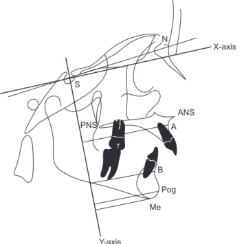
Materials and Methods: This retrospective cohort study evaluated the postoperative stability of mandibular advancement in class II skeletal sub- jects who underwent bilateral sagittal split osteotomy. Radiographs taken preoperatively, immediately postoperatively and 1 year postoperatively were traced and analyzed using linear and angular measurements. To determine horizontal and vertical relapse, an X-Y coordinate system was established in which the X-axis was constructed by rotating S-N downward by 7° (approximation of the Frankfort horizontal plane) and the Y-axis was defined as a line perpendicular to the X-axis and passing through the point Sella. For certain reference points including point A, point B, pogonion and menton, the perpendicular distance between each point and both axes was determined and cephalometric variables were recorded as X and Y coordinates.
Results: Twenty-five subjects were studied. A significant correlation between the amount of mandibular advancement and relapse in the B point (ver- tical and horizontal) and the pogonion point was observed (vertical and horizontal, P<0.001). Evaluation of data demonstrated a positive correlation between the mandibular plane angle (SN/ML) change and vertical relapse in the B point (P<0.05). A simple regression model demonstrated that 74%
of horizontal relapse and 42.3% of vertical relapse in the B point was related to the amount of mandibular advancement. The receiver operating charac- teristic test showed that 8.5 mm mandibular advancement is related to a relapse rate of 1 mm or more in the pogonion, vertically or horizontally.
Conclusion: The magnitude of mandibular advancement is a stronger surgical predictor for horizontal rather than vertical relapse at the B point.
Changes in mandibular plane angle (SN/ML) during surgery affect vertical, but not horizontal relapse at the B point.
Key words: Mandible, Orthognathic surgery, Osteotomy, Mandibular advancement
[paper submitted 2016. 9. 24 / revised 2016. 11. 4 / accepted 2016. 11. 16]
Copyright Ⓒ 2017 The Korean Association of Oral and Maxillofacial Surgeons. All rights reserved.
cephalograms. Patients were stabilized in the ProMax cepha- lometric unit (Planmeca, Helsinki, Finland) using a cephalo- stat. This positions the patient with the head oriented at a 90°
angle relative to the x-ray beam at a distance of 5 ft from the tube. The jaws were in maximum intercuspation, with the tip of the tongue behind the upper incisor teeth and the lips in repose. The receptor (CR; Konica Minolta Medical Imaging, Maitland, FL, USA) was placed 38.1 cm from the head (this is the standard under which all cephalometric radiographs are taken and ensures that radiographs taken at different time points are directly comparable).
Radiographic exposure was at 60-80 kVp and 10-15 mA (23-61 seconds) and was repeated for each case. The radio- graphs were processed in the laser readout processor of the mentioned CR system. A DICOM PACS system was used to save and transfer images.
All lateral cephalograms were traced by hand, digitized, superimposed and evaluated by the same examiner. Trac- ings were then retraced and reanalyzed by another examiner.
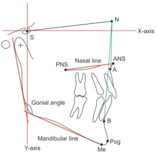
Cephalometric points included: Go (gonion), Pog (pogonion), Me (menton), point B, S (sella), and Na (nasion).(Fig. 1, Table 1)
Two angular parameters were determined using the points previously mentioned cephalometric reference. These angular parameters consisted of the gonial angle and SN/ML (man- dibular line to sella turcica-nasion).
To determine horizontal and vertical relapse, an X-Y coor- resorption, which leads to loss of height of the condyle and
mandibular ramus5,8.
Postoperative skeletal relapse is attributed to either bio- logical factors, such as further mandibular growth, or factors related to the surgical procedure and postoperative patient care2. These factors include the magnitude and direction of skeletal movement, method of fixation, use of bone graft, condylar positioning9,10, and achievement of postoperative occlusion9. However, other factors such as the magnitude of mandibular movement10 and the mandibular plane angle the effects of which on relapse have yet to be elucidated8,11. Other variables such as age and sex may affect the new mandibular position after surgery.
In this study, we sought to assess if any correlation exists between the magnitude of mandibular movement and relapse in BSSO and if changes of the mandibular plane angle affect stability following surgery.
II. Materials and Methods
This retrospective cohort study aimed to evaluate the post- operative stability of mandibular advancement in class II skeletal subjects who underwent BSSO at Chamran Medical Center in Shiraz University of Medical Sciences, from 2008 to 2012. The study was approved by the medical ethics com- mittee at Shiraz University. Subjects eligible for inclusion had class II skeletal deformities that required mandibular advancement without maxillary osteotomy. Each subject was checked for adequate records, including date and type of surgery. All patients had completed growth prior to surgery.
Subjects were excluded if they had a history of trauma, or- thognathic surgery, any augmentation with alloplastic or au- togenous materials during or after surgery, temporomandibu- lar joint (TMJ) surgery before or after orthognathic surgery, temporomandibular disorder before surgery or asymmetric mandible.
Twenty-five subjects were assessed. All subjects were surgically treated by BSSO for mandibular advancement ac- cording to the Obwegeser/Dal Pont method. Rigid internal fixation was obtained with miniplates and four monocortical screws bilaterally.
By using lateral cephalograms taken on three occasions (preoperatively, immediately after surgery, and 1 year postop- eratively) as a raw data base, skeletal points were determined and digitized to evaluate two-dimensional skeletal changes during and after surgery and to determine the amount of re- lapse. The same x-ray machine and settings were used for all
X-axis
Y-axis
PNS
ANS
Pog Me
N
S
Gonial angle
Mandibular line Nasal line
A
B
Fig. 1. Skeletal landmarks used in cephalometric analysis. Refer to Table 1 for the definition of landmarks.
Reza Tabrizi et al: Skeletal stability following mandibular advancement: is it influenced by the magnitude of advancement or changes of the mandibular plane angle? J Korean Assoc Oral Maxillofac Surg 2017
eration and conclusion of orthodontic treatments.
1. Tracing technique
All cephalograms were traced by a single operator on Garware matte acetate tracing paper of 0.003-inch thickness with a 3-H microlead pencil. Cephalometric landmarks were located, identified, and marked. The tracings were rechecked by another examiner.
dinate system was established. The X-axis was constructed by rotating S-N downward by 7° (an approximation of the Frankfort horizontal plane), and the Y-axis was defined as the line perpendicular to the X-axis and passing through the point sella. For certain reference points including point A, point B, pogonion, and menton, the perpendicular distance between each point and both axes was determined and the cephalo- metric variables were recorded as X- and Y-coordinates.(Fig.
2, 3) All measurements were performed 1 year after the op-
Table 1. Cephalometric landmarks
Landmark Definition
Point A (subspinale) Point B (supramentale) Me (menton)
Pog (pogonion)
ANS (anterior nasal spine) PNS (posterior nasal spine) NL (nasal line)
ML (mandibular line/plane) SN (sella-nasion)
S N
Gonial angle
The most posterior point in the concavity between ANS and Prosthion (most inferior point overlying maxillary incisors)
The most posterior midline point in the concavity of the mandible between the most superior point on the alveolar bone overlying the mandibular incisors (infradentale) and Pog
The most inferior point on the symphysis The most anterior point on the symphysis
Anterior tip of the sharp bony process of the maxilla Posterior spine of palatal bone
Line connecting ANS and PNS
Line tangent to inferior border of mandible Line connecting sella and nasion
Geometric center of pituitary fossa Most anterior point on fronto-nasal suture
Angle between two lines along inferior border of mandible and posterior border of ramus.
Reza Tabrizi et al: Skeletal stability following mandibular advancement: is it influenced by the magnitude of advancement or changes of the mandibular plane angle? J Korean Assoc Oral Maxillofac Surg 2017
X-axis
Y-axis PNS
ANS
Pog Me
N
S
A
B
Fig. 2. Horizontal measurements. Refer to Table 1 for the defini- tion of landmarks.
Reza Tabrizi et al: Skeletal stability following mandibular advancement: is it influenced by the magnitude of advancement or changes of the mandibular plane angle? J Korean Assoc Oral Maxillofac Surg 2017
X-axis
Y-axis
PNS ANS
Pog Me
N
S
A
B
Fig. 3. Vertical measurements. Refer to Table 1 for the definition of landmarks.
Reza Tabrizi et al: Skeletal stability following mandibular advancement: is it influenced by the magnitude of advancement or changes of the mandibular plane angle? J Korean Assoc Oral Maxillofac Surg 2017
2. Surgical technique
After induction of general anesthesia and nasotracheal in- tubation, lidocaine plus epinephrine 1:80,000 (persocaine-E;
Darupakhsh, Tehran, Iran) was submucosally injected into the surgical site to control bleeding during the procedure.
Bilateral mucosal incisions were made at the retromolar area to provide access to the bone. BSSOs were performed according to the abovementioned method using splitting forceps and elevators (curved Smith Sagittal Split Separa- tors; Walter Lorentz Surgical, Biomet, FL, USA). After sub- periosteal dissection, bone cuts were performed using a saw.
The bone was cut laterally at the mesial area of the second molar, and a medial cut was created horizontally above the lingula approximately 1 cm above the occlusal plane. Two cuts were connected through the alveolar ridge. Splitting was performed with the elevator positioned in the vertical bone cut and the forceps in the sagittal bone cut. Once the superior part of the mandible began to split, the elevator was reposi- tioned at the inferior end of the vertical cut, and the splitting was completed. After complete mobilization of the mandible and detachment of the medial pterygoid attachments on the proximal and distal segments, it was advanced into the de- sired position using a thin wafer. Intermaxillary wire fixation and elastics were not used postoperatively. In all subjects, condyles were positioned in the superior and posterior part of the glenoid fossa by manually guiding the proximal segment.
Titanium miniplates (four holes) were used for fixation of the fragments.
3. Statistical analysis
Statistical analyses were performed using the IBM SPSS Statistics, version 19.0 (IBM Co., Armonk, NY, USA). Pear- son correlation test was used to find correlations between
age, amount of advancement, and study outcomes. A linear general model was applied to predict possible relapse. Inde- pendent t-test was used to determine the possible difference between genders. The receiver operating characteristic (ROC) test was used to identify a cut-off point in anatomical size.
Inter-examiner reliability analysis was performed using the Kappa statistic to determine consistency between examiners.
III. Results
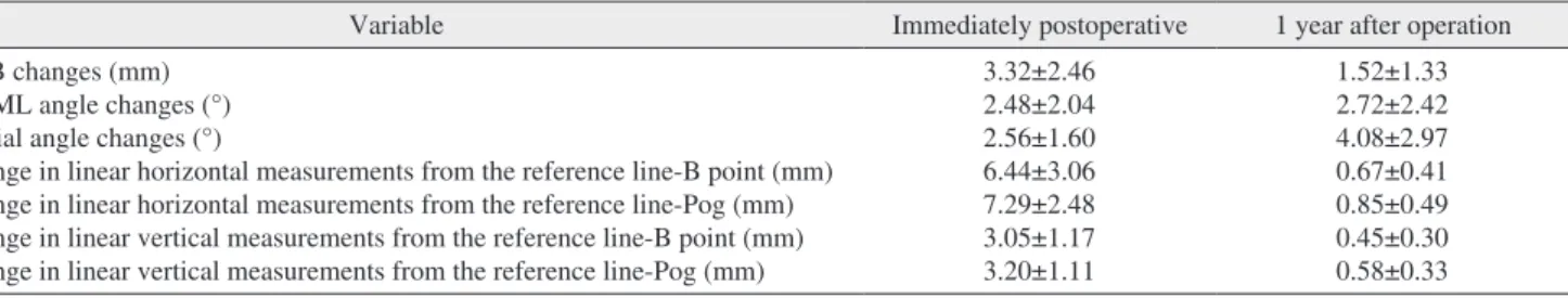
Twenty-five subjects (10 males, 15 females) were studied.
The mean age was 23.64±4.41 years. Mean mandibular ad- vancement was 6.44±3.06 mm at the B point. Mean relapse was 0.67±0.41 mm in the horizontal vector at the B point and 0.45±0.30 mm in the vertical vector. Mean relapse in the po- gonion point was 0.85±0.49 mm in the horizontal and 0.58±
0.33 mm in the vertical vector.(Table 2)
Data analysis by Pearson correlation test showed signifi- cant correlation between the amount of mandibular advance- ment and relapse in the B point (vertical and horizontal) and pogonion point (vertical and horizontal) (P<0.001). The re- sults did not show any correlation between relapse in SN/ML and amount of mandibular advancement (P>0.05).
Mean SN/ML change after the surgeries was 4.40°±3.48°.
Data evaluation demonstrated a positive correlation between the SN/ML change and vertical relapse in the B point and the mandibular plane (P<0.05). There was no correlation among SN/ML change, horizontal relapse in the B point, and relapse in the pogonion (vertical and horizontal) (P>0.05).
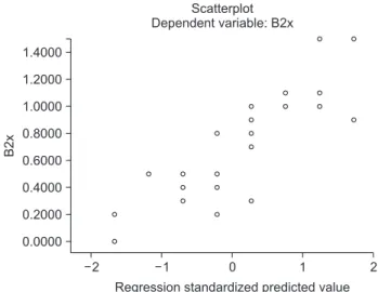
A simple regression model demonstrated that 74% of the horizontal relapse in B point was accounted for by the amount of mandibular advancement. The results showed that when the mandible was advanced 1 mm, horizontal relapse increased by 0.17 mm.(Fig. 4) For one unit standard devia- tion (SD) increase in mandibular advancement, the horizontal
Table 2. Changes in reference points immediately after operation and 1 year after surgery
Variable Immediately postoperative 1 year after operation
ANB changes (mm) SN/ML angle changes (°) Gonial angle changes (°)
Change in linear horizontal measurements from the reference line-B point (mm) Change in linear horizontal measurements from the reference line-Pog (mm) Change in linear vertical measurements from the reference line-B point (mm) Change in linear vertical measurements from the reference line-Pog (mm)
3.32±2.46 2.48±2.04 2.56±1.60 6.44±3.06 7.29±2.48 3.05±1.17 3.20±1.11
1.52±1.33 2.72±2.42 4.08±2.97 0.67±0.41 0.85±0.49 0.45±0.30 0.58±0.33 Refer to Table 1 for the definition of landmarks.
Values are presented as mean±standard deviation.
Reza Tabrizi et al: Skeletal stability following mandibular advancement: is it influenced by the magnitude of advancement or changes of the mandibular plane angle? J Korean Assoc Oral Maxillofac Surg 2017
mandibular plane angle change. When mandibular plane an- gle increased by one degree, vertical relapse increased by 0.04 mm.(Fig. 6) For one unit SD increase in mandibular plane angle, the vertical relapse is expected to increase by 0.49 of a SD unit (R2=0.24, β=0.49, P=0.001).
The ROC test showed that mandibular advancement by 8.5 mm had a relapse rate of 1 mm or more in the pogonion ver- tically and horizontally.(Fig. 7)
The inter-examiner reliability for examiners was found to be Kappa=0.69 (P<0.001) with 95% confidence interval, demonstrating substantial agreement between examiners.
relapse is expected to increase by 0.86 of a SD unit (R2=0.74, β=0.86, P=0.001). Data analysis showed that 42.3% of the vertical relapse in B point could be predicted by the amount of mandibular advancement. When the mandible was ad- vanced 1 mm, vertical relapse increased by 0.09 mm.(Fig. 5) For one unit SD increase in mandibular advancement, verti- cal relapse increased by 0.65 of a SD unit (R2=0.42, β=0.65, P=0.001).
The general linear model revealed that 24.2% of vertical relapse in the B point was accounted for by the amount of
B2x
2 1.4000 1.2000 1.0000 0.8000 0.6000 0.4000 0.2000
2 Regression standardized predicted value 0.0000
Scatterplot Dependent variable: B2x
1 0
1
Fig. 4. Horizontal relapse in the B point (B2x: dependent factor) related to the amount of mandibular advancement (predictive fac- tor).
Reza Tabrizi et al: Skeletal stability following mandibular advancement: is it influenced by the magnitude of advancement or changes of the mandibular plane angle? J Korean Assoc Oral Maxillofac Surg 2017
B2y
Regression standardized predicted value 1.0
0.8
0.6
0.4
0.2
0.0
Scatterplot Dependent variable: B2y
2 1 0 1 2
Fig. 5. Vertical relapse in the B point (B2y: dependent factor) related to the amount of mandibular advancement (predictive fac- tor).
Reza Tabrizi et al: Skeletal stability following mandibular advancement: is it influenced by the magnitude of advancement or changes of the mandibular plane angle? J Korean Assoc Oral Maxillofac Surg 2017
Expectedcumulativeprob
Observed cumulative prob 1.0
0.8
0.6
0.4
0.2
0.0
Normal P-P plot of regression standardized residual Dependent variable: B2y
1.0 0.8 0.6 0.4 0.2 0.0
Fig. 6. Vertical relapse in the B point (B2y: dependent factor) re- lated to mandibular plane angle change (predictive factor).
Reza Tabrizi et al: Skeletal stability following mandibular advancement: is it influenced by the magnitude of advancement or changes of the mandibular plane angle? J Korean Assoc Oral Maxillofac Surg 2017
Sensitivity
1-Specificity
Diagonal segments are produced by ties 1.0
0.8
0.6
0.4
0.2
0.0
ROC curve
1.0 0.8
0.6 0.4
0.2 0.0
Fig. 7. Receiver operating characteristic (ROC) test indicates the specificity and sensitivity of 1-mm relapse due to the amount of mandibular advancement. Diagonal segments are produced by ties.
Reza Tabrizi et al: Skeletal stability following mandibular advancement: is it influenced by the magnitude of advancement or changes of the mandibular plane angle? J Korean Assoc Oral Maxillofac Surg 2017
surrounding soft tissue, larger advancements have a smaller interface of bone at the osteotomy site. This may make them more susceptible to short-term relapse13.
Eggensperger et al.11 concluded that magnitude of relapse correlates to magnitude of surgical movements in 2004. In a later study8 analyzing short- and long-term skeletal relapse, they found no significant relationship between the amount of initial surgical advancement and skeletal relapse. This result is probably because the amount of surgical movement in their study was lower than that in other investigations (4.1 at the B point and 4.9 at the pogonion)8.
According to many studies, when advancement exceeds 6 or 7 mm measured at the B point and pogonion, the risk of horizontal relapse increased14,18.
In a study by Arpornmaeklong et al.19, mandibular ad- vancement greater than 10 mm was associated with a signifi- cant increase in the relapse rate.
Our results are consistent with the findings of other au- thors. Our study had an average mandibular advancement of 6.44±3.06 mm, and significant increase in relapse was observed when the magnitude of advancement exceeded 8.5 mm. However, some previous studies did not report the same results. Perrott et al.6 reported an average of 6.34 mm man- dibular advancement and found no correlation between the amount of mandibular advancement and relapse.
In another study with B point advancement of 4.4 mm (range 1-10 mm), no patient showed a clinically significant relapse20.
A number of other investigations evaluated long-term re- lapse15,21. Van Sickels15 noted considerable long-term relapse for advancements beyond 13 mm.
The effect of preoperative mandibular plane angle was not investigated in our survey. However, several studies have investigated the correlation between mandibular plane angle and horizontal and vertical relapse14.
Different patterns of relapse were observed among high- angle and low-angle subjects. High-angle cases were associ- ated with more horizontal relapse, whereas patients with a low mandibular plane angle had increased vertical relapse.
Moreover, low-angle mandibular plane angle was associated with short-term relapse. In high-angle cases, the majority of relapses occurred late in the follow-up period14,22.
Eggensperger et al.11 reported that cases with high man- dibulo-nasal plane angle (hyper divergence) had 30% higher relapse rates. In another investigation of short- and long- term relapse, Eggensperger8 concluded that preoperative high mandibulo-nasal plane (ML-NL) angle was correlated with
IV. Discussion
Relapse is a known short- and long-term complication of BSSO, and it has been studied extensively for more than three decades. It is a multifactorial phenomenon affected by many variables4; the extent to which each factor contributes to total relapse is not yet known12. The amount of mandibular movement along with other factors has been shown to play a role in the magnitude of relapse5,13,14.
We assessed the effect of the magnitude of mandibular ad- vancement, change of mandibular plane angle during surgery, age and sex on postoperative stability.
Since a number of factors, including TMJ pathologies re- lated to greater advancements, are involved in postoperative relapse, certain considerations should be taken into account for interpretation of results. To establish a reliable study, measurement error should be considered. Many variables and analytical errors were eliminated. All patients under- went only one orthognathic surgical procedure by the same surgical team at the same institution, and all cephalometric measurements were made by one operator. Because they all had the same type of osteotomy and were operated on under similar conditions allows general conclusions to be drawn.
The B point and pogonion were selected as identifiable an- terior references. These two points are the most widely used parameters to assess mandibular movements because of their stability and reliability7.
Our results confirmed that significant correlation existed between the amount of advancement and vertical and hori- zontal relapse in both the B point and pogonion. The current study indicates that although the magnitude of mandibular advancement significantly affects degree of relapse in the B point and pogonion, it does not correlate with the degree of postoperative change in mandibular plane angle (SN/ML).
The results showed that there was a positive correlation be- tween the change in SN/ML during surgery and postopera- tive relapse of SN/ML. Changes in SN/ML affect vertical but not horizontal relapse at the B point. According to the results, the magnitude of mandibular advancement was a stronger surgical predictor for horizontal rather than vertical relapse at the B point. In addition, mandibular plane angle (SN/ML) changes during surgery affected the vertical relapse at the B point.
Previous studies have mostly found similar results in terms of short-term stability. Their findings indicated that magni- tude of advancement was a major contributor to relapse12,15-17. In addition to placing increasing amounts of stretch on the
long-term skeletal relapse. These findings were in accordance with other studies reporting more frequent relapse in BSSO patients with high mandibular plane angle19,23.
Mobarak et al.22 reported that low-angle patients are at higher risk of short-term relapse due to early postoperative movement at the osteotomy site.
Alteration of mandibular plane angle during surgery is considered another surgical factor that affects postoperative relapse. Together with large mandibular advancement, it in- creases the relapse rate24. However, few studies have evaluat- ed the effects of differing mandibular plane angles on relapse rate.
Frey et al.12 concluded that alteration of mandibular plane angle with counterclockwise rotation of mandibular plane angulation was associated with late vertical and horizontal relapse. The results of our study suggest a positive correlation between change in mandibular plane angle during surgery and postoperative relapse. Changes in mandibular plane angle during surgery affected vertical but not horizontal relapse at the B point.
The mandibular plane angle changes according to position of the proximal segment relative to the distal segment. If the proximal segment is rotated upwards and forwards, this alone decreases the mandibular plane angle regardless of it is a high-angle case or a low-angle case.
Age has been described as another significant factor in postoperative stability, with younger subjects showing a higher probability of late relapse11. The reason we did not find a correlation between age and relapse in this study is likely because a longer follow-up period is necessary.
There are some limitations about this study. The aim of this investigation was to evaluate relapse for one year. A longer period of observation is necessary to assess the results of or- thognathic surgery. Our study did not evaluate other possible contributing factors such as condylar change, and we did not have a comparison group. Stronger statements could have been made by considering contributing factors with a com- parison group. Because the majority of relapses occur early during the first 6 months after surgery, movement at the oste- otomy site or failure to properly seat the condyle are strongly implicated. However, condylar position was not studied here.
Open bite and deep bite subjects were not designated or com- pared in terms of changes in mandibular plane angle during surgery and postoperative stability. Surgery stability depends on occlusion, but analysis of occlusion was not conducted in this study.
Other factors such as postoperative patient management,
which may influence the outcome of surgery, were also not taken into account. The relatively small sample size in this study does not permit discussion of predictable patterns of mandibular movement. A larger sample size is required to draw general conclusions. The results of different studies are not fully comparable because there are differences in patient populations, cephalometric analyses, magnitudes of advance- ment, use of occlusal splints, fixation techniques and follow- up periods.
V. Conclusion
The following conclusions can be made on the basis of findings from a sample of 25 class II subjects characterized by mandibular retrognathism who received BSSO to correct the class II relationship:
1. Magnitude of mandibular advancement is a major con- tributor to relapse; it is a stronger surgical predictor for hori- zontal than vertical relapse at the B point.
2. With average mandibular advancement of 6.44±3.06 mm at the B point, significantly increased relapse was observed when the magnitude of advancement exceeded 8.5 mm.
3. Changes in mandibular plane angle during surgery affect early vertical but not horizontal relapse at the B point.
4. No significant relationship was found between sex and relapse.
5. There was no significant correlation between age and postoperative stability.
Conflict of Interest
No potential conflict of interest relevant to this article was reported.
ORCID
Reza Tabrizi, http://orcid.org/0000-0001-7204-7746 Mahsa Nili, http://orcid.org/0000-0003-2572-6257 Ehsan Aliabadi, http://orcid.org/0000-0003-0760-6200 Fereydoun Pourdanesh, http://orcid.org/0000-0003-4957- 9693
References
1. Oba Y, Yasue A, Kaneko K, Uchida R, Shioyasono A, Moriyama K.
Comparison of stability of mandibular segments following the sag- ittal split ramus osteotomy with poly-l-lactic acid (PLLA) screws and titanium screws fixation. Orthodc Waves 2008;67:1-8.
2. Moen K, Wisth PJ, Skaale S, Bøe OE, Tornes K. Dental or skeletal relapse after sagittal split osteotomy advancement surgery? Long- term follow-up. J Oral Maxillofac Surg 2011;69:e461-8.
3. Pangrazio-Kulbersh V, Berger JL, Kaczynski R, Shunock M. Sta- bility of skeletal Class II correction with 2 surgical techniques:
the sagittal split ramus osteotomy and the total mandibular subapical alveolar osteotomy. Am J Orthod Dentofacial Orthop 2001;120:134-43.
4. Berger JL, Pangrazio-Kulbersh V, Bacchus SN, Kaczynski R.
Stability of bilateral sagittal split ramus osteotomy: rigid fixa- tion versus transosseous wiring. Am J Orthod Dentofacial Orthop 2000;118:397-403.
5. Hoffmannová J, Foltán R, Vlk M, Klíma K, Pavlíková G, Bulik O.
Factors affecting the stability of bilateral sagittal split osteotomy of a mandible. Prague Med Rep 2008;109:286-97.
6. Perrott DH, Lu YF, Pogrel MA, Kaban LB. Stability of sagittal split osteotomies. A comparison of three stabilization techniques.
Oral Surg Oral Med Oral Pathol 1994;78:696-704.
7. Rao SH, Selvaraj L, Lankupalli AS. Skeletal stability after bilateral sagittal split advancement and setback osteotomy of the man- dible with miniplate fixation. Craniomaxillofac Trauma Reconstr 2014;7:9-16.
8. Eggensperger N, Smolka K, Luder J, Iizuka T. Short- and long- term skeletal relapse after mandibular advancement surgery. Int J Oral Maxillofac Surg 2006;35:36-42.
9. den Besten CA, Mensink G, van Merkesteyn JP. Skeletal stability after mandibular advancement in bilateral sagittal split osteotomies during adolescence. J Craniomaxillofac Surg 2013;41:e78-82.
10. Oguz Y, Saglam H, Dolanmaz D, Uckan S. Comparison of stability of 2.0 mm standard and 2.0 mm locking miniplate/screws for the fixation of sagittal split ramus osteotomy on sheep mandibles. Br J Oral Maxillofac Surg 2011;49:135-7.
11. Eggensperger N, Smolka W, Rahal A, Iizuka T. Skeletal relapse after mandibular advancement and setback in single-jaw surgery. J Oral Maxillofac Surg 2004;62:1486-96.
12. Frey DR, Hatch JP, Van Sickels JE, Dolce C, Rugh JD. Alteration of the mandibular plane during sagittal split advancement: short- and long-term stability. Oral Surg Oral Med Oral Pathol Oral Ra- diol Endod 2007;104:160-9.
13. Van Sickels JE, Larsen AJ, Thrash WJ. A retrospective study of
relapse in rigidly fixated sagittal split osteotomies: contributing factors. Am J Orthod Dentofacial Orthop 1988;93:413-8.
14. Joss CU, Vassalli IM. Stability after bilateral sagittal split osteoto- my advancement surgery with rigid internal fixation: a systematic review. J Oral Maxillofac Surg 2009;67:301-13.
15. Van Sickels JE. A comparative study of bicortical screws and suspension wires versus bicortical screws in large mandibular ad- vancements. J Oral Maxillofac Surg 1991;49:1293-8.
16. Kierl MJ, Nanda RS, Currier GF. A 3-year evaluation of skeletal stability of mandibular advancement with rigid fixation. J Oral Maxillofac Surg 1990;48:587-92.
17. Scheerlinck JP, Stoelinga PJ, Blijdorp PA, Brouns JJ, Nijs ML.
Sagittal split advancement osteotomies stabilized with miniplates.
A 2-5-year follow-up. Int J Oral Maxillofac Surg 1994;23:127-31.
18. Kallela I, Laine P, Suuronen R, Iizuka T, Pirinen S, Lindqvist C.
Skeletal stability following mandibular advancement and rigid fixation with polylactide biodegradable screws. Int J Oral Maxil- lofac Surg 1998;27:3-8.
19. Arpornmaeklong P, Shand JM, Heggie AA. Skeletal stability fol- lowing maxillary impaction and mandibular advancement. Int J Oral Maxillofac Surg 2004;33:656-63.
20. Bouwman JP, Tuinzing DB, Kostense PJ, van Teeseling RA, Mokhtari H. The value of long-term follow-up of mandibular ad- vancement surgery in patients with a low to normal mandibular plane angle. Mund Kiefer Gesichtschir 1997;1:311-5.
21. Emshoff R, Scheiderbauer A, Gerhard S, Norer B. Stability after rigid fixation of simultaneous maxillary impaction and mandibular advancement osteotomies. Int J Oral Maxillofac Surg 2003;32:137- 22. Mobarak KA, Espeland L, Krogstad O, Lyberg T. Mandibular ad-42.
vancement surgery in high-angle and low-angle class II patients:
different long-term skeletal responses. Am J Orthod Dentofacial Orthop 2001;119:368-81.
23. Ow A, Cheung LK. Skeletal stability and complications of bilateral sagittal split osteotomies and mandibular distraction osteogenesis:
an evidence-based review. J Oral Maxillofac Surg 2009;67:2344- 24. Yoshida K, Rivera GA, Matsuo N, Takaishi M, Inamoto H, Kurita 53.
K. Long-term prognosis of BSSO mandibular relapse and its rela- tion to different facial types. Angle Orthod 2000;70:220-6.