• 검색 결과가 없습니다.

Clinical evaluation of mandibular angle fracture

N/A
N/A
Protected

Academic year: 2021

Share "Clinical evaluation of mandibular angle fracture"

Copied!
6
0
0

로드 중.... (전체 텍스트 보기)

전체 글

(1)

Introduction

The oral and maxillofacial areas can be readily exposed to trauma. Among the facial bones, the mandible is an area that is relatively protrusive in comparison with other bones. Th us, together with nasal bone fractures, the mandible is one of the areas where fractures occur frequently (Ellis et al., 1985).

Among the mandibular areas, the mandibular angle is curved, which is where the pressure is most concentrated; it has been

shown to be an area that can be readily fractured dynamically (Weiss, 1965). Recent studies have reported that, in cases that retain the mandibular 3rd molar, mandibular angle fractures are increased (Lee & Dodson, 2000; Ugboko et al., 2000;

Fuselier et al., 2002). In particular, it has been reported that in cases in which the 3rd molar is partially erupted, the risks for mandibular angle fracture are higher(Meisami et al., 2002).

If the mandibular 3rd molar is an impacted tooth, depending on whether it is erupted or not as well as at which level, it may affect the development of a mandibular angle fracture.

In addition, in the reduction of bone fragments that contain the mandibular 3rd molar, the condition of an impacted mandibular 3rd molar and whether it is extracted or not may affect postsurgical infection. In particular, in cases with mandibular fractures containing teeth, the injured teeth are necrotized or react in combination with existing periodontitis or periapical lesions because of pulpal or periodontal problems;

Clinical evaluation of mandibular angle fracture

Moon-Seob Kim, Su-Gwan Kim*, Ji-Su Oh

Department of Oral and Maxillofacial Surgery, School of Dentistry, Chosun University, Gwangju, Korea

ABSTRACT

Purpose: The mandibular angle is the most frequent site of mandibular fractures, and has the highest rate of complications.

An anatomical reduction of the fracture together with functionally stable fixationis required to minimize the complications and achieve an immediate, active function. This study evaluated the genderdistribution, etiology, relationships between tooth extraction from the line of the mandibular angle fracture and the infection rate, location of the fracture, number of plates, and post-operative complications related to mandibular angle fractures.

Materials and Methods: One hundred and twenty-eight patients were enrolled in this study from 2008 to 2010.

Results: Mandibular angle fractures were more common in males and teenagers. The most common causes were assault and automobile accidents. Extractions were performed in 43 patients (33.6%), and postoperative infections occurred in 2 patients (4.6%). Eighty-fi ve patients were in the non-extraction group, and postoperative infections occurred in 5 patients (5.9%). No signifi cant differences were observed between the 2 groups. The mandibular angle was the only fracture in 61 cases (47.7%). The remaining cases had fractures occurring at other locations. Twenty-seven patients (21%) were treated with trocars. Of these, 2 patients suffered facial nerve damage.

Conclusion: An indication for tooth extraction may exist in cases showingmore than 2° movement, cases with periodontitis or infl ammation around the root, cases with tooth caries, and cases in which the tooth impedes mandibular reduction.

Key Words: Complication, Fracture, Mandible, Open reduction

Received Jan 23, 2012

Revised version received 1st, Jan 30, 2012; 2nd, Feb 15, 2012 Accepted Mar 6, 2012

Corresponding author: Su-Gwan Kim

Department of Oral and Maxillofacial Surgery, School of Dentistry, Chosun University, 375 Seosuk-dong, Dong-gu, Gwangju 501-759, Korea

Tel: 82-62-220-3815, Fax: 82-62-228-7316

E-mail: [email protected]

(2)

they may cause infection in the fracture area and, thus, delay healing (Rubin et al., 1990). In the past, if a fracture line was present at the mandibular angle, it was believed that it would be better to extract a tooth that was present at the fracture line (Schneider & Stern, 1971). However, recently, for cases in which the fracture line at the mandibular angle contains the mandibular 3rd molar, elective extraction of the tooth is recommended if the tooth impedes the reduction of the mandible, if the tooth is deviated severely, if the root apex is exposed, or if pericoronitis or periodontitis is already present (Shetty & Freymiller, 1989).

In the past, in cases in which intermaxillary fixation alone was performed aft er surgery for fractures or osteosynthesis, the application of wires was performed, and intermaxillary fi xation was required for approximately for 4-6 weeks to obtain bone fusion mediated by the primary callus. However, because strong fixation of fracture fragments can be achieved by the use of the miniplates introduced by Champy and Lodde (1976). Th e need for intermaxillary fixation and the duration of fixation have decreased. Nonetheless, the standard for the duration of intermaxillary fi xation, as well as the need for it, is not clear.

As described above, in mandibular angle fractures, tooth extraction, and intermaxillary fixation are still controversial, and opinions are diverse. Based on maxillary angle fracture patients who visited our department from January 2008 to December 2010 and who underwent reduction, mandibular angle fractures were clinically evaluated.

Materials and Methods

This study was conducted on 128 patients who visited the Department of Oral and Maxillofacial Surgery at Chosun University Dental Hospital from 2008 to 2010 and who underwent invasive reduction under general anesthesia.

Patients with pathological fractures caused by jaw diseases were excluded. In addition, patients with serious generalized trauma that may have had immediate eff ects on the prognosis of the fracture and patients with systemic diseases, such as osteoporosis or diabetes, were also excluded for the consistency of the study subjects and outcomes. In addition, patients with comminuted fractures that may have had immediate eff ects on the infection rate were also excluded from our study. For all hospitalized patients, prior to performing invasive reduction,

intermaxillary fixation was performed, using wire splints or wires. For cases in which the stabilization of bone fragments could not be achieved with one miniplate using a trocar, an additional miniplate was fixated in the inferior margin.

Postsurgical intermaxillary fixation was maintained for 2-3 weeks.

During invasive reduction, extraction of the tooth in the fracture line was determined based on the presurgical clinical examination and on the radiological examination, as well as on additional tests that were performed during surgery. Th e tooth was extracted according to the criteria that the tooth impeded reduction of the mandible, the tooth was deviated severely, the root apex was exposed, and pericoronitis or periodontitis was already present. Th e evaluation materials were presurgical and postsurgical radiographs; medical records, such as operation notes, progress notes, the discharge summary, and the record of the collaborators; and discussions with other departments.

As evaluation categories, the distribution of the fractures according to sex and age, the presence or absence of a tooth in the fracture line, infection, the fracture site, and the number of miniplates were assessed according to surgical methods.

Based on the clinical symptoms, the presence or absence of infection was evaluated. Cases with abscesses associated with edema in the mandibular angle and the presence of a fistula were considered to be cases of infection.

Based on the study materials and results, a statistical analysis was performed. Statistical signifi cance (p-value less than 0.05) was evaluated by a chi-squared test, using the IBM SPSS ver.

20.0 (IBM Co., Armonk, NY, USA).

Results

Of the total 128 patients, 116 patients (90.6%) were male, and

12 patients (9.4%) were female. Numerous fractures occurred

in patients in their teens and 20s (Table 1). As the cause of

fracture, assault was the cause in 46 patients (35.9%), traffic

accidents were the causes in 30 patients (23.4%), and both

of these causes constituted a high proportion of the cohort

(Table 2). There were 75 patients with a tooth in the fracture

line. Th e number of cases in which teeth were detected during

invasive reduction was 43 (57.3%). In patients whose teeth were

extracted, infection developed in 2 patients (4.6%). In patients

without tooth extraction, infection developed in 5 patients

(3)

(5.9%). Th e rates of infection in tooth extraction patients and non-extraction patients were not statistically significantly different (p>0.05) (Table 3). Postsurgical infection developed between 3 days and 2 weeks after surgery. The formation of abscesses associated with edema and the formation of fi stulae were detected. For patients with infections, drainage was performed by intraoral incision in the mandibular angle area, and dressings and antibiotics were administered every day. In all 7 patients, the infections resolved without other complications or recurrences. Th ere were only 61 patients with fractures in the mandibular angle (47.7%). Th e number of cases with fractures in the mandibular symphysis was also 43 (33.6%), and they constituted a high proportion of the cohort (Table 4). There

were 27 patients in whom the stabilization of bone fragments could not be achieved with one miniplate; thus, an additional miniplate was fixated in the inferior margin using a trocar (21%) (Table 5). Among them, two patients demonstrated facial nerve injuries caused by the trocar. Nevertheless, both patients recovered immediately with hot packs and physical therapy.

As described above, in 7 patients who developed infections, the infections were resolved using daily dressings and the administration of antibiotics (Table 6).

Discussion

Among the structural components of the facial bones, only the mandible is separated from the cranium. In addition, it is composed of dense bones in various thicknesses and of cancellous bones, and it is surrounded by thick, soft tissues and skin. Because the mandible should absorb the strong pressure generated by the masticatory muscles and the teeth, it is structured to withstand this pressure well (Ralph & Caputo, 1975). Nevertheless, when the mandible receives diverse patterns of pressures and impacts from outside sources, it cannot distribute and absorb them effectively. Because the mandible protrudes, it is exposed to external stress frequently, it does not have the ability to absorb the impacts that are connected to adjacent structures by sutures, such as the Table 1. Gender and Age Distribution

Characteristic Number (%)

Gender Male Female Age (yr) 0-9 10-19 20-29 30-39 40-49 50-59 60-69 70-79 Total

116 (90.6) 12 (9.4)

0 (0) 46 (35.9) 40 (31.2) 19 (14.8) 12 (9.3) 7 (5.4) 3 (2.3) 1 (0.7) 128 (100)

Table 3. The Relationship between Tooth Extraction in the Line of Mandibular Angle Fracture and Infection Rate

Patients (n) Infection patients (n)

Infection rate (%) Extraction

Non-extraction

43 85

2 5

4.6 5.9 Table 2. Etiology

Etiology Number (%)

Trauma Traffi c accident Slip down Falling down Sports Total

46 (35.9) 30 (23.4) 17 (13.3) 29 (22.7) 6 (4.7) 128 (100)

Table 4. Location of the Fracture

Location Number (%)

Angle

Angle and parasymphysis Angle and subcondyle Angle and body Angle and symphysis

Angle and subcondyle and parasymphysis Angle and zygomaticomaxillary complex Angle and condyle neck

Total

61 (47.7) 43 (33.6) 5 (3.9) 3 (2.3) 12 (9.4) 2 (1.5) 1 (0.8) 1 (0.8) 128 (100)

Table 5. Number of Plates

Plate Number (%)

One plate

2 plates with trocar (Sup. border and Inf. border)

101 (79)

27 (21)

Sup.: superior, Inf.: inferior.

(4)

maxilla and the malar bone, and it has an arched shape; hence, stress is concentrated in many areas (Fridrich et al., 1992). In particular, because of its location and its anatomical structure, the mandibular angle is curved, and this angle is the area where stress is concentrated, so it is vulnerable to fracture (Weiss, 1965).

2

Even in cases in which force is delivered to the mandi- bular mentum or to the subcondylar area, that stress is readily delivered and concentrated in the mandibular angle, and thus, it is vulnerable to fractures. In addition to the anatomical characteristics of the mandibular angle, numerous studies have been conducted on the eff ect of the presence or absence of the lower 3rd molar and its eruption state on mandibular angle fractures. In particular, it has been reported that an impacted 3rd molar renders the mandibular angle vulnerable to fracture and raises the incidence of fracture. Moreover, as the volume of the mandibular 3rd molar increases, mandibular angle fractures occur more readily (Safdar & Meechan, 1995; Metin et al., 2007).

Similarly, in our study composed entirely of mandibular angle fracture patients, there were 75 patients with mandibular 3rd molars (59%), and patients without that tooth numbered 53 (41%). It appears that in cases with the mandibular 3rd molar, the probability of developing a mandibular angle fracture was high.

In addition, in comparison with the mandibular symphysis, anatomically strong fusion of bone fragments is diffi cult in the mandibular angle area; thus, the incidence of complications is higher than in other areas (James et al., 1981; Ellis et al., 1985).

Thus, Choi et al. (2005) have reported that in cases in which reduction and fixation were performed using forceps for the reduction of the mandibular angle, satisfactory postsurgical outcomes and low infection rates were seen.

Surgical treatment methods for the mandibular angle area have been revised, while numerous experimental studies have been performed. In the past, osteosynthesis was performed

using wires. In 1973, Michelet et al. revised and developed uni-cortical plate osteosynthesis. Champy et al. (1976) applied the procedure in the clinic. Champy described that, in the reduction and fixation of mandibular fractures, fixation using a single miniplate in the superior margin fixes the gap between bone fragments. In addition, a study conducted on the distribution of tension reported that the ideal bone fusion line is present in the vicinity of the external oblique ridge, and thus, normal osteosynthesis can be obtained, even by fixation with non-compression miniplates only (Champy et al., 1976). In addition, Ellis (1999) compared 8 diff erent surgical procedures for the reduction and fixation of mandibular angle fractures, and the lowest infection rate was observed in patients who were treated by a surgical procedure using a single miniplate in the superior margin of the mandibular angle.

In fixation using miniplates by an intraoral approach, an incision is initiated from the anterior surface of the ascending ramus and extended to the vestibule of the 1st molar to expose the fracture lines. Subsequently, the shape of the miniplate is prepared in parallel with the external oblique ridge, and screw fi xation is performed. At that time, if stabilization of the fracture fragments could not be achieved, miniplate fixation is performed on the buccal side in addition. In such cases, in the area approximately 1 cm above the mandibular superior margin, a puncture incision matching the fracture line is made.

A percutaneous trocar is inserted into one side of the puncture incision, and in the lateral cortical bone, holes are made for screw fixation. Each bone fragment is tightened with bone- holding forceps, miniplates are placed by an intraoral approach in the fl at upper side of the inside of the external oblique ridge, and fixation is performed (Nishioka & Van Sickels, 1988). In our study, similarly, fixation of the superior margin, using a single miniplate, was performed as the basic surgical method.

For cases in which reduction and fi xation could not be achieved using a trocar, an additional miniplate was fixated in the inferior margin.

Of the patients on whom a trocar was used, due to excessive traction during the procedure, 2 patients showed symptoms of temporary facial nerve paralysis. Nonetheless, they recovered immediately aft er heat pack application and physical therapy.

With regard to the tooth in the fracture line, it is still controversial whether it should be extracted or preserved (Shetty & Freymiller, 1989). In the past, because fractures that Table 6. Complications after Surgery

Complication Number

Infection

Mandibular displacement Malocclusion

Temporomandibular joint disorder Nerve damage

7

3

2

1

5

(5)

passed through the dental socket showed patterns of complex fracture and the tooth included in the fracture line may have induced osteomyelitis, nonunion, delayed union, and other complications, extraction of the teeth prior to reduction was recommended (Hamill et al., 1964). In contrast, Choung (1983) reported that of 327 cases with mandibular fractures in which 109 cases were mandibular angle fractures. Among them, in 38 cases in which the mandibular 3rd molar was extracted, 5 cases developed infection, which is a 13.15% infection rate. In 50 cases retaining the mandibular 3rd molar, 5 cases developed infection, which is a 10% infection rate. Because the diff erence was not statistically significant between the infection rates of extraction cases and non-extraction cases, extraction is not preferable. Wagner et al. (1979) reported that, with regard to a tooth included in the fracture line, the highest infection rate was seen in cases in which the tooth was extracted while performing invasive reduction.

In addition, numerous studies have reported that, with the administration of antibiotics, a tooth included in the fracture line could be preserved, and postsurgical infection could be reduced (Neal et al., 1978; Kahnberg & Ridell, 1979;

de Amaratunga, 1987). Recent studies have reported that the presence of a healthy tooth in the fracture line does not impede fracture healing and, rather, that it could stabilize bone fragments; thus, extraction of the tooth aft er the completion of the bone healing process is recommended (Freitag & Landau, 1996). Ellis et al. (2002) reported 402 patients with mandibular fractures, and the infection rate in relation to the status of the extraction of the tooth in the fracture was examined. It was observed that the overall postsurgical infection rate was 19%.

In mandibular angle fracture patients, when a tooth in the fracture line was not extracted, the infection rate was 19.5%, and when a tooth was extracted, the infection rate was 19.0%.

The difference between the two cohorts was not statistically signifi cant.

In addition, extraction of a tooth present in the fracture line has been suggested in the following situations: cases with an infection in the tooth, the crown, or the periodontium; cases with overall tooth caries; cases with the severe levels of tooth movement; cases in which more than half of the root was exposed; and cases in which reduction could not be performed without tooth extraction. James et al. (1981) also suggested extraction of a tooth in the fracture line during reduction. Th ey

recommended extraction if there is severe movement of the tooth, if the root is fractured, if lesions are present in the root apex, and if the tooth in the fracture line impedes the reduction of bone fragments. In our study, similarly, the infection rate in non-extraction cases was 5.9% and 4.6% in extraction cases, and a statistically significant difference was not shown. It appears that the incidence of infection in cases in which a tooth included in the fracture line was extracted was not different from non-extracted cases. Therefore, it is concluded that it is better to perform elective tooth extraction in limited cases in which the tooth impedes mandibular reduction or for cases with fractures in the root or infl ammation in the root apex.

Summarizing the above results, it was observed that fractures in the mandibular angle occur frequently in male patients in their teens and 20s, and impacted mandibular 3rd molars act as causes of mandibular angle fractures. Th erefore, for males in their teens and 20s, as a prophylactic measure, it may be better to extract the mandibular 3rd molar.

Between the cases in which a tooth in the fracture line was extracted and the non-extraction cases, statistically signifi cant differences were not found. Thus, an indication for the extraction of the tooth may exist in cases that show movement greater than 2

o

, cases with periodontitis or inflammation around the root, cases with tooth caries, and cases in which the tooth impedes mandibular reduction.

Discussion

This study was supported by research fund from Chosun University, 2012.

References

Champy M, Lodde JP: Mandibular synthesis. Placement of the synthesis as a function of mandibular stress. Rev Stomatol Chir Maxillofac 77:971-976, 1976.

Choi BH, Kim HJ, Kim MK, Han SG, Huh JY, Kim BY, Zhu SJ, Jung JH: Management of mandibular angle fractures using the mandibular angle reduction forceps. Int J Oral Maxillofac Surg 34:257-261, 2005.

Chong R, Donoff RB, Guralnick WC: A retrospective analysis of 327 mandibular fractures. J Oral Maxillofac Surg 41:305-309, 1983.

Ellis E 3rd, el-Attar A, Moos KF: An analysis of 2,067 cases of

zygomatico-orbital fracture. J Oral Maxillofac Surg 43:417-

(6)

428, 1985.

Ellis E 3rd: Treatment methods for fractures of the mandibular angle. Int J Oral Maxillofac Surg 28:243-252, 1999.

Ellis E 3rd: Outcomes of patients with teeth in the line of mandi- bular angle fractures treated with stable internal fixation. J Oral Maxillofac Surg 60:863-865, 2002.

de Amaratunga NA: Th e eff ect of teeth in the line of mandibular fractures on healing. J Oral Maxillofac Surg 45:312-314, 1987.

Freitag V, Landau H: Healing of dentate or edentulous mandibular fractures treated with rigid or semirigid plate fixation--an experimental study in dogs. J Craniomaxillofac Surg 24:83-87, 1996.

Fridrich KL, Pena-Velasco G, Olson RA: Changing trends with mandibular fractures: a review of 1,067 cases. J Oral Maxillofac Surg 50:586-589, 1992.

Fuselier JC, Ellis EE 3rd, Dodson TB: Do mandibular third molars alter the risk of angle fracture? J Oral Maxillofac Surg 60:514- 518, 2002.

Hamill JP, Owsley JQ Jr, Kauffman RR, Blackfield HM: The treatment of fractures of the mandible. Calif Med 101:184- 187, 1964 .

James RB, Fredrickson C, Kent JN: Prospective study of mandibular fractures. J Oral Surg 39:275-281, 1981.

Kahnberg KE, Ridell A: Prognosis of teeth involved in the line of mandibular fractures. Int J Oral Surg 8:163-172, 1979.

Lee JT, Dodson TB: Th e eff ect of mandibular third molar presence and position on the risk of an angle fracture. J Oral Maxillofac Surg 58:394-398, 2000.

Meisami T, Sojat A, Sàndor GK, Lawrence HP, Clokie CM:

Impacted third molars and risk of angle fracture. Int J Oral Maxillofac Surg 31:140-144, 2002.

Metin M, Sener I, Tek M: Impacted teeth and mandibular fracture.

Eur J Dent 1:18-20, 2007.

Michelet FX, Deymes J, Dessus B: Osteosynthesis with miniaturized screwed plates in maxillo-facial surgery. J Maxillofac Surg 1:79-84, 1973.

Neal DC, Wagner WF, Alpert B: Morbidity associated with teeth in the line of mandibular fractures. J Oral Surg 36:859-862, 1978.

Nishioka GJ, Van Sickels JE: Transoral plating of mandibular angle fractures: a technique. Oral Surg Oral Med Oral Pathol 66:531-535, 1988.

Ralph JP, Caputo AA: Analysis of stress patterns in the human mandible. J Dent Res 54:814-821, 1975.

Rubin MM, Koll TJ, Sadoff RS: Morbidity associated with incompletely erupted third molars in the line of mandibular fractures. J Oral Maxillofac Surg 48:1045-1047, 1990.

Safdar N, Meechan JG: Relationship between fractures of the mandibular angle and the presence and state of eruption of the lower third molar. Oral Surg Oral Med Oral Pathol Oral Radiol Endod 79:680-684, 1995.

Schneider SS, Stern M: Teeth in the line of mandibular fractures. J Oral Surg 29:107-109, 1971.

Shetty V, Freymiller E: Teeth in the line of fracture: a review. J Oral Maxillofac Surg 47:1303-1306, 1989.

Ugboko VI, Oginni FO, Owotade FJ: An investigation into the relationship between mandibular third molars and angle fractures in Nigerians. Br J Oral Maxillofac Surg 38:427-429, 2000.

Wagner WF, Neal DC, Alpert B: Morbidity associated with extraoral open reduction of mandibular fractures. J Oral Surg 37:97-100, 1979.

Weiss L: Static loading of the mandible. Oral Surg Oral Med Oral

Pathol 19:253-262, 1965.

수치

Table 3. The Relationship between Tooth Extraction in the Line of  Mandibular Angle Fracture and Infection Rate

참조

관련 문서

Although the possibility that the pain was caused by the pressure applied on the bone and the tooth adjacent to the mandibular third molar during extraction could not be

Purva Vijay Sinai Khandeparker et al: Transbuccal versus transoral approach for management of mandibular angle fractures: a prospective, clinical and radiographic study.

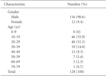
Results: According Sanders classification, type II was 37 cases (31%), type III 66 cases (55%), and type IV 17 cases (14%). On plane radiography, the Böhler angle improved to