J Korean Soc Coloproctol Vol. 20, No. 3, 2004
133
The Early Experience of Virtual Colono- scopy on Colorectal Cancer
Han Il Lee, M.D., Tae Soon Lee, M.D., Soon Jai Jung, M.D., Ki Hyuk Park, M.D., Dong Rack Choi, M.D., Dae Hyun Joo, M.D., Sung Hwan Park, M.D., Yong Oon Yoo, M.D., Ki Ho Park, M.D., Young Hwan Lee, M.D.1, Jin Cheon Kim, M.D.2
Departments of Surgery and 1Radiology, Catholic University of Daegu School of Medicine, Daegu, 2Department of Surgery, University of Ulsan, Asan Medical Center, Seoul, Korea Purpose: Virtual colonoscopy (VC) is a newly developing non-invasive technique used to detect polyps and cancers of the colon. The aim of this study is to assess the efficacy of VC in the detection of synchronous polyps or cancers in preoperative patients as well as metachronous polyps of postoperative colorectal cancer patients.
Methods: Both VC and conventional colonoscopy (CFS) were performed on 40 patients with colorectal cancer (10 cases of preoperative state and 30 cases of post- operative follow-up) during Sep. 2002 to June 2003 in Daegu Catholic Medical Centre, Catholic University of Daegu, Republic of Korea. The success rate and the detection rate of polyps or cancers along with the loca- tions and sizes of masses and the findings of anastomotic site were compared between VC and CFS.
Results: The entire colon was clearly visualized by CFS in all cases. In the preoperative group, VC was success- fully performed in 8 out of 10 cases (80%). 8 out of 10 cancers, 4 out of 4 polyps (5 mm or more in diameter) and 3 out of 6 polyps (5 mm or less in diameter) were identified. The success rate of VC in the postoperative group were 58% of low anterior resection (LAR) from cecum to hepatic flexure, 89% of LAR, 45% of right hemicolectomy (RHC) from hepatic flexure to splenic flexure, 63% of LAR, 45% of RHC from splenic flexure to sigmoid colon, and 53% of LAR, 72% of RHC in rectum. The causes of failure were inadequate bowel
distension and retained fluid. In postoperative group, VC identified only 3 of 7 polyps(5 mm or more in diameter), 1 of 10 polyps (5 mm or less in diameter) and 1 of 1 recurrent cancer. The anastomotic site was clearly seen by VC in 9 of 19 cases (47%) of LAR and 3 of 11 cases (27%) of RHC. VC also identified 28 extracolonic find- ings.
Conclusions: Although the efficacy of VC in postopera- tive colorectal cancer follow up seems to be disappoint- ing, but it can be used as an alternative method for pa- tients with incomplete conventional colonoscopy due to anastomotic site stricture or for other failed cases. Fur- ther technological advancement of VC is needed in order for it to replace conventional colonoscopy as a postopera- tive follow-up test. J Korean Soc Coloproctol 2004;20:
133-137
Key Words: Virtual colonoscopy, Colorectal neoplasm 가상대장내시경, 대장직장종양
ꠏꠏꠏꠏꠏꠏꠏꠏꠏꠏꠏꠏꠏꠏꠏꠏꠏꠏꠏꠏꠏꠏꠏꠏꠏꠏꠏꠏꠏꠏꠏꠏꠏꠏꠏꠏꠏꠏꠏꠏꠏꠏꠏꠏꠏꠏꠏꠏꠏ
서 론
대장암은 우리나라에서 4번째로 호발하는 암으로 해마 다 증가하는 추세이다.1 대부분의 대장암은 이전에 존재 하는 용종에서 발생하므로 암의 전구가 되는 용종의 발 견과 조기 제거는 대장암의 발생빈도를 낮출 수 있을 뿐 만 아니라 암으로 인한 사망률 역시 낮출 수 있다. 지금까 지 분변잠혈검사, 대장조영술, 에스결장경검사, 대장내시 경검사 등을 통해 대장암 조기 발견을 시행하였으나, 각 각의 검사들은 한계를 가지고 있으며, 보다 비침습적이 며 확실한 검사법에 대한 필요성은 여전히 남아 있다.
가상대장내시경(virtual colonoscopy)은 CT 혹은 MRI를 이용하여 환자의 영상정보를 얻은 후 소프트웨어를 이용 하여 대장내강에 대한 3차원 이미지를 재구성하여 용종 혹은 암을 진단하는 최근에 개발된 비침습적인 방법으 로,2-5 에스결장경 검사에서 종양의 발견으로 상부의 추가 검사가 필요하거나 대장내시경에 실패한 경우 혹은 대장 암의 선별 검사의 하나로 시도될 수 있을 것으로 보고되 고 있다.6-10 그러나 대장암으로 진단받았거나 대장암 수
대장암에서 가상대장내시경의 초기경험
대구가톨릭대학교 의과대학 외과학교실, 1방사선과학교실, 2울산대학교 의과대학 외과학교실
이한일․이태순․정순재․박기혁․최동락․주대현․박성환․유용운․박기호․이영환1․김진천2
책임저자: 이한일, 대구광역시 남구 대명 4동 3056-6 대구가톨릭대학교 의과대학 외과학교실 (우편번호: 705-718)
Tel : 053-650-4061, Fax: 053-624-7185 E-mail: [email protected]
본 논문의 요지는 2003년 대한대장항문학회 춘계학술대회에서 구연 발표되었음.
방 법
2002년 9월부터 2003년 6월까지 대구가톨릭대학병원
를 소디움 포스페이트(Solin , Pharma, Korea) 혹은 Polye- thylene glycol (ColyteⓇ, Tae-Joon, Korea)을 이용해 시행하 였으며, 검사 직전에 약 1,000∼1,500 cc의 공기를 항문을 통해 주입하여 대장 전체가 팽창되게 하였다.
가상대장내시경은 전산화단층촬영술(CT)을 이용하여 시행하였으며, 나선형 CT (Lightspeed plus, GE)로 횡경막 부터 항문까지 2.5 mm 간격으로 촬영 후 workstation을 이 용하여 3차원 영상으로 재구성하였다.
결 과
남녀의 수는 각각 22, 18명, 평균 나이는 59.2±10.7세, 62±9.1세로 나타났으며 수술 전 환자는 10예, 수술 후 추 적관리환자는 30예로 수술의 종류는 저위전방절제술 19 예, 우반결장절제술 11예였다. 대장내시경은 모든 환자에 서 별문제 없이 시행되었으나, 가상대장내시경은 술 전 환자의 80% (8/10)에서 만족할 만한 검사가 이루어진 반 면, 술 후 환자에서는 대장을 맹장-간곡, 간곡-비장곡, 비장
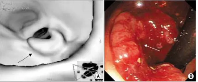
Fig. 2. VC image demonstrating the anastomotic site (arrows) of low anterior resection (A). Same site (arrows) seen at conventional colonoscopy (B).
Fig. 1. Sectional success rate of virtual colonoscopy in pos- toperative colon cancer patients (CE = cecum; HF = hepatic flexure; SF = splenic flexure; SC = sigmoid colon; RE = rectum;
LAR = low anterior resecion; RHC = right hemicolectomy).
Fig. 5. Comparison of the detection rate of cancers and polyps between conventional colonoscopy and virtual colonoscopy (CFS = conventional colonoscopy; VC = virtual colonoscopy;
Preop = preoperative; Postop = postoperative).
곡- 에스결장, 직장부로 나누어 관찰하였을 때 저위전방 절제술은 각 11 (58%), 17 (89%), 12 (63%), 10예(53%)에 서, 우반결장절제술은 맹장-간곡을 제외하고 5 (45%), 5 (45%), 8예(72%)에서 절대 만족 혹은 상대적으로 만족할 만하였다(Fig. 1).
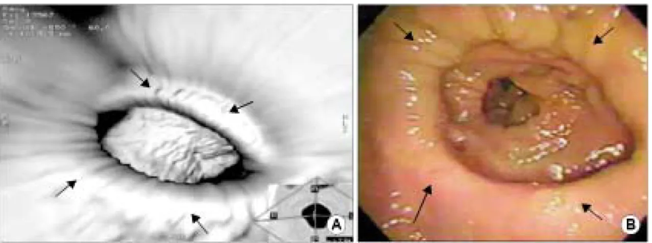
실패 이유로는 충분한 팽창의 실패와 과도한 대장내용 물이 지적되었다. 문합부(Fig. 2)의 확인은 저위전방절제 술 19예 중 9예(47%), 우반결장절제술 11예 중 3예(27%) 에서만 성공적으로 관찰되었다. 가상대장내시경에서 술 전 환자는 대장내시경에서 관찰된 10개의 종양(Fig. 3) 가 운데 8개를 확인할 수 있었고, 용종(Fig. 4)은 5 mm 이상 크기의 4개의 용종 모두 관찰되었으며, 5 mm 미만은 6개 중 3개를 확인하였으나, 수술 후 환자는 5 mm 이상은 7개 중 3개, 5 mm 미만은 10개 중 1개에서만 관찰되었다(Fig. 5).
대장 외 병변은 모두 28예의 병변이 관찰되었으며, 재 발을 나타내는 병변은 관찰되지 않았다(Fig. 6).
Fig. 3. VC image demonstrating a mass (arrows) in the rectum (A). Same mass (arrows) seen at conventional colonoscopy (B).
Fig. 4. VC image demonstrating a 6 mm polyp (arrow) in the sigmoid colon (A). Same polyp (arrow) seen at conventional colonoscopy (B).
고 찰
가상대장내시경은 1994년 Vining 등2이 처음 제시한 이 후, 급속한 기술의 발달과 더불어 그 유용성에 대해 많은 연구가 이루어져 왔다. 기존의 대장내시경은 아직까지 대장암의 검사에 가장 효과적인 검사이지만, 시술에 따 른 환자의 불편함, 5∼15%의 검사 실패율 및 24%에 이르 는 용종성 병변의 발견 실패율로 인해 이상적인 검사로 는 부족한 것으로 알려져 있다.11-15 가상대장내시경은 기 존의 대장내시경의 단점을 극복하면서 내시경과 유사한 2차원 및 3차원 영상을 나선 CT를 통해 나타내어 대장암 의 이상적인 진단의 도구로서 제시되었다.
지금까지의 연구에 따르면 10 mm 이상의 용종에서 발 견의 민감도는 73∼91%, 6∼9 mm의 용종에서는 65∼
82%로 보고되어 아직까지는 연구마다 편차가 있으나,3-10 최근의 두 연구에서는,3-4 10 mm 이상의 용종에서 발견의 민감도는 90%, 89%로 보고하여 대장암의 선별검사의 가 능성을 제시하였다. 하지만 아직까지도 6∼9 mm의 용종 은 발견 민감도가 80% 미만으로 알려져 선별검사로서 적 용하는 데 한계점으로 지적되고 있으며,8-10 그 외에 평편 병변에 대한 낮은 민감도,16 잔변 등에 의한 위양성, 대장 내 잉여 수분 등으로 인한 위음성 등이 한계점으로 지적 되고 있다.8,10 본 연구에서는 비록 적은 수의 환자이지만 술 전 환자에서 5 mm 이상의 용종은 4개 모두 발견이 가 능했으나, 5 mm 미만의 용종은 가상대장내시경에서 50%
만이 관찰되어 앞의 연구와 유사한 결과를 나타내었다.
현재까지 보고된 가상대장내시경의 장점은 대장 전반 을 검사할 수 있으며, 비침습적이며, 환자의 진정이 필요 없으며, 빠른 검사 시간과 사회 복귀 등이 있다. 특히 가
대장내시경은 국소재발의 확인, 이시성 병변의 확인, 문 합부 술 후 협착, 그리고 다른 대장질환의 확인 등에 사용 되고 있으나, 대부분의 국소 재발은 대장내강에서 발생 하지 않으므로 내시경검사에서 불분명할 경우가 많
다.17,18 혈청 CEA 검사는 재발성 대장암에서 가장 민감한
혈청 표지자이지만, 높은 위음성 및 위양성률의 제한점 이 있다.19,20 정기적 컴퓨터단층 촬영은 복막전이, 간전이 및 림프절 전이 등을 확인하는 데 이용되나 국소 재발에 는 낮은 민감도를 나타낸다.17
하지만 가상대장내시경은 컴퓨터단층촬영으로 이루어 지기 때문에 대장 외 병변을 동시에 확인할 수 있으며,5-8 내시경과 같이 대장 내강을 직접적으로 관찰하면서 문합 부, 대장점막부위, 대장벽 및 대장 주위조직을 함께 확인 할 수 있다. 본 연구에서도 28개의 대장 외 병변이 관찰되 었으나, 추적기간이 짧고 적은 환자수로 인해 의미 있는 재발성 병변은 관찰되지 않았다. 하지만 이 점은 가상대 장내시경이 추적검사로서 가질 수 있는 중요한 장점이라 고 저자들은 생각한다. 그리고 결과에 서술하지 않았지 만 본 연구 중 3예에서 가장대장내시경에서만 용종이 확 인되었는데, 이는 가상대장내시경이 기존의 내시경 검사 에 보조적 역할을 할 수 있다는 가능성을 제시한다고 생 각한다.
Fletcher 등21은 근치적 수술을 받은 진행성 대장암 환자 에서 조영제 추가 가상대장내시경이 국소 재발, 이시성 병변 및 원격재발의 확인에 유망한 검사로 제시하였으 나, 수술 후 해부학적 변화로 인한 부적절한 대장 팽창, 잔변 등에 의한 위양성 및 위음성 등을 극복해야 할 문제 점으로 제시하였다. 본 연구의 결과에서도 술 후 환자에 서의 가상대장내시경의 결과는 술 전 환자에서와 달리 성공률이 매우 저조하였는데, 이는 술 후 나타난 해부학 적 변형, 즉 저위전방절제술에서의 항문괄약근의 기능저 하, 방사선 치료 또는 수술로 인한 조직의 섬유화, 우반절 제술에서의 회맹판의 소실 등이 적절한 대장 팽창을 하 Fig. 6. Extracolonic findings of virtual colonoscopy.
는 데 제약이 되어 낮은 성공률을 보이게 된 것으로 생각 된다.21
그리고, 몇몇 보고에서 나타났듯이 가상대장내시경의 적절한 판독을 위해서는 일정한 학습기간이 필요하며 특 히 기존의 대장내시경에 대한 방사선과 의사의 이해가 중요하다고 하였다.9,10 따라서 기술의 발달과 환자의 자 세 변경, 적절한 공기주입, 적절한 대장청소 및 대장 팽창 의 직접적인 확인 등을 추가하여 경험을 증가시킨다면 보다 나은 결과가 나타날 것으로 생각된다.
결 론
경험의 증가와 함께 충분한 전처치 및 안전한 대장의 팽창을 유도한다면 가상대장내시경은 대장내시경을 거 부하는 환자 혹은 선별 검사의 하나로 조심스럽게 시도 될 수 있을 것으로 보이나, 술 후 추적검사로는 대장암의 수술 후 항문괄약근의 기능저하 혹은 회맹판의 손실로 인해 충분한 대장의 팽창이 어려우므로 문합부 협착 혹 은 과도한 복강 내 유착으로 인해 대장내시경이 불가능 할 때 대장 내시경의 보조적 수단으로 사용될 수 있을 것 으로 생각된다.
REFERENCES
1. Annual report of Korea central cancer registry program, Headquater of Korea Central Cancer Registry, Ministry of Health and Welfare, Republic of Korea, 2002.
2. Vining DJ, Gelfand DW, Bechtold RE, Scharling ES, Gri- shaw EK, Shifrin RY. Technical feasibility of colon imaging with helical CT and virtual reality. AJR 1994;162:Suppl:104.
3. Hara AK, Johnson CD, MacCarty RL, Welch TJ, Mc- Collough CH, Harmsen WS. CT colonography: single-versus multi-detector row imaging. Radiology 2001;219:461-5.
4. Yee J, Akerkar GA, Hung RK, Steinauer-Gebauer AM, Wall SD, McQuaid KR. Colorectal neoplasia: performance charac- teristics of CT colonography for detection in 300 patients.
Radiology 2001;219:685-92.
5. Hara AK, Johnson CD, MacCarty RL, Welch TJ. Incidental extracolonic findings at CT colonography. Radiology 2000;
215:353-7.
6. Hopper KD, Khandelwal M, Thompson C. CT colonoscopy:
experience of 100 cases using volumetric rendering. Proc SPIE 2001;12:489-94.
7. Morrin MM, Kruskal JB, Farrell RJ, Goldberg SN, McGee JB, Raptopoulos V. Endoluminal CT colonography after an incomplete endoscopic colonoscopy. AJR 1999;172:913-8.
8. Laghi A, Iannaccone R, Carbone I, Catalano C, Di Giulio
E, Schillaci A, et al. Detection of colorectal lesions with virtual computed tomographic colonography. Am J Surg.
2002;183:124-31.
9. Gluecker TM., Fletcher JG. CT colonography (virtual colono- scopy) for the detection of colorectal polyps and neoplasms.
current status and future developments. Eur J Cancer 2002;
8:2070-8.
10. Dachman AH. Diagnostic performance of virtual colono- scopy. Abdom Imaging 2002;27:260-7.
11. Fenlon HM, McAneny DB, Nunes DP, Clarke PD, Ferrucci JT. Occlusive colon carcinoma: virtual colonoscopy in the preoperative evaluation of the proximal colon. Radiology 1999;210:423-8.
12. Macari M, Berman P, Dicker M, Milano A, Megibow AJ.
Usefulness of CT colonography in patients with incomplete colonoscopy. AJR 1999;173:561-4.
13. Rex DK. Colonoscopic withdrawal technique is associated with adenoma miss rates. Gastrointest Endosc 2000;51:33-6.
14. Morrin MM, Farrell RJ, Raptopoulos V, McGee JB, Bleday R, Kruskal JB. Role of virtual computed tomographic colonography in patients with colorectal cancers and ob- structing colorectal lesions. Dis Colon Rectum 2000;43:
303-11.
15. Rex DK, Cutler CS, Lemmel GT, Rahmani EY, Clark DW, Helper DJ, et al. Colonoscopic miss rates of adenomas determined by back-to-back colonoscopies. Gastroenterology 1997;112:24-8.
16. Gluecker T, Johnson CD, Fletcher JG, et al. Understanding errors at CT colonography: a blinded comparison of missed and identified colon lesions. SGR Meeting, 25 March 2001 (abstr).
17. Wenebo HJ, Llaneras M, Martin T, Kaiser D. Prospective monitoring trial for carcinoma of the colon and rectum after surgical resection. Surg Gynecol Obstet 1989;169:479-85.
18. Barkin JS, Cohen ME, Flaxman M, Lindblad AS, Mayer RJ, Kalser MH, et al. Value of a routine follow-up endoscopy program for the detection of recurrent colorectal carcinoma.
Am J Gastroenterol 1988;83:1355-60.
19. Kelly CJ, Daly JH. Colorectal cancer: principles of post- operative follow-up. Cancer 1992;70:1397-408.
20. Moertel CG, Fleming TR, Macdonald JS, Haller DG, Laurie JA, Tangen C. An evaluation of the carcino-embryonic antigen (CEA) test for monitoring patients with resected colon cancer. JAMA 1993;270:943-7.
21. Fletcher JG, Johnson CD, Krueger WR, Ahlquist DA, Nel- son H, Ilstrup D, et al. Contrast-enhanced CT colonography in recurrent colorectal carcinoma: feasibility of simul- taneous evaluation for metastatic disease, local recurrence, and metachronous neoplasia in colorectal carcinoma. AJR 2002;178:283-90.