백두구 추출물의 항산화 및 항비만 효과
박정애1, 진경숙1, 이지영1, 권현주1,2, 김병우1,2*
1동의대학교블루바이오소재개발및실용화지원센터
2동의대학교생명응용학과
Received: March 6, 2014 / Revised: May 26, 2014 / Accepted: May 29, 2014
서 론
비만은신체에너지소비량보다과잉으로에너지를섭취 하였을때점차적으로체지방이피하조직이나, 장간막에축 적되어체중이증가하는상태로유전적, 영양적, 환경적및 사회적요인등다양한원인들이관여하는복합증후군이다
[7]. 비만의발생원인으로는과도한식이섭취, 신체활동부
족, 그릇된생활습관, 내분비기능이상질환및유전적인 소인등이있으며비정상적인지방세포의크기및숫자증 가로지방축적을일으키게된다[3, 15]. 이는 adipokine의생
산과분비를통하여 adipogenesis 과정으로분화가일어나
게하며 지방세포의 크기 증가(hypertrophy)와숫자 증가 (hyperplasia)는비만뿐만아니라염증, 당뇨등만성대사질 환에영향을미치게된다[2, 8, 14, 23]. 따라서, 체중감소를 위해서는운동, 식이요법, 약물투여, 외과적수술등의방법 이수행되고있으며이중식이요법은비만의예방과치료에 가장근본적이며, 중요한방법이다[16].
비만은지방전구세포의분화및지방생성과정에의하여지 방세포의세포내중성지방(triglyceride, TG)의축적으로발 생하므로이러한지방생성기전을조절하는것이비만억제
의효과적인치료방법으로알려져있다[4, 5]. 잘알려진대
표적인비만치료제인 orlistat의경우췌장및위장에서분
비되는지방분해효소인 lipase의활성을억제하여섭취한
지방중약 30%의흡수를차단함으로서체중의감소에도
움을주는것으로알려져있다[13]. 이에최근천연물을이
Anti-Oxidative and Anti-Obesity Effects of Amomum Cardamomum L. Extract Jung Ae Park1, Kyong-Suk Jin1, Ji Young Lee1, Hyun Ju Kwon1,2, and Byung Woo Kim1,2*
1Blue-Bio Industry Regional Innovation Center, 2Department of Life Science and Biotechnology, College of Natural Science, Dong-Eui University, Busan 614-714, Republic of Korea
In this study, the anti-oxidative and anti-obesity activities of Amomum cardamomum L. methanol extract (ACME) were evalu- ated using DPPH radical scavenging activity assay, pancreatic lipase enzyme inhibition assay, and the cell culture model sys- tem. ACME exhibited DPPH radical scavenging activities dose-dependently, with IC50 of DPPH radical scavenging activities of ACME being 25.15 μg/ml. Furthermore, ACME effectively suppressed pancreatic lipase enzyme activity dose-dependently.
ACME also significantly suppressed adipocyte differentiation, lipid accumulation, triglyceride (TG) contents, and triggered lip- olysis activity on 3T3-L1 preadipocytes in a dose-dependent manner, without cytotoxicity. Their anti-obesity effect was modu- lated by the cytidine-cytidine-adenosine-adenosine-thymidine (CCAAT)/enhancer binding proteins α (C/EBPα), C/EBPβ and the peroxisome proliferator-activated receptor γ (PPARγ) gene and protein expressions. Taken together, these results provide an important new insight that A. cardamomum L. possesses anti-oxidative and anti-obesity activities such as pancreatic lipase inhibition, anti-adipogenic, and lipolysis effects. There is therefore potential for its use as a promising component in the field of nutraceuticals and the identification of the active compounds that confer the anti-oxidative and anti-obesity activities of ACME might be an appropriate next step.
Keywords: Amomum cardamomum L. methanol extract, anti-oxidative activity, anti-obesity activity, lipase inhibition activity, anti-adipogenic and lipolysis effects
*Corresponding author
Tel: +82-51-890-2900, Fax: +82-51-890-2914 E-mail: [email protected]
© 2014, The Korean Society for Microbiology and Biotechnology
용한유용비만소재개발의많은연구가천연유래 lipase 활성저해제로서의작용가능성탐색에초점을맞추고있다. 지방세포의분화는세포형태및유전자발현의다양한변 화가동반되는복합적인과정을통하여일어나게된다. 지방 세포는몸에필요한에너지를축적하여, 필요시중성지방을 분해시켜이용한다. 하지만최근연구에의하여지방조직은 에너지축적의기능뿐만아니라, 내분비기관으로지방대사, 당대사를포함한체내에너지대사를조절하는기능을수행
하는역할을하는것으로도알려져있다[9, 11, 17]. 지방세
포는 mesenchymal precursor에서 preadipocyte를거쳐지 방세포로분화되며, 분화과정동안형태적, 생화학적변화를 통하여체내지방을축적하며, 지방조직은크기의증가, 새
로운 preadipocyte로부터분화되고이는지방조직세포에특
징적으로 발현되는 유전자들의 조절부위에 작용하는 transcription factor들이지방세포로분화되는과정에서발 현된다[10].
3T3-L1 지방전구세포가지방세포로분화될때 cytidine-
cytidine-adenosine-adenosine-thymidine (CCAAT)/enhancer binding protein의 종류인 C/EBPβ, C/EBPδ가 insulin, dexamethasone (DEX), 3-isobutyl-1-methylxanthine (IBMX) 와같은호르몬자극으로부터초기분화가시작되며이는상 호작용 또는 단독으로 peroxisome proliferator-activated receptor γ (PPARγ) 및 C/EBPα의발현을조절하게된다[4, 6, 21, 22, 27-30]. C/EPBα는 PPARγ와분화초기에발현이
유도되어분화후기가되면다양한 adipogenic 유전자들의
발현을유도하여, 분화된지방세포에서발현양이현저하게 증가되는양상을나타낸다[19, 23, 24].
한편전세계적으로비만치료제의개발을위한다양한연 구가수행되고있는가운데현재시판되고있는비만치료 제들의부작용들이보고되면서사용기준이강화되는등의 논란이일고있어우수한효능과함께안전성이높은물질 의개발이요구되고있다. 이에특히천연유래소재로부터 독성및부작용이없는항비만효능보유소재를발굴하기 위한많은노력이집중되고있다[12, 16, 19, 22].
백두구(Amomum cardamomum L.)는생강과에속하는
다년생초본인백두구와자바백두구의과실을지칭하는것 으로, 다골(多骨), 백구(白寇), 각박(殼泊)이라고도불리며주 로성숙한과실의과피를제거한것을백두구라고한다. 백 두구의과실은길이 1-2 cm, 지름 5-10 mm 크기로거의원 구형을이루고있으며바깥면은엷은황백색내지황갈색이 고 3줄의둔한능과여러개의세로줄이있으며, 윗쪽은움 푹들어갔고아랫쪽은과병이붙었던흔적이있다. 가을에 황녹색을띤성숙한과실을채취하여과병과과피를제거하 고햇빛에말려사용한다. 백두구는캄보디아, 베트남, 태국 이주산지이며, 자바백두구는인도네시아가주산지이나, 상
기 2종모두중국의남부지방에서재배한다. 소화기계질환 에자주쓰이며, 몸안의습을없애고몸을따뜻하게하여기 를잘통하게하고구토를멈추는효과가있는것으로알려 져있다. 또한위액분비를촉진하고위장의운동을강화하여 장내이상발효를억제하고가스를잘나가게하여입맛을돋 구고소화불량및식적등을없앤다. 만성위염으로인한위 통이나트림, 구토등의증상에좋으며, 술독을제거하는작 용이있다. 보고된생리활성으로는항암[1, 20] 및항산화[18]
효과등이있으나백두구의항비만활성에대해서는보고된 바없다.
이에본연구에서는한방에서약재로사용되고있는백두 구추출물의항비만효과및그기전을알아보고기능성소 재로서의활용가능성을확인해보고자하였다.
재료 및 방법
백두구 추출물의 제조
실험에사용한백두구는부산광역시소재㈜대한생약제품 에서구입하여사용하였다. 백두구 10 g을측량하여분말로 파쇄한 후 시료 부피 5배의 methanol (MeOH)을 가하여 75oC에서 3회반복추출하였다. 추출한시료는여과후감 압농축기(N-1000S-W, EYELA, Japan)로농축하고동결건 조(FDU2100, EYELA, Japan)한후중량법으로각각의수 율을계산하였다.
DPPH radical 소거 활성 측정을 통한 백두구 추출물의 항 산화능 분석
전자공여작용은인체내에서생성되는 free radical의전
자를공여하여 free radical에의한노화와질병을억제한
다. 따라서전자공여능은항산화작용의지표로서사용되고 있으며특히식물추출물의항산화능측정에많이사용되고 있다. 이러한전자공여능은 1,1-diphenyl-2-picryl hydrazyl (DPPH) radical 소거능분석을이용하여측정하였다. DPPH 는비교적안정한 free radical로써, ascorbic acid, tocopherol, polyhydroxy 방향족화합물, 방향족아민류등에의해환원 되어짙은자색이탈색되는원리를이용하여항산화활성을 간단히측정할수있는동시에식물체의항산화활성과도연
관성이매우높기때문에많이이용되고있는방법이다[15].
백두구추출물을농도별(12.8-1,600 μg/ml)로 MeOH에녹 여준비한후, 96 well plate에 MeOH에용해된 1.5×10⁻⁴ M DPPH 40 μl와각시료 160 μl를분주한혼합액을실온에서 30분간반응시키고, multi-plate reader (Paradigm, Beckman, CA, USA)를이용하여 520 nm에서흡광도를측정하였다. 시
료를첨가하지않은음성대조군과비교하여처리농도별 free
radical 소거 정도를 백분율로 나타내고, 50% 저해 농도
(Inhibitory Concentration, IC50)를계산하였다. 대표적인항
산화제로 DPPH radical 소거활성측정시양성대조군으
로주로사용되는 ascorbic acid를함께비교분석하였으며
측정값은 3회반복실험의평균값으로나타내었다.
백두구 추출물의 pancreatic lipase 효소 활성 저해능 분석 Lipase는주로췌장에서분비되어 TG를 glycerol과 fatty acid로가수분해하는효소로서지방소화효소인 pancreatic
lipase의활성저해능은시료가보유한항비만활성을예측
하기에매우유용한시험법이다[9]. 본연구에서는백두구의 항비만활성을세포실험계에서분석하기에앞서시료가보 유한 lipase 효소활성저해능을다음과같이측정하였다. 먼 저 1.5 ml tube에 0.25 M Tris (pH 7.7), 250 mM CaCl2, 5 mM 4-nitrophenyl dodecanoate (PNPD)로 구성된효소 액과기질을넣고잘섞어준후 37oC에서 5분간예열하고, 0.25 M Tris (pH 7.7)에녹인 lipase와시료를넣어 37oC에 서 10분간반응시킨후 20% sodium dodecyl sulfate (SDS) 를첨가하여반응을종료하였다. 반응액을 4oC, 15,000 rpm 에서 20분간원심분리하여, 분리한상층액을 96-well tissue culture plate에 분주하고 multi-plate reader를 이용하여
412 nm에서흡광도를측정한후, 10분간반응시킨시료의
흡광도로부터 0분반응시료의흡광도를뺀값을 control 대 비백분율로나타내었다. 측정값은 3회반복실험의평균값 으로나타내었다.
세포 배양 및 시료 처리
백두구의항비만활성을세포실험계에서분석하기위해항 비만활성분석에사용되는대표적인 cell model system인 3T3-L1 preadipocyte를 American Type Culture Collection (ATCC, VA, USA)로부터 구입하여 10% fetal bovine serum (FBS) 및 penicillin/streptomycin이포함된 DMEM 배지에서배양하였다. 0.5 μM 3-isobutyl-1-methylxanthine (IBMX), 1 μM dexamethasone (DEX), 10 μg/ml의 insulin (이하 MDI)를처리하여 adipogenesis를유도하고백두구추 출물에의한항비만활성을분석하였다[25].
세포 독성 유무 분석
항비만활성분석수행전시료가세포생존율에미치는영 향을확인함과동시에세포독성을유발하지않는시료의처 리농도를결정하기위해 water soluble tetrazolium (WST) assay를수행하였다. 1×10⁵ cell을 24-well tissue culture
plate에분주하여 24시간동안부착시킨후시료를농도별
로처리하여 72시간동안배양하였다. 시료처리후 WST 시
약이든배지로교체하여한시간동안반응시킨후 multi-
plate reader를이용하여 450 nm에서흡광도를측정하였다.
측정값은 3회반복실험의평균값으로나타내었으며독성을 유발하지않는농도범위에서이후실험을수행하였다. Oil Red O staining을 통한 지방세포 분화 및 TG 생성 저해능 분석
3T3-L1 지방전구세포의지방세포로의분화는상기의 MDI
를 처리하여 유도하였다. 12-well tissue culture plate에 well 당 2×10⁵개의세포를분주하고 2일후 10% FBS가든 배지로교체하였다. 2일경과후 MDI가든배지로교체하 면서 시료를 농도 별로 처리하고 2일 간격으로 총 4회 insulin과시료를처리하였다. 마지막시료를처리하고 2일 경과후위상차현미경을이용하여지방세포분화정도및 시료에의한분화억제정도를 200배배율로관찰하여촬영 한후, 지방세포분화억제능및 TG 생성저해능을 Oil Red O staining을통해분석하였다. 지방세포분화및시료처리 가 완료된 세포를 1× phosphate buffered saline (PBS)로
씻어준 다음 10% formalin으로 고정하고 Oil Red O
staining solution을처리한후 30분간염색하였다. 염색완 료후 100% isopropanol을사용하여염색된지방을추출하 고 multi-plate reader를이용하여 500 nm에서흡광도를측 정하였다.
Reverse transcription-polymerase chain reaction (RT-PCR)을 통한 지방세포 분화 관련 유전자 발현 조절능 분석
백두구가지방세포분화에중요한역할을담당하는유전자 의발현에미치는영향을알아보기위해 6-well tissue culture plate에 3×10⁵개의세포를분주하고지방생성억제능분석 과동일한방법으로시료를처리한후 RNA를분리하여각유
전자의발현을 RT-PCR로분석하였다. 먼저시료처리가완료
된배양세포의 total RNA를 TRIzol® (Invitrogen, CA, USA) 을 사용하여 추출한 후 NanoVue plus spectrophotometer (GE healthcare, WI, USA)를이용하여정량하고 SuperScriptTM First-Strand Synthesis System (Invitrogen)을 이용하여 cDNA를합성한후 PCR을수행하였다. 유전자발현분석의 internal control로는 housekeeping gene인 glyceraldehydes-3- phosphate dehydrogenase (GAPDH)를사용하였으며실험 에사용한대상유전자의염기서열은 Table 1에제시한바 와같다.
Western blot hybridization을 통한 지방세포 분화 관련 단백질 발현 조절능 분석
상기의 RT-PCR 분석에서와같은방법으로실험을수행
한후시료처리가끝난세포에서단백질을분리하여지방 생성관련단백질의발현변화를 Western blot hybridization으
로분석하였다. 먼저시료처리가끝난배양세포에서 cell lysate를추출하여 Bradford assay로단백질농도를결정한 후 50μg의 단백질을 10% SDS-polyacrylamide gel electrophoresis로 전기영동하고 nitrocellulose membrane 에 blotting한후대상단백질의일차항체와 hybridization 하였다. 실험에사용한 C/EBPα와 C/EBPβ의일차항체는 Cell Signaling Technology (Beverly, MA, USA)로부터구 입하였고, PPARγ 및 actin의 일차 항체와 horse radish peroxidase가부착된 anti-goat, anti-rabbit, anti-mouse등 의이차항체는 Santa Cruz Biotechnology Inc. (Santa Cruz, CA, USA)로부터구입하여사용하였다. Membrane 수세후 이차항체로한시간동안반응시키고 chemiluminescence detection system (FluoChem® FC2, AlphaInnotech, USA)을 이용하여각단백질의발현을분석하였다.
지방세포내 중성지방 제거량 측정(lipolysis assay)
백두구가보유한지방세포내중성지방제거능(lipolysis
activity)은 아래와 같이 수행하였다. Confluence 상태의
3T3-L1 지방전구세포를 2일간배양한다음 MDI를첨가한
DMEM 배지로 2일간배양하고 10 μg/ml이든 DMEM 배지 에 4일간추가배양하였다. 이러한과정을통해지방세포분
화가완료된 3T3-L1 cell에서백두구추출물을농도별로처
리한다음 48시간후중성지방분해에의해배지에방출된 glycerol 양을 glycerol-3-phosphateoxidase (GPO)-TRINDER kit (Sigma, St. Louis, MO, USA)를사용하여 540 nm에서 흡광도를측정하였다. 측정값은 3회반복실험의평균값으 로나타내었다.
통계 분석
각실험의결과는평균(mean)± 표준편차(standard deviation, SD)로 나타내었고, 각 데이터의 통계 분석은 unpaired Student’s t-test를통해 p 값이 0.05 미만(p < 0.05)인경우 유의성이있는것으로판단하였다.
결과 및 고찰
백두구 추출물의 제조 및 항산화능 분석
백두구 10 g으로부터 MeOH 추출을통해 0.26 g의추출
물을확보하여그수율은 2.6%로나타났다. 또한항산화능
의지표인 DPPH radical 소거능분석을통해백두구추출
물의항산능을분석한결과 Table 2에제시된바와같이농
도의존적인 radical 소거능을보였으며 radical을 50% 억제
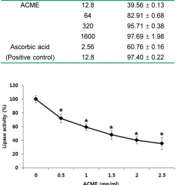
Table 2. DPPH radical scavenging activity by ACME. Extract Concentration
(μg/ml)
Inhibition rate (%)
ACME 12.8 39.56 ± 0.13
64 82.91 ± 0.68
320 95.71 ± 0.38
1600 97.69 ± 1.98
Ascorbic acid 2.56 60.76 ± 0.16
(Positive control) 12.8 97.40 ± 0.22
Fig. 1. Lipase enzyme inhibition activity of ACME. The effects of ACME on pancreatic lipase activity. Values are represented as the mean ± SD (n = 3) *p < 0.01 vs vehicle control (0).
Table 1. Primer sequences used for RT-PCR.
Gene name Sequence
C/EBPα Sense
Antisense
5'-GTG TGC ACG TCT ATG CTA AAC CA-3' 5'-GCC GTT AGT GAA GAG TCT CAG TTT G-3'
C/EBPβ Sense
Antisense
5'-GTT TCG GGA GTT GAT GCA ATC-3' 5'-AAC AAC CCC GCA GGA ACA T-3'
PPARγ Sense
Antisense
5'-CGC TGA TGC ACT GCC TAT GA-3' 5'-TGC GAG TGG TCT TCC ATC AC-3'
GAPDH Sense
Antisense
5'-GGG AGT CAA CGG ATT TGG TCG TAT-3' 5'-AGC CTT CTC CAT GGT GGT GAA GAC-3'
시키는농도를의미하는 IC50값은 25.15 μg/ml으로나타나 양성대조군으로사용한 ascorbic acid에비해서는활성이낮 았으나단일물이아닌추출물상태인점을감안할때높은 항산화능을보유하는것으로판단된다.
백두구 추출물의 pancreatic lipase 활성 억제능 분석
백두구의 lipase 활성억제능보유유무를알아보기위해
각추출물의농도별처리에따른 lipase의활성변화를분석
하였다. 그결과 Fig. 1에서 보는바와 같이 0.25, 0.5, 1.0, 1.5, 2.0, 2.5 mg/ml의처리에의해시료비처리대조군대비 백두구 추출물은 84.2, 65.3, 59.0, 53.1, 44.7, 25.5%의 lipase 활성을나타내며 lipase의활성을농도의존적으로유 의적으로억제시키는것을확인하였다. 이러한결과는백두 구가 lipase inhibitor로작용하여항비만활성을보유할가 능성을시사하였다.
백두구 추출물이 3T3-L1 preadipocyte의 세포 생존율에 미치는 영향
시료의지방생성억제능의평가를위해먼저백두구추출 물이 3T3-L1 preadipocyte의세포생존율에미치는영향을 WST assay를이용하여분석하였다. 그결과 Fig. 2A에서제 시된바와같이 50-500 μg/ml의시료처리에서세포독성을전 혀유발하지않음을확인하였다. 이러한결과는백두구가세 포독성을유발하지않는낮은독성의안전성이높은소재임 을의미한다. 이에백두구추출물의지방생성억제능의분석 을독성이없는것으로확인된농도범위에서수행하였다. 백두구 추출물이 MDI로 유도한 3T3-L1 preadipocyte의 adipogenesis에 미치는 영향
백두구추출물의항비만활성보유유무를알아보기위해 MDI로분화를유도한 3T3-L1 preadipocyte의 adipogenesis Fig. 2. Effect of ACME on 3T3-L1 cell proliferation (A), morphological change and lipid accumulation (B), and TG contents (C).
(A) Cells were treated with the indicated concentrations of ACME for 72 h and viability was determined by WST assay. Data are expressed as the mean ± SD of triplicate experiments. (B) Differentiation of confluent 3T3-L1 preadipocytes was initiated with MDI treatment and maintained in DMEM containing 5% FBS in presence and absence of ACME. After day 8, cells were fixed and stained with Oil red O.
The morphological change and lipid droplet accumulation were visualized using by inverted microscopy (×200). (C) TG contents were determined by Oil red O staining after treatment of ACME. TG contents was measured at 500 nm by multi-plate reader. Data are expressed as the mean ± SD of triplicate experiments. *, #Significantly different from the undifferentiated cell control (Con) and untreated cell control (0), respectively (p < 0.05).
에백두구추출물이미치는영향을살펴보았다. 그결과 Fig.
2B와 2C에서제시된바와같이농도의존적인지방세포분 화억제능이관찰되었고특히 200 μg/ml에서강한활성을보 여 Oil Red O staining 결과염색된지방의수가급격히감 소됨을확인할수있었다. 지방생성의억제정도를정량적 으로평가하기위해염색된지방을추출하여 TG 생성량의 정도를측정한결과 50에서 200 μg/ml의시료처리범위에 서농도의존적으로감소됨을보여 200 μg/ml의처리에의한 억제능이 33%로나타났다.
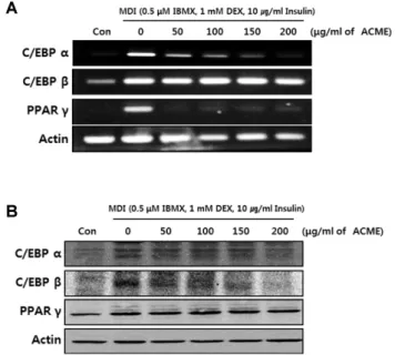
백두구 추출물이 adipogenesis 관련 유전자 및 단백질 발 현에 미치는 영향
지방전구세포가지방세포로분화되는 adipogenesis의과 정에는초기, 중기, 후기의각단계별로중요한분화조절자 들이관여한다. MDI 처리초기에는가장먼저 c-fos, c-jun, c-myc 등의유전자발현이증가하며이와함께 C/EBPβ와 δ 의발현또한유도된다. C/EBPβ와δ는 preadipocyte가 MDI 등의분화인자에노출되었을때분화초기에가장먼저작 용하는 전사 인자(transcription factor)로 MDI의경우 C/
EBPβ는 DEX에, C/EBPδ는 IBMX에의해활성이유도되는
것으로알려져있다. 이러한 C/EBPβ와δ의활성은분화중
기및후기에서작용하는 PPARγ와 C/EBPα의발현매개인
자로서, 활성화된 PPARγ와 C/EBPα는단독혹은상호작용 을통해지방세포특이적유전자의발현(adipocyte specific gene expression)을유도하여지방세포분화및지방형성
을완성한다[15]. 따라서이와같은지방세포분화에관여
하는주요인자들의발현조절유무는소재가보유한지방 생성억제능및그작용기전을판단하는주요지표중하나 이다.
본연구에서는백두구가보유한지방생성억제능의작용
기전을알아보기위하여백두구추출물이 adipogenesis에관
여하는 주요 핵심 조절자인 C/EBPα, C/EBPβ, 그리고
PPARγ의유전자및단백질발현에미치는영향을분석하였
다. 그결과 Fig. 3에제시된바와같이 MDI 처리에의해지 방세포분화가일어난대조군에서는세인자의유전자및단 백질발현이유의적으로증가되었으며추출물의처리에의 해농도의존적인감소를보였으나그정도및양상은각인
자에따라차이를보였다. 먼저 C/EBPα의경우유전자발
현은 50 μg/ml의처리에서부터 농도의존적으로억제되어
200μg/ml에서는미분화대조군과유사한정도의낮은발현
을 보였다. 이는 단백질 발현에서도 유사한 양상을 보여
200μg/ml의처리에서단백질발현또한미분화대조군과유
사한정도로억제되었다. C/EBPβ의경우, 유전자발현의억 제정도는낮았으나단백질발현에서농도의증가에따른발 현저해가관찰되었다. 한편 PPARγ의경우유전자발현은
모든시료처리군에서급격한감소를보여 50 μg/ml에서부 터미분화대조군과유사한정도로억제됨을보였으나단백 질발현은큰변화를보이지않았다. 이러한결과를바탕으 로 백두구 추출물이 보유한 지방세포분화 억제능이
adipogenesis에관여하는핵심인자의유전자및단백질발
현저해를통해나타나며그중에서도특히세인자중가장 늦은시점에발현되어지방세포분화조절자로서작용하는
C/EBPα의발현조절을통해나타나는것으로판단된다. 한
편, 백두구에의한세인자의유전자와단백질발현양상이 일부차이를나타내는데이는통상적으로유전자와 단백질 의발현양상이유사한형태를보이기는 하나항상일치하 는것은아니며 또한유전자의 발현및활성 시간과단백 질발현이유도되는시점의차이에서기인할가능성을시 사한다[26].
백두구 추출물의 중성지방 제거능(lipolysis activity) 백두구가지방세포분화를억제할뿐만아니라생성된지 방의제거에도효과적인지를판단하기위해지방세포내중 성지방제거능을 lipolysis activity를통해분석하였다. 그결
과 Fig. 4에제시한바와같이 MDI로분화시킨지방세포에
백두구추출물을처리한결과세포내축적되어있던중성
지방의분해를통해배지로방출된 glycerol의양이농도의
존적으로증가되는것으로나타났다. 배지를제거한후 Oil
Fig. 3. Effect of ACME on adipogenesis related gene and protein expressions. (A) Modulation of adipogenic transcription factors by ACME was evaluated by RT-PCR. GAPDH was used as an internal control. (B) Modulation of adipogenesis related protein expressions by ACME was evaluated by Western blot analysis. Actin was used as an internal control. The data are rep- resentative of three independent experiments.
Red O staining을수행한후현미경관찰및남아있는 TG
양을측정한결과방출된 glycerol의양과비례적으로세포
내에남아있는 TG의양이감소되는것으로나타나백두구 추출물이지방세포내에축적되어있는중성지방을농도의 존적으로제거하는것을확인하였다.
이러한결과를통해백두구추출물이높은항산화능을보
유함과동시에 lipase 효소활성억제능, 지방세포분화억제
능, 지방세포내중성지방제거능을통한항비만활성을보 유함을확인하였다. 이러한결과는백두구추출물의항비만 활성을처음으로밝혀낸것으로그의미가크다. 백두구의 주요성분으로α-borneol, α-camphor, α-numulene, numulene epoxide, 1.8-cineol, α-terpinene, β-terpinene, α-pinene, β-pinene, caryophyllene, myrcenal, carvone, terpinele-4- o, sabinene 등의정유, 방향성분이알려지고있으나이에 대한구체적인문헌및연구결과는알려진것이없으며상 기에제시한단일물질의항비만활성또한규명된바는없 다. 다만앞서언급한백두구의항암[1], 항염증[18], 면역조
절[20] 등의활성성분이대부분정유, 방향성분임을감안
할때, 항비만활성의주요성분또한유사할것으로생각된 다. 추후계속적인연구를통해백두구가보유한항비만활 성물질의규명이필요할것으로판단된다.
요 약
본연구에서는백두구(A. cardamomum L.) 메탄올추출 물(ACME)의항산화및항비만활성을 DPPH radical 소거 능과췌장 lipase 효소활성억제능, 그리고세포실험계를이 용하여분석하였다. 그결과 ACME는 DPPH radical을농도 의존적으로소거하였으며 DPPH radical 소거능의 50% 저 해농도(IC50)는 25.15 μg/ml로나타났다. 또한 ACME는농 도의존적으로 lipase 효소활성을유의적으로억제시켰으며, 3T3-L1 preadipocyte를이용하여지방세포분화및지방생
성, 생성된지방의분해에미치는영향을분석한결과 ACME
는지방세포분화, 세포내지방축적, TG 함량등을독성없 Fig. 4. Stimulatory effect of ACME on glycerol release in MDI-induced 3T3-L1 adipocytes (A), morphological change and lipid accumulation (B), and TG contents (C). (A) Amount of released glycerol in culture media was measured after ACME treatment. Glycerol contents were measured 540 nm by multi-plate reader. (B) Differentiation of confluent 3T3-L1 preadipocytes was initiated with MDI treat- ment and maintained in DMEM containing 5% FBS. After day 8, MDI-induced adipocytes were treated with ACME for 48 h. Cells were fixed and stained with Oil red O. The morphological change and lipid droplet accumulation was visualized using by inverted microscopy (×200). (C) TG contents were determined by Oil red O staining after treatment of ACME. TG contents was measured at 500 nm by multi-plate reader. Data are expressed as the mean ± SD of triplicate experiments. *,#Significantly different from the undifferentiated cell control (Con) and untreated cell control (0), respectively (p < 0.05).
이농도의존적으로억제하였으며지방세포내중성지방을 유의적으로분해시키는것으로나타났다. 이러한백두구의
지방세포 분화 억제능은 핵심 작용 인자인 C/EBPα, C/
EBPβ, 그리고 PPARγ의유전자및단백질발현조절에서기 인함을확인하였다. 이러한결과는백두구가보유한항산화 능과췌장 lipase 활성저해능, 지방세포분화억제능, 지방 세포내지방분해능을통한항비만활성을처음으로밝혀 낸것이며추후계속적인연구를통해활성물질의규명이 필요할것으로판단된다.
Acknowledgments
This work was supported by Blue-Bio Industry Regional Innova- tion Center (RIC08-06-07) at Dong-Eui University as a RIC pro- gram under Ministry of Trade, Industry & Energy and Busan city.
References
1. Acharya A, Das I, Singh S, Saha T. 2010. Chemopreventive properties of indole-3-carbinol, diindolylmethane and other constituents of cardamom against carcinogenesis. Recent Pat. Food Nutr. Agric. 2: 166-177.
2. Attie AD, Scherer PE. 2009. Adipocyte metabolism and obe- sity. J. Lipid Res. 50: 395-399.
3. Bae CR, Kwon DY, Cha YS. 2013. Anti-obesity effects of salted and unsalted doenjang supplementation in C57BL/6J mice fed with high fat diet. J. Korean Soc. Food Sci. Nutr. 42:
1036-1042.
4. Cao Z, Umek RM, McKnight SL. 1991. Regulcated express- tion of three C/EBP isoforms during adipose conversion of 3T3-L1 cells. Genes Dev. 5: 1538-1552.
5. Chen HC, Farese RV Jr. 2005. Inhibition of triglyceride syn- thesis as a treatment strategy for obesity: lessons from DGAT1-deficient mice. Arterioscler. Thromb. Vasc. Biol. 25:
482-486.
6. Chen Z, Torrens JI, Anand A, Spiegelman BM, Friedman JM.
2005. Krox20 stimulates adipogenesis via C/EBPbeta-depen- dent and –independent mechanisms. Cell Metab. 1: 93-106.
7. Chua SC Jr. 1997. Monogenic models of obesity. Behav.
Genet. 27: 277-284.
8. Després JP, Lemieux I. 2006. Abdominal obesity and meta- bolic syndrome. Nature. 444: 881-887.
9. Flier JS, Maratos Flier E. 1998. Obesity and the hypothala- mus: novel peptides for new pathways. Cell. 92: 437-440.
10. Gesta S, Tseng YH, Kahn CR. 2007. Developmental origin of fat: Tracking obesity to its source. Cell. 131: 242-256.
11. Gregoire FM, Smas CM, Sul HS. 1998. Understanding adipo- cyte differentiation. Physiol. Rev. 78: 783-809.
12. Gupta R, Rathi P, Gupta N, Bradoo S. 2003. Lipase assays for conventional and molecular screening: an overview. Biotech- nol. Appl. Biochem. 37: 63-71.
13. Heck AM, Yanovski JA, Calis KA. 2000. Orlistat, a new lipase inhibitor for the management of obesity. Pharmacotherapy.
20: 270-279.
14. Hotamisligil GS. 2006. Inflammation and metabolic disorders.
Nature. 444: 860-867.
15. Kedare SB, Singh RP. 2011. Genesis and development of DPPH method of antioxidant assay. J. Food Sci. Technol. 48:
412-422.
16. Kim SH, Kim JY, Ryu KA, Sohn CM. 2007. Evaluation of the dietary diversity and nutrient intakes in obese adults. Korean J. Community Nutr. 12: 583-591.
17. Kim EJ, Kim GY, Kim YM, Choi KH, Jang SJ. 2009. Anti-obe- sity effect of mulberry leaves extraction in obese rats high-fat diet. Korean J. Oriental Physiol. Pathol. 23: 831-836.
18. Lee DH, Kang SS, Chang IM, Mar W. 1997. Detection of anti- inflammatory agents from natural products as inhibitors of cyclooxygenase I and II. Nat. Prod. Sci. 3: 19-28.
19. Liu F, Kim J, Li Y, Liu X, Li J, Chen X. 2001. An extract of Lagerstroemia speciosa L. has insulin-like uptake-stimulatory and adipocyte differentiation-inhibitory activities in 3T3-L1 cells. J. Nutr. 131: 2242-2247.
20. Majdalawieh AF, Carr RI. 2010. In vitro investigation of the potential immunomodulatory and anti-cancer activities of black pepper (Piper nigrum) and cardamom (Elettaria carda- momum). J. Med. Food. 13: 371-381.
21. Morrison RF, Farmer SR. 2000. Hormonal signaling and tran- scriptional control of adipocyte differentiation. J. Nutr. 130:
3116-3121.
22. Ntambi JM, Kim YC. 2000. Adipocyte differentiation and gene expression. J. Nutr. 130: 3122-3126.
23. Park JA, Park C, Han MH, Kim BW, Chung YH, Choi YH.
2011. Inhibition of adipocyte differentiation and adipogenesis by aged black garlic extracts in 3T3-L1 preadipocytes. J. Life Sci. 21: 720-728.
24. Rosen ED, Macdougald OA. 2006. Adipocyte differentiation from the inside out. Nat. Rev. Mol. Cell Biol. 7: 885-896.
25. Suzuki R, Tanaka M, Takanashi M, Hussain A, Yuan B, Toyoda H, et al. 2011. Anthocyanidins-enriched bilberry extracts inhibit 3T3-L1 adipocyte differentiation via the insulin pathway. Nutr. Metab. (Lond). 8: 8-14.
26. Vogel C, Marcotte EM. 2012. Insights into the regulation of protein abundance from proteomic and transcriptomic analy- ses. Nat. Rev. Genet. 13: 227-232.
27. Wu Z, Bucher NL, Farmer SR. 1996. Indeuction of peroxi- some proliferator-activated receptor gamma during the con- version of 3T3 fibroblasts into adipocytes is mediated by C/
EBP beta, C/EBP delta, and glucocorticoids. Mol. Cell Biol.
16: 4128-4136.
28. Wu Z, Xie Y, Bucher NL, Farmer SR. 1995. Conditional etopic expression of C/EBP beta in NIH-3T3 cells induces PPAR gamma and stimulates adipogenesis. Genes Dev. 9: 2350- 2363.
29. Yeh WC, Cao Z, Classon M, McKnight SL. 1995. Cascade
regulation of terminal adipocyte differentiation by three mem- bers of the C/EBP family of leucine zipper proteins. Genes Dev. 9: 168-181.
30. Zhang JW, Klemm DJ, Vinson C, Lane MD. 2004. Role of
CREB in transcriptional regulation of CCAAT/enhancer-bind- ing protein beta gene during adipogenesis. J. Biol. Chem.
279: 4471-4478.