60
Introduction
Sinus of Valsalva aneurysm (SVA) is a rare cardiac anomaly which may be acquired or congenital.1) SVA is caused by sepa- ration of the aortic media of the sinus from the media adjacent to the hinge line of the aortic valve cusp.2) Although unrup- tured SVA usually remains asymptomatic and undetected, with the advent of echocardiography, unruptured SVA is diag- nosed more frequently. However, SVA is still usually diag- nosed in the setting of rupture. In this report, we describe an asymptomatic unruptured giant SVA, which was confused with a pericardial cyst.
Case
A 46-year-old female was referred to our hospital for evalua- tion of a cardiac mass which was incidentally detected during health assessment. She was asymptomatic. Her blood pressure was 110/70 mmHg and her pulse rate, 66 beats per minute.
Physical examination did not reveal any significant abnormal- ities and routine laboratory tests were within normal range.
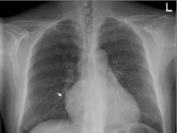
The electrocardiogram showed normal sinus rhythm. Chest radiography revealed prominent right heart border with mild cardiomegaly (Fig. 1). Transthoracic echocardiography (TTE) obtained in a parasternal long axis showed aneurysmal struc- ture (arrows) arising from unknown origin (Fig. 2A). Apical 4-chamber view revealed a balloon-like mass (arrows), occupy- ing almost completely the right atrium (Fig. 2B). There was no significant hemodynamic effect of the mass on right atri- um (RA) or right ventricle (RV). The round shaped cystic mass was in contact with the tricuspid annulus and RA. The aortic valve was competent. The left ventricular systolic func- tion and wall motion were normal. Initially, the mass was thought to be a pericardial cyst because the cystic mass was superiorly located and extended to the pericardial space. Un- ruptured coronary SVA was not suspected in first TTE because of its very large size. For confirmation, a thoracic angio-com- puted tomography (CT) and coronary angiography were per- formed. Thoracic angio-CT showed a 7 × 7.5 cm sized large saccular lesion arising from the right coronary sinus of Valsal- va, with the right coronary artery arising from the aneurysm (Fig. 3). After reviewing the TTE images, we recognized the
pISSN 1975-4612/ eISSN 2005-9655 Copyright © 2012 Korean Society of Echocardiography www.kse-jcu.org http://dx.doi.org/10.4250/jcu.2012.20.1.60
CASE REPORT J Cardiovasc Ultrasound 2012;20(1):60-62
A Giant Unruptured Right Coronary Sinus of Valsalva Aneurysm
Seok-Jae Huh, MD1, Tae-Ho Park, MD1, Dong-Yeol Lee, MD1, Hyojin Kang, MD1, Bo-Sung Kim, MD1, Yong-Rack Cho, MD1, Moo-Hyun Kim, MD1,
Young-Dae Kim, MD1 and Sun-Mi Lee, MD2
1Departments of Cardiology, 2Family Medicine, Dong-A University College of Medicine, Busan, Korea
There have been few case reports on giant sinus of Valsalva aneurysm (SVA). We report a case of a giant unruptured right coro- nary SVA that was confused with a pericardial cyst by transthoracic echocardiography.
KEY WORDS: Sinus of Valsalva · Aortic aneurysm · Echocardiography.
• Received: October 31, 2011 • Revised: January 5, 2012 • Accepted: February 16, 2012
• Address for Correspondence: Tae-Ho Park, Department of Cardiology, Dong-A University College of Medicine, 26 Daesingongwon-ro, Seo-gu, Busan 602-715, Korea Tel: +82-51-240-2964, Fax: +82-51-244-1713, E-mail: [email protected]
• This is an Open Access article distributed under the terms of the Creative Commons Attribution Non-Commercial License (http://creativecommons.org/licenses/by-nc/3.0) which permits unrestricted non-commercial use, distribution, and reproduction in any medium, provided the original work is properly cited.
Fig. 1. Chest radiography showed prominent right heart border (arrow) with mild cardiomegaly.
Giant Aneurysm of the Sinus of Valsalva | Seok-Jae Huh, et al.
61 giant cystic mass as a very large sinus of Valsalva aneurysm,
which was bulging out from the proximal aortic root (Fig.
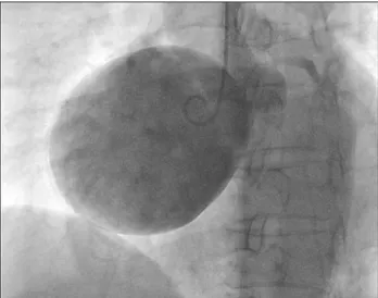
2C). Coronary angiography revealed normal coronary arteries and a large unruptured aneurysm in the sinus of Valsalva aris- ing from the right coronary sinus (Fig. 4). The patient under- went elective surgery for SVA repair. Aneurysmectomy and right coronary artery reimplantation were done. Five days af- ter surgery, coronary angiography revealed good anastomosis and normal flow in the right coronary artery. The patient had an uneventful postoperative course, and she was discharged without complications.
Discussion
The incidence of SVA is 0.14 to 0.96% of patients undergo- ing open heart surgery.3) As the aneurysms are frequently si- lent, their exact prevalence is unknown, however an autopsy study of 8138 individuals suggests a prevalence of 0.09% in the general population.4) Although most SVAs are congenital in origin, acquired aneurysm is caused by conditions affecting the aortic wall, such as infection, degenerative disease, or tho- racic trauma.5)6) Congenital SVAs occur more often in males than female. SVAs are found most often in right coronary si- nus, less often in the noncoronary sinus, and least often in left
coronary sinus.7) The size of SVA is variable from 1 cm to 10 cm.8)9) Associated abnormalities include ventricular septal de- fect (11.6%), biscuspid aortic valve (16.3%), and aortic regur- gitation (44.2%).3) Unruptured SVAs are usually asymptom- atic. Symptoms develop when the enlarging aneurysm ruptures or compresses adjacent structures. Exertional dsypnea, palpita- tions, and chest pain have been reported in patients with un- ruptured aneurysm of the sinus of Valsalva.10) Surgical treat- ment is advised in the presence of rupture, compression of ad- jacent structures or in the presence of significant dilatation or aortic regurgitation. The natural history of unruptured aneu- rysm has not been clearly defined, and no guidelines were pro- vided regarding the timing of surgical intervention.10) How- ever, early surgery is usually recommended because surgery produces excellent outcomes in such cases. The operative mor- tality of SVA is generally low (approximately 1%) in patients with noninfected SVAs.11)
Prior to the advent of noninvasive imaging modalities, SVA was diagnosed by cardiac catheterization. Currently, TTE is the initial imaging technique of choice to delineate ruptured or unruptured SVA. It allows accurate imaging of SVA as well as the presence of associated abnormalities. Parasternal short axis views demonstrate finger-like windsock extending from
Fig. 2. Transthoracic echocardiography obtained in a parasternal long axis (A) showed aneurysmal structure (arrows) arising from unknown origin.
Apical 4-chamber view (B) revealed a balloon-like mass (arrows), occupying almost completely the right atrium. Parasternal short axis view (C) showed that origin (arrow) of aneurysm was visualized from right coronary sinus with blood flow from aortic root to aneurysm.
A B C
Fig. 3. Thoracic CT showed a large saccular lesion, arising from the
right coronary sinus of Valsalva (arrow). Fig. 4. Coronary angiography revealed a large unruptured aneurysm in the sinus of Valsalva arising from the right coronary sinus.
Journal of Cardiovascular Ultrasound 20 | March 2012
62
the base of the sinus to right ventricle or right atrium. When the diagnosis of the ruptured SVA is uncertain, coronary angi- ography, magnetic resonance imaging, or CT can be useful.11)
In our patient, TTE allowed prompt detection of the huge cystic mass. However, it was difficult to differentiate from a pericardial cyst because the aneurysmal dilatation was seen as balloon-like cystic mass which is not usual finding of SVA, and extended to the pericardial space. SVA was confirmed by car- diac CT. After reviewing the TTE images, origin of aneurysm was visualized from right coronary sinus with blood flow from aortic root to aneurysm. Therefore, for the diagnosis of the SVA, it is important to determine that the aneurysm origi- nates from the aortic sinus. This case could be accurately diag- nosed by TTE, if we had used multiple views including para- sternal short axis view of aortic root. This is a rare case of a giant right coronary SVA, confused with pericardial cyst.
References
1. Feldman DN, Roman MJ. Aneurysms of the sinuses of Valsalva. Cardi- ology 2006;106:73-81.
2. Edwards JE, Burchell HB. The pathological anatomy of deficiencies be- tween the aortic root and the heart, including aortic sinus aneurysms. Tho- rax 1957;12:125-39.
3. Takach TJ, Reul GJ, Duncan JM, Cooley DA, Livesay JJ, Ott DA, Frazier OH. Sinus of Valsalva aneurysm or fistula: management and out- come. Ann Thorac Surg 1999;68:1573-7.
4. Smith WA. Aneurysm of the sinus of Valsalva, with report of 2 cases.
JAMA 1914;62:1878-80.
5. Batiste C, Bansal RC, Razzouk AJ. Echocardiographic features of an unruptured mycotic aneurysm of the right aortic sinus of Valsalva. J Am Soc Echocardiogr 2004;17:474-7.
6. Greiss I, Ugolini P, Joyal M, Bouchard D, Mercier LA. Ruptured an- eurysm of the left sinus of Valsalva discovered 41 years after a decelerational injury. J Am Soc Echocardiogr 2004;17:906-9.
7. Fishbein MC, Obma R, Roberts WC. Unruptured sinus of Valsalva aneurysm. Am J Cardiol 1975;35:918-22.
8. Sen T, Guray Y, Hajro E, Demirkan BM. Giant unruptured noncoro- nary sinus of Valsalva aneurysm with ascending aorta dissection. Eur J Cardiothorac Surg 2009;36:187.
9. Viktorsson TV, Arnorsson T, Sigurdsson MI, Sverrisson JT, Gudb- jartsson T. A giant unruptured aneurysm of the sinus of Valsalva together with ectasia of the left coronary artery. Ann Thorac Surg 2011;92:354-6.
10. Liau CS, Chu IT, Ho FM. Unruptured congenital aneurysm of the sinus of Valsalva presenting with pulmonary stenosis. Catheter Cardiovasc Interv 1999;46:210-3.
11. Ott DA. Aneurysm of the sinus of valsalva. Semin Thorac Cardiovasc Surg Pediatr Card Surg Annu 2006:165-76.