gain of clinical attachment depending on the mode of interproximal bone resorption
Yoon Mi Choi1, Ju-Youn Lee1,2, Jeomil Choi1,2, Ji-Young Joo1,*
1Department of Periodontology, Pusan National University Dental Hospital, Dental Research Institute, Yangsan, Korea
2Department of Periodontology, Pusan National University School of Dentistry, Yangsan, Korea
Research Article
J Periodontal Implant Sci 2015;45:184-189 http://dx.doi.org/10.5051/jpis.2015.45.5.184
Purpose: The purpose of the present study was to evaluate the effect of root planing on the reduction of probing pocket depth and the gain of clinical attachment depending on the pattern of bone resorption (vertical versus horizontal bone loss) in the interproximal aspect of premolar teeth that showed an initial probing pocket depth of 4–6 mm.
Methods: In this study, we analyzed 68 teeth (15 from the maxilla and 53 from the mandi- ble) from 32 patients with chronic periodontitis (17 men and 15 women; mean age, 53.6 years). The probing pocket depth and clinical attachment level at all six sites around each tooth were recorded before treatment to establish a baseline value, and then three months and six months after root planing.
Results: The reduction in interdental pocket depth was 1.1 mm in teeth that experienced horizontal bone loss and 0.7 mm in teeth that experienced vertical bone loss. Interdental attachment was increased by 1.0 mm in teeth with horizontal bone loss and by 0.7 mm in teeth with vertical bone loss. The reduction of probing pocket depth and the gain of clini- cal attachment occurred regardless of defect patterns three and six months after root planing.
Conclusions: The reduction of pocket depth and gain in the clinical attachment level were significantly larger in horizontally patterned interproximal bone defects than in vertical bone defects.
Keywords: Alveolar bone loss, Periodontal pocket, Root planing.
Received: Sep. 20, 2015 Accepted: Oct. 16, 2015
*Correspondence:
Ji-Young Joo
Department of Periodontology, Pusan National University Dental Hospital, Dental Research Institute, 20 Geumo-ro, Beomeo-ri, Mulgeum-eup, Yangsan 50612, Korea E-mail: [email protected] Tel:+82-55-360-5203 Fax:+82-55-360-5194
INTRODUCTION
Periodontitis is an inflammatory disease caused by a subset of the bacteria present in dental plaque that attack vulnerable periodontal tissue, leading to periodontal pocket depth formation, gingival recession, loss of connective tissue attachment, and progressive destruc- tion of the alveolar bone and periodontal ligament accompanied by a wide range of symp- toms [1-3]. The radiographic pattern of bone loss surrounding infected teeth manifests ei- ther horizontally or vertically [4].
The initial treatment of periodontitis involves controlling its causes, reducing the bacterial load on the supragingival and subgingival surfaces through nonsurgical periodontal treat- ment (root planing), drug treatment if needed, and reinforcing oral hygiene education [5].
These nonsurgical treatments are considered the gold standard for controlling periodontitis [6]. A long-term observational study reported that attachment loss increased by 0.05–0.3 mm
This is an Open Access article distributed under the terms of the Creative Commons Attribution Non-Commercial License (http://creativecommons.org/licenses/by-nc/3.0/).
yearly in patients who did not undergo periodontal treatment [7].
According to Heitz-Mayfield et al. [1] and Lindhe et al. [8], the critical probing depth represents a threshold value of the probing depth value, above which the outcome of therapy will result in at- tachment gain and below which the outcome of therapy will result in clinical attachment loss. According to Lindhe et al. [8], the critical probing depth of root planing is 2.9±0.3 mm; if the pocket depth is greater than this, gain of clinical attachment is optimally achieved through treatment with root planing. If the pocket depth is greater than 4.2±0.2 mm, clinical attachment gain does occur through sur- gical treatment. If it is deeper than 5.4 mm, the gain of clinical at- tachment level is greater with periodontal surgery than with root planing. Moreover, according to Heitz-Mayfield et al. [1], root plan- ing is generally preferable when the pocket depth is 1–3 mm, al- though periodontal surgery is superior in terms of pocket depth re- duction. However, root planing is superior in terms of gain of clinical attachment level when the pocket depth is 4–6 mm, and periodon- tal surgery is superior overall when the pocket depth is >6 mm.
Clinically, the response to periodontal treatment of teeth with periodontitis that have similar pocket depths differs depending on the pattern of bone loss. Bone loss can be categorized as horizon- tal or vertical. Vertical bone loss is evaluated based on the slope angle between the root and bone wall using radiographs. Accord- ing to Linares et al. [9], an angle less than 25° is narrow and an angle greater than 37° degrees is wide. Highly predictable results can be estimated in the regenerative treatment of cases with a slope angle of 25°–37°, when the slope angle is estimated from pretreatment radiographs. According to Steffensen and Weber [10], most defects with an angle <45° showed a greater extent of bone acquisition. Furthermore, according to Lang [11], when the defect angle is <45° in a single root, the mean bone acquisition was 1.22 mm after Widman flap surgery without bone resection, whereas only 0.05 mm of bone was acquired when the angle was between 45° and 90°. That is, when the defect angle was more acute, the results were better. Therefore, the defect angle corresponding to vertical loss was fixed at 25°–37° in the present study, and pocket depth and clinical attachment level were evaluated after treat- ment. Moreover, since defect shapes are classified based on radio- graphic images, this study focused on interdental bone. Only sin- gle-rooted premolars were used in this experiment in order to ex- clude the effect of treatment on the furcation area.
Although several studies [12,13] have described gains of clinical attachment level after root planing according to pocket depth, few have reported gains of clinical attachment level after root planing according to the pattern of interdental bone resorption.
The purpose of the present study was to compare the effect of root planing on the reduction of probing pocket depth and the gain of clinical attachment depending on the pattern of bone re- sorption (vertical versus horizontal bone loss) in the interproximal aspects of premolar teeth that showed an initial probing pocket depth of 4–6 mm.
MATERIALS AND METHODS
Study design
Sixty-eight teeth were included from 32 patients (17 men and 15 women; mean age, 53.6 years) who visited Pusan National University Dental Hospital for periodontal treatment between March 2014 and April 2014 and were diagnosed with chronic periodontitis. The inclu- sion criteria were as follows: (1) chronic periodontitis, (2) horizontal/
vertical bone loss in different quadrants (contralateral pattern), (3) pocket depth of 4–6 mm, (4) premolars, and (5) the presence of ad- jacent teeth and normal occlusion with opposing dentition.
The exclusion criteria were as follows: (1) systemic disease, (2) smoking, (3) prior periodontal treatment, (4) pregnancy, (5) alcohol or drug addiction, and (6) lack of cooperativeness or failure to maintain good oral hygiene.
The study protocol was approved by the Institutional Review Board (IRB # PNUDH-2014-012), Pusan National University Dental Hospital, Yangsan, Korea. All patients received a detailed descrip- tion of the proposed study protocol and provided written informed consent to participate in the study.
Study methods
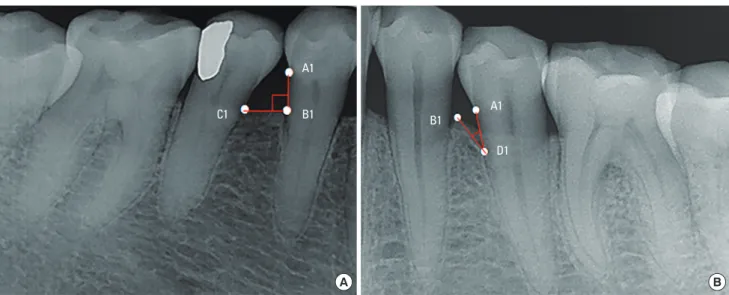
A clinical examination was conducted. Each tooth was divided into six parts (mesiobuccal, midbuccal, distobuccal, mesiolingual, midlingual, and distolingual), and the pocket depth and clinical at- tachment level were measured. Probing pocket depths and attach- ment levels were measured by a single experienced periodontist using a periodontal probe (Periodontal probe PFG-W, OSUNG Co., Gimpo, Korea). Bone loss at interdental sites was categorized as horizontal or vertical. The classification of a defect as involving horizontal or vertical bone loss was determined by the slope angle between the root and the interdental bone wall on radiographs. If the slope angle between the root and interdental bone wall was 90°±10° on radiographs, the defect was designated as horizontal bone loss, whereas if the angle of bone loss was between 25°–37°, the defect was designated as vertical bone loss (Figure 1).
Nonsurgical periodontal treatment (root planing)
After the clinical examination, supragingival scaling was per- formed in all teeth included in the study, and the subjects received oral hygiene instructions. Subgingival curettage and root planing were performed under local anesthesia with an ultrasonic instru- ment and/or a hand instrument in the quadrant that included the subject tooth, preferentially after an interval of two weeks. Sup- portive treatment and clinical examinations were performed three and six months after root planing.
Measuring clinical indices
Probing pocket depth and clinical attachment level were mea- sured at the time of root planing, as well as three and six months after treatment.
Probing pocket depth and attachment level on the mesiobuccal,
midbuccal, distobuccal, mesiolingual, midlingual, and distolingual sites of each tooth were measured using a constant-force peri- odontal probe with 1-mm markings. The values were rounded up to the nearest millimeter.
Statistical analysis
After root planing, the average changes in each clinical index of each tooth overall and at the buccal/labial and lingual/palatal in- terdental areas were determined. Additionally, the changes in each measurement between the first visit and after treatment were de- termined.
Statistical analysis was performed using SPSS (version 20 for Windows; SPSS Inc., Chicago, IL, USA), and the paired t-test and the Student’s t-test were used to compare the changes in the two experimental groups. P<0.05 were considered to indicate statisti- cal significance.
RESULTS
This study evaluated 68 teeth from 32 patients who were diag- nosed with chronic periodontitis, evenly divided between teeth with horizontal and vertical bone loss defects. Fifteen defects were in the maxilla and 53 defects were in the mandible. Pre-treatment interdental pocket depth and clinical attachment level varied be- tween the groups, but total pocket depth and clinical attachment level were not significantly different between the groups (Table 1).
Probing pocket depth
In both groups of teeth, pocket depth was found to be signifi- cantly decreased three and six months after the first visit. However, the pocket depth did not decrease significantly between three and
six months. After treatment, a greater decrease in pocket depth was observed in teeth with horizontal bone loss than in teeth with vertical bone loss (Table 2).
Furthermore, a significant decrease in interdental pocket depth was observed both three and six months after treatment, with a greater decrease observed in teeth with horizontal bone loss than in those with vertical bone loss. Both groups of teeth showed sta- tistically significant differences (P <0.01) in total pocket depth both three and six months after the first visit. However, no signifi- cant difference was observed between three and six months after treatment.
Clinical attachment level
The attachment level at all sides of the teeth showed a statisti- cally significant gain three and six months after root planing, but did not show a statistically significant difference between three Table 1. Comparison of the average initial probing pocket depth (PD) and clinical attachment level (CAL) (mm) between groups.
Site Defect Mean SD Sample size P-value
Total Vertical PD 3.63 0.41 34 0.085
CAL 3.76 0.05 0.049
Horizontal PD 3.82 0.48 34 0.085
CAL 4.06 0.05 0.049
Interdental Vertical PD 4.34 0.47 34 0.002
CAL 4.46 0.56 0.003
Horizontal PD 4.74 0.55 34 0.002
CAL 4.94 0.74 0.003
SD: standard deviation.
Figure 1. Bone loss at interdental sites was categorized as horizontal or vertical bone loss. (A) If the slope angle between the root and interdental bone wall was 90°±10° on radiographs, the defect was designated as horizontal bone loss. (B) If the angle of bone loss was between 25° and 37°, the defect was desig- nated as vertical bone loss. A1, CEJ; B1, the top of the crest; C1, the top of the proximal bone crest of the adjacent tooth; D1, the bottom of the bone defect.
A1 B1 C1
A
B1 A1
D1
B
and six months. Both three and six months after root planing, a significantly greater gain of clinical attachment level was observed for horizontal than for vertical bone loss (P<0.01). Similar results were observed for the interdental attachment level at the buccal/
labial and palatal/lingual interfaces (Table 3). Similar results were also observed for total and interdental attachment levels.
DISCUSSION
In this study, the outcome of periodontal treatment was com- pared between different quadrants in each person, by limiting the sample to a single-root premolar with a pocket depth of 4–6 mm pocket depth and determining the clinical attachment level after root planing depending on the pattern of interdental bone resorp- tion. Reduction of pocket depth was statistically significant (P<0.01) three and six months after root planing in areas of both horizontal and vertical bone loss. Moreover, the gain of clinical attachment level was statistically significant (P<0.01). Of note, greater reduc- tion of pocket depth and gain of clinical attachment level were ob- served in teeth with horizontal bone loss than in teeth with vertical bone loss.
Although a significant reduction of pocket depth and gain of clinical attachment level were observed three and six months after root planing and supportive periodontal therapy, no statistically significant pocket depth reduction or gain of clinical attachment level occurred in the period from three to six months.
Tunkel et al. [14], Torfason et al. [15], and Hallmon and Rees [16]
found no significant differences between the results of non-surgi- cal periodontal therapy depending on whether a hand instrument or ultrasound scaler was used; thus, this study excluded the effect of the instrument used in root planing. Moreover, Badersten et al.
[17] showed that the number of root planing sessions did not sig- nificantly affect the reduction of pocket depth or gain of clinical attachment level; thus, in this study, probing pocket depth and gain of clinical attachment level were measured after root planing was performed once.
The average reduction of interdental probing pocket depth was 1.1 mm in teeth with horizontal bone loss and 0.7 mm in teeth with vertical bone loss, which is similar to the results reported by Lang [11]. According to Lang [11], after motivating patients to fol- low oral hygiene instructions for one month and performing sub- gingival scaling and root planing, regions with a pocket depth of Table 2. Comparison of the changes in probing depth between treatment and three and six months of follow-up.
Defect Site n Baseline to three months Three months to six months Baseline to six months
Mean±SD P-value Mean±SD P-value Mean±SD P-value
Horizontal Interdental PDC 68 0.67±0.3 <0.001 0.04±0.24 0.285 0.72±0.36 <0.001
Total PDC 204 1.07±0.83 <0.001 0.16±0.54 0.075 1.24±0.95 <0.001
Vertical Interdental PDC 68 0.41±0.42 <0.001 0.07±0.3 0.186 0.48±0.42 <0.001
Total PDC 204 0.71±0.69 <0.001 0.02±0.64 0.832 0.72±0.75 <0.001
Horizontal Interdental PDCD 68 1.07±0.83 <0.006 0.16±0.59 0.164 1.24±0.95 <0.001
Total PDCD 204 0.67±0.79 <0.001 0.04±0.56 0.659 0.72±0.90 <0.006
Vertical Interdental PDCD 68 0.71±0.69 <0.006 0.02±0.64 0.164 0.72±0.75 <0.001
Total PDCD 204 0.41±0.82 <0.001 0.07±0.57 0.659 0.48±0.84 <0.006
SD: standard deviation, PDC: probing depth change, PDCD: difference in probing depth change.
Table 3. Comparison of the changes in clinical attachment level between treatment and three to six months of follow-up.
Defect Site n Baseline to three months Three months to six months Baseline to six months
Mean±SD P-value Mean±SD P-value Mean±SD P-value
Horizontal Interdental CAL 68 0.70±0.36 <0.001 0.05±0.24 0.244 0.75±0.38 <0.001
Total CAL 204 1.02±0.61 <0.001 0.16±0.53 0.086 1.18±0.68 <0.001
Vertical Interdental CAL 68 0.39±0.38 <0.001 0.06±0.31 0.239 0.46±0.39 <0.001
Total CAL 204 0.72±0.51 <0.001 0.02±0.47 0.856 0.74±0.46 <0.001
Horizontal Interdental CALD 68 1.02±0.92 <0.039 0.16±0.61 0.172 1.18±1.01 <0.005
Total CALD 204 0.70±0.81 <0.001 0.05±0.55 0.791 0.75±0.88 <0.001
Vertical Interdental CALD 68 0.72±0.71 <0.039 0.02±0.64 0.172 0.74±0.77 <0.005
Total CALD 204 0.39±0.78 <0.001 0.06±0.57 0.791 0.46±0.81 <0.001
SD: standard deviation, CAL: clinical attachment level, CALD: difference in clinical attachment level changes.
4–6 mm showed a 1 mm decrease, while regions with a pocket depth of 7–12 mm showed a 2 mm decrease. Of this reduction, 50% was due to gingival recession followed by the relief of gingi- val swelling, while the other 50% was due to gain of clinical at- tachment level followed by tightening of soft tissue at the base of the lesion. Moreover, Lindhe et al. [12] and Badersten et al. [17,18]
have reported similar findings. However, the gain of clinical at- tachment level was greater in the study by Pihlstrom et al. [19].
Lindhe et al. [12] showed that a single-root tooth with an initial pocket depth of 4–6 mm showed a 0.7±0.4 mm gain of clinical attachment level 12 months after root planing therapy, while Pihl- strom et al. [19] reported a 0.34-mm gain.
Overall, cases teeth horizontal bone loss showed a greater re- duction of pocket depth and gain of clinical attachment level than those with vertical bone loss.
Our results were most similar to those of Lang et al. [11], but dif- fered from the results of other studies [11,12,17-19] that did not differentiate between horizontal and vertical bone loss, instead fo- cusing only on pocket depth. In particular, the interdental area showed the greatest reduction of pocket depth and gain of clinical attachment level compared to the tooth overall, possibly because of the inclusion of 3 mm of pocket depth in the mid-buccal and mid-lingual area due to the focus on interdental bone loss. More- over, a single clinician performed all the measurements, which may have been another reason for this result.
This study found that a greater reduction of pocket depth and gain of clinical attachment level was observed in teeth with hori- zontal bone loss than in those with vertical bone loss.
The sample of teeth included in this study was chosen consider- ing radiographic bone loss and pocket depth alone. In order to confirm the factors that affect the outcomes of root planing, a more systemic study is necessary. Such a study would include a larger sample of teeth, classify them as maxillary and mandibular teeth, compare single-root teeth and multiple-root teeth, and de- termine treatment effects depending on the initial pocket depth.
CONFLICT OF INTEREST
No potential conflict of interest relevant to this article was re- ported.
ACKNOWLEDGEMENTS
This study was supported by a clinical research grant, Pusan Na- tional University Dental Hospital (2016).
ORCID
Yoon Mi Choi http://orcid.org/0000-0002-2925-2565 Ju-Youn Lee http://orcid.org/0000-0002-0772-033X Jeomil Choi http://orcid.org/0000-0002-7491-6711 Ji-Young Joo http://orcid.org/0000-0002-4050-5797
REFERENCES
1. Heitz-Mayfield LJ, Trombelli L, Heitz F, Needleman I, Moles D. A systematic review of the effect of surgical debridement vs non- surgical debridement for the treatment of chronic periodontitis.
J Clin Periodontol 2002;29 Suppl 3:92-102.
2. Socransky SS, Haffajee AD. The bacterial etiology of destructive periodontal disease: current concepts. J Periodontol 1992;63:
322-31.
3. Renvert S, Persson GR. A systematic review on the use of residual probing depth, bleeding on probing and furcation status follow- ing initial periodontal therapy to predict further attachment and tooth loss. J Clin Periodontol 2002;29 Suppl 3:82-9.
4. Papapanou PN, Wennström JL. The angular bony defect as indi- cator of further alveolar bone loss. J Clin Periodontol 1991;18:
317-22.
5. Van der Weijden GA, Timmerman MF. A systematic review on the clinical efficacy of subgingival debridement in the treatment of chronic periodontitis. J Clin Periodontol 2002;29 Suppl 3:55-71.
6. Ishikawa I, Baehni P. Nonsurgical periodontal therapy--where do we stand now? Periodontol 2000 2004;36:9-13.
7. Brown LJ, Löe H. Prevalence, extent, severity and progression of periodontal disease. Periodontol 2000 1993;2:57-71.
8. Lindhe J, Socransky SS, Nyman S, Haffajee A, Westfelt E. “Critical probing depths” in periodontal therapy. J Clin Periodontol 1982;
9:323-36.
9. Liñares A, Cortellini P, Lang NP, Suvan J, Tonetti MS; European Re- search Group on Periodontology. (ErgoPerio). Guided tissue regen- eration/deproteinized bovine bone mineral or papilla preservation flaps alone for treatment of intrabony defects. II: radiographic predictors and outcomes. J Clin Periodontol 2006;33:351-8.
10. Steffensen B, Webert HP. Relationship between the radiographic periodontal defect angle and healing after treatment. J Periodon- tol 1989;60:248-54.
11. Lang NP. Focus on intrabony defects–conservative therapy. Peri- odontol 2000 2000;22:51-8.
12. Lindhe J, Westfelt E, Nyman S, Socransky SS, Heijl L, Bratthall G.
Healing following surgical/non-surgical treatment of periodontal disease. A clinical study. J Clin Periodontol 1982;9:115-28.
13. Badersten A, Nilvéus R, Egelberg J. Effect of nonsurgical periodon- tal therapy. I. Moderately advanced periodontitis. J Clin Periodon- tol 1981;8:57-72.
14. Tunkel J, Heinecke A, Flemmig TF. A systematic review of efficacy of machine-driven and manual subgingival debridement in the treatment of chronic periodontitis. J Clin Periodontol 2002;29 Suppl 3:72-81.
15. Torfason T, Kiger R, Selvig KA, Egelberg J. Clinical improvement of gingival conditions following ultrasonic versus hand instru- mentation of periodontal pockets. J Clin Periodontol 1979;6:165- 76.
16. Hallmon WW, Rees TD. Local anti-infective therapy: mechanical and physical approaches. A systematic review. Ann Periodontol
2003;8:99-114.
17. Badersten A, Nilveus R, Egelberg J. Effect of nonsurgical periodon- tal therapy. III. Single versus repeated instrumentation. J Clin Peri- odontol 1984;11:114-24.
18. Badersten A, Nilveus R, Egelberg J. Effect of nonsurgical peri-
odontal therapy. II. Severely advanced periodontitis. J Clin Peri- odontol 1984;11:63-76.
19. Pihlstrom BL, Oliphant TH, McHugh RB. Molar and nonmolar teeth compared over 6 1/2 years following two methods of peri- odontal therapy. J Periodontol 1984;55:499-504.