Amyloid
β Protein (25-35) 유도 배양신경세포 독성에 대한 목단피의 억제효과
김주연*·주현수*·반주연**·송경식***·성연희*†
*충북대학교 수의과대학, **경희대학교 의과대학, ***경북대학교 농업생명과학대학
Moutan Cortex Extract Inhibits Amyloid β Protein (25-35)-induced Neurotoxicity in Cultured Rat Cortical Neurons
Joo Youn Kim*, Hyun Soo Ju*, Ju Yeon Ban**, Kyung Sik Song***, and Yeon Hee Seong*†
*College of Veterinary Medicine, Chungbuk National University, Cheongju, Chungbuk 361-763, Korea.
**Department of Pharmacology, College of Medicine, Kyung Hee University, Seoul 130-701, Korea.
***College of Agriculture and Life-Sciences, Kyungpook National University, Daegu 702-701, Korea.
ABSTRACT : Moutan cortex, the root bark of Paeonia suffruticosa Andrews (Paeoniaceae), has pharmacological effects such as anti-inflammatory, antiallergic, analgesic and antioxidant activities. We investigated a methanol extract of Moutan cortex for neuroprotective effects on neurotoxicity induced by amyloid β protein (Aβ) (25-35) in cultured rat cortical neu- rons. Exposure of cultured cortical neurons to 10µM Aβ (25-35) for 24 h induced neuronal apoptotic death. Moutan cortex inhibited 10µM Aβ (25-35)-induced neuronal cell death at 30 and 50 ㎍/㎖, which was measured by a 3-[4,5-dimethylthia- zol-2-yl]-2,5-diphenyl-tetrazolium bromide (MTT) assay and Hoechst 33342 staining. Moutan cortex inhibited 10µM Aβ (25-35)-induced elevation of intracellular calcium concentration ([Ca2+]i), and generation of reactive oxygen species (ROS) which were measured by fluorescent dyes. Moutan cortex also inhibited glutamate release into medium induced by 10µM Aβ (25-35), which was measured by HPLC. These results suggest that Moutan cortex prevents Aβ (25-35)-induced neuronal cell damage by interfering with the increase of [Ca2+]i, and then inhibiting glutamate release and ROS generation. Moutan cortex may have a therapeutic role in preventing the progression of Alzheimer’s disease.
Key Words : Moutan Cortex, Amyloid β protein (25-35), Neurotoxicity, Alzheimer’s Disease, Neuroprotection
INTRODUCTION
Alzheimer’s disease (AD) is a neurodegenerative disorder characterized clinically by cognitive impairment and patholo- gically by the appearance of senile plaques and neurofi- brillary tangles (Price et al., 1998). Amyloid β protein (Aβ), a 39- to 43-amino-acid peptide fragment derived from amyloid precursor protein, is thought to be closely related to the pathogenesis of AD as the major component of the senile plaques that characterize this disease (Ivins et al., 1999). In cultures, Aβ can directly induce neuronal cell death (Ueda et al., 1994) and can render neurons vulnerable to excitotoxicity (Koh et al., 1990) and oxidative insults (Goodman and Mattson, 1994). Although the precise mechanism of Aβ-induced cell death is not well understood,
Aβ neurotoxicity has been speculated to be due to various factors, including oxidative stress, excessive increases in intracellular Ca2+ ([Ca2+]i) and glutamate accumulation, and induction of neurotoxic cascades (Gray and Patel, 1995;
Ueda et al., 1997; Ekinci et al., 2000).
Moutan cortex, the root bark of Paeonia suffruticosa Andrews (Paeoniaceae), has been used extensively as a traditional Chinese medicine in eastern Asian countries.
Moutan cortex is used to treat a plethora of disease classes such as atherosclerosis, infection and inflammation. Various pharmacological activities of Moutan cortex include anti- hepatotoxic, anti-inflammatory, anti-allergic, anti-oxidant and analgesic effects (Shon and Nam, 2004; Tatsumi et al., 2004; Rho et al., 2005; Chun et al, 2007; Jiang et al., 2007). In the CNS, a possible neuroprotective effect of a
†Corresponding author: (Phone) +82-43-261-2968 (E-mail) [email protected] Received October 10, 2008 / Revised November 24, 2008 / Accepted December 2, 2008
traditional Chinese medical formulation containing Moutan cortex against rat brain ischemia/reperfusion injury has been reported (Le et al., 2007). Paeonol, a common component of Moutan cortex, reduced cerebral infarction in ischemia/
reperfusion injured rats (Hsieh et al., 2006). Furthermore, Moutan cortex was effective to prevent oxidative stress-induced neuronal death and generation of reactive oxygen species in cultured neurons (Shimada et al., 2004; Rho et al., 2005).
These reports indicate that Moutan cortex can be a potential protective agent against neurodegenerative diseases such as AD and stroke. The aim of our study was to determine whether Moutan cortex had a protective effect against Aβ (25-35)- induced neuronal damage in cultured rat cortical neurons.
MATERIALS AND METHODS
1. Plant material and extraction
Moutan cortex was purchased from a market in Daegu, Korea and identified by Professor K.S. Song, Kyungpook National University. The voucher specimen (KNUNPC-MC1) is stored at the Natural Products Chemistry Lab., Division of Applied Biology and Chemistry, Kyungpook National University, Daegu, Korea. The dried material (1㎏) was refluxed in 2 L methanol twice at room temperature and the residue was filtered off using a filter paper. The filtrate was evaporated to dryness under 45℃ to obtain methanol extract (189.33 g).
2. Experimental animals
Pregnant Sprague-Dawley rats for primary neuronal culture were supplied by Daehan BioLink Co., Ltd. (Chungbuk, Korea) and housed in an environmentally controlled room at 22± 2℃, with a relative humidity of 55 ± 5%, a 12-h light/
dark cycle, and food and water ad libitum. The procedures involving experimental animals complied with the animal care guidelines of the National Institutes of Health and the animal ethics committee of Chungbuk National University.
3. Induction of neurotoxicity in primary cultures of rat cerebral cortical neurons
Primary cortical neuron cultures were prepared using embryonic day 15 to 16 Sprague-Dawley rat fetuses, as described previously (Ban et al., 2005; Lee et al., 2007).
Neurotoxicity experiments were performed on neurons after 3-4 days in culture. Cultured neurons were treated with 10
µM Aβ (25-35) (Bachem, Bubendorf, Switzerland) in serum- free DMEM (Sigma) at 37℃ for 24 h (unless otherwise indicated) to produce neurotoxicity. An Aβ (25-35) stock solution of 2 mM was prepared in sterile distilled water, stored at −20℃, and incubated for more than 2 days at 37
℃ to aggregate before use. Moutan cortex was dissolved in DMSO at concentrations of 50㎎/㎖ and further diluted in experimental buffers. The final concentration of DMSO was
≤ 0.1%, which did not affect cell viability. For each experiment, Moutan cortex was applied 15 min prior to treatment with 10µM Aβ (25-35). It was also present in the medium during Aβ (25-35) incubation.
4. Measurements of Aβ (25-35)–induced neuronal death and intracellular biochemical changes
A 3-(4,5-dimethylthiazol-2-yl)-2,5-diphenyl-tetrazolium bromide (MTT; Sigma) assay and Hoechst 33342 (Molecular Probes, Eugene, OR, USA) staining were performed to measure neuronal death 24 h after exposure of cultured neurons to 10µM Aβ (25-35), as described previously (Ban et al., 2005; Lee et al., 2007). Changes in [Ca2+]i were measured with Fluo-4 AM (Molecular Probes), a calcium-sensitive fluorescent dye, using a laser scanning confocal microscope (LSM 510, Carl Zeiss, Oberkochen, Germany) with a 488-㎚ excitation argon laser and 515-㎚ longpass emission filters (Ban et al., 2005; Lee et al., 2007). To measure glutamate secreted into the medium, cells were treated with 10µM Aβ (25-35) in a HEPES buffer containing 8.6 mM HEPES, 154 mM NaCl, 5.6 mM KCl, 2.3 mM CaCl2 and 10 mM glucose at pH 7.4, and glutamate secreted over 6 h was quantified by HPLC with an electrochemical detector (MF series, BAS, IN, USA) (Ban et al., 2005; Lee et al., 2007). The microfluorescence of 2',7'-dichlorofluorescein, the fluorescent product of 2',7'-dichlorodihydrofluorescein diacetate (H2DCF-DA; Molecular Probes), and a laser scanning confocal microscope (MRC1024ES, Bio-Rad, Maylands, UK) with 488-㎚ excitation and 510-㎚ emission filters were used to monitor the generation of reactive oxygen species (ROS) in neurons treated with 10µM Aβ (25-35) for 24 h (Ban et al., 2005).
5. Statistical analysis
Data are expressed as mean± SEM and statistical signifi- cance was assessed by one-way analysis of variance (ANOVA) and Tukey’s tests. P < 0.05 was considered significant.
RESULTS
1. Moutan cortex inhibits Aβ (25-35)–induced neuronal cell death
In previous experiments (Ban and Seong, 2005), we have demonstrated that Aβ (25-35) over the concentration range of 5-20µM produced a concentration-dependent reduction of cell viability in cultured cortical neuron. Therefore, the concentration of 10µM was used for the determination of Aβ (25-35)-induced neuronal cell damage in the present experiments. When cortical neurons were exposed to 10µM Aβ (25-35) for 24 h, absorbance in the MTT assay was 68.3± 1.2% of that of the untreated controls (Fig. 1), indicating that Aβ (25-35) caused neuronal cell death. In cultures treated with Moutan cortex (30 or 50㎍/㎖), the Aβ (25-35)-induced neuronal death was significantly reduced (absorbance, 112.5± 3.2% of control with 50㎍/㎖ Moutan cortex).
Hoechst 33342 staining was used to detect condensed or fragmented DNA, which is indicative of Aβ (25-35)-induced neuronal apoptotic death. Treatment of neurons with 10µM Aβ (25-35) induced apoptosis in 31.2 ± 1.9% of cultured cortical neurons, compared with 6.6± 0.8% in control cultures. The addition of Moutan cortex (50㎍/㎖) signifi- cantly decreased Aβ (25-35)-induced apoptotic cell death to 10.2± 1.2% of all neurons (Fig. 2).
2. Moutan cortex inhibits Aβ (25-35)–induced [Ca2+]i
elevation
Increases in [Ca2+]i have been associated with Aβ-induced cell death. In our cell cultures, treatment with 10µM Aβ (25-35) produced a slow but gradual increase in [Ca2+]i, with the maximum fluorescence intensity (ca. 180, compared to a basal level of 100) observed about 5 min after Aβ (25-35) application. In contrast, pretreatment with Moutan cortex (50㎍/㎖) significantly inhibited the increase of [Ca2+]i induced by 10µM Aβ (25-35) throughout the measurement period (Fig. 3). Moutan cortex did not affect basal [Ca2+]i.
3. Moutan cortex inhibits Aβ (25-35)–induced elevation of glutamate release
We next quantified the glutamate released into the extracellular medium after treatment with 10µM Aβ (25-35) for 6 h. As shown in Fig. 4, 10µM Aβ (25-35) elevated the basal glutamate level from 0.39± 0.02 µM in control neurons to 0.78± 0.10 µM. Moutan cortex (50㎍/㎖) signi- ficantly blocked the Aβ (25-35)-induced elevation of gluta- mate release, resulting in maximal values of 0.40± 0.05 µM.
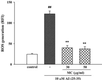
4. Moutan cortex inhibits Aβ (25-35)–induced ROS generation
To clarify the involvement of oxidative stress in Aβ Fig. 1. Inhibitory effect of Moutan cortex (MC) on Aβ (25-35)-
induced neuronal cell death in cultured cortical neurons.
Neuronal cell death was measured using the MTT assay.
The MTT absorbance from untreated cells was normali- zed to 100%. Results are expressed as mean ± S.E.M. of data obtained from 3 independent experiments. ##P < 0.01 vs control; *P < 0.05, **P < 0.01 vs 10 µM Aβ (25-35).
Fig. 2. Inhibitory effect of Moutan cortex (MC) on Aβ (25-35)- induced apoptosis of cultured cortical neurons. Apoptotic cells measured by Hoechst 33342 staining were counted in 5 to 6 fields per well. Results are apoptotic cells as a percentage of the total number of cells expressed as mean ± S.E.M. of data obtained from 4 independent experiments.
##P < 0.01 vs control, **P < 0.01 vs 10 µM Aβ (25-35).
neurotoxicity, we measured the accumulation of ROS after the exposure of the cells to Aβ (25-35) for 24 h. In H2DCF-DA-loaded cerebral cortical neurons, 10µM Aβ (25- 35) increased the fluorescence intensity, indicating that ROS were generated. In neurons treated with 10µM Aβ (25-35), the relative fluorescence increased approximately 3-fold to 121.7± 7.1 compared with the value in control neurons (24.5± 1.9). The Aβ (25-35)-induced increase in ROS
generation was significantly inhibited by Moutan cortex (30 and 50㎍/㎖) (Fig. 5).
DISCUSSION
As an active partial fragment of Aβ, Aβ (25-35) forms a β-sheet structure and induces neuronal cell death, neuritic atrophy, synaptic loss, and memory impairment, although it is not found in the AD brain (Pike et al., 1995; Grace et al., 2002; Tohda et al., 2004). The present study demo- nstrated that Aβ (25-35) causes [Ca2+]i increase, glutamate release, ROS generation, and neuronal cell death in cultured cortical neurons, all of which were blocked by treatment with Moutan cortex. In our previous reports, Aβ (25-35)- induced [Ca2+]i increase, glutamate release, ROS generation, and neuronal apoptotic death were blocked by treatment with MK-801, an N-methyl-D-aspartate (NMDA) antagonist;
verapamil, an L-type Ca2+ channel blocker; and NG-nitro-L- arginine methyl ester (L-NAME), a nitric oxide synthase (NOS) inhibitor (Ban and Seong, 2005; Lee et al., 2005).
These results supporting the involvement of NMDA glutamate receptor activation, increased Ca2+ influx, and generation of ROS in Aβ (25-35)-induced neurotoxicity in cultured neurons are consistent with the results of other studies (Harkany et al., 1999; Ekinci et al., 2000).
Regardless of the relative contribution of these events to Aβ (25-35)-induced neurotoxicity, the primary event following Fig. 3. Inhibitory effect of Moutan cortex (MC) on Aβ (25-35)-
induced [Ca2+]i elevation in cultured cortical neurons.
[Ca2+]i was monitored using Fluo-4 AM dye and a confocal laser scanning microscope. All images were processed to analyze changes in [Ca2+]i at the single cell level. Results are expressed as the relative fluorescence intensity (RFI). Each trace shows a single cell that is representative of at least 3 independent experiments.
Fig. 4. Inhibitory effect of Moutan cortex (MC) on Aβ (25-35)- induced glutamate release in cultured cortical neurons.
The amount of glutamate released over 6 h was measured by HPLC with an electrochemical detector.
Results are expressed as mean ± S.E.M. of data obtained from 3 independent experiments. #P < 0.05 vs control;
*P < 0.05 vs 10 µM Aβ (25-35).
Fig. 5. Inhibitory effect of Moutan cortex (MC) on Aβ (25-35)- induced ROS generation in cultured cortical neurons.
ROS was monitored using H2DCF-DA dye and a confocal laser scanning microscope. Results are expressed as mean ± S.E.M. of RFI obtained from 3 independent experiments.
##P < 0.01 vs control; **P < 0.01 vs 10 µM Aβ (25-35).
Aβ (25-35) treatment of cultured neurons has been sugges- ted to be Ca2+ influx, apparently via L-type voltage-depen- dent Ca2+ channels (L-VDCC), because blockade of this channel or Ca2+ chelation prevents other consequences (Ueda et al., 1997; Mattson and Chan, 2003). Furthermore, Aβ (25-35)-induced elevation of [Ca2+]i and neurotoxicity were inhibited by MK-801, suggesting that Ca2+ influx through NMDA receptor-coupled L-VDCC plays a critical role in the neurotoxicity (Harkany et al., 1999; Ban and Seong, 2005; Lee et al., 2005). In the present study, Aβ (25-35) elicited gradual and significant [Ca2+]i increase, which was blocked by Moutan cortex. Moutan cortex also significantly inhibited the Aβ (25-35)-induced glutamate elevation. These findings indicate that the sustained inhibition on [Ca2+]i
elevation by Moutan cortex resulted in the decrease of the Aβ (25-35)-induced glutamate release.
A variety of events occur downstream of neuronal Ca2+
overloading, including cytosolic ROS generation due to the influx of Ca2+ (Pereira et al., 2000). Many reports have demonstrated the role of ROS formation in Aβ-induced neurotoxicity (Yatin et al., 1999; Parks et al. 2001).
Although researchs show that Ca2+ signals activate enzymes associated with ROS generation, ROS can also facilitate [Ca2+]i increases by damaging [Ca2+]i regulatory mechanisms and by activating Ca2+ release from intracellular Ca2+ stores (Butterfield et al., 2007). We found that Moutan cortex inhibited an Aβ (25-35)-induced increase in ROS generation, but we have not determined whether Moutan cortex suppresses ROS generation via inhibition of a [Ca2+]i
increase or vice versa. It can be suggested that Moutan cortex might prevent Ca2+ entry through VDCC- and/or NMDA-receptor-coupled channels to inhibit glutamate release and ROS generation, and then Aβ (25-35)-induced neuronal death, although the mechanism by which Moutan cortex blocks the channels is not clear. A variety of compounds such as paeonoside, paeoniflorin, paeonolide and paeonol have been identified and determined in Moutan cortex (Chen et al., 2006). Paeonol inhibited NMDA receptor- coupled Ca2+ influx to protect oxygen-glucose deprivation- induced neuronal death in cultures and protected against myocardial injury due to its VDCC blocking effect (Zhang et al., 2003; Wu et al., 2008). Paeoniflorin also inhibited VDCC in neuronal cell lines to produce neuronal or neuroendocrine function. The inhibition by Moutan cortex on Aβ (25-35)-induced [Ca2+]i increase might be due to these
compounds, which could stabilize membranes in a manner that blocks Ca2+ influx via VDCC. Further, recent pharma- cological studies revealed that Moutan cortex can inhibit the production of ROS and some paeonol glycosides exhibited radical scavenging effects (Yoshikawa et al., 1992; Matsuda et al., 2001; Rho et al., 2005), suggesting that inhibition of Aβ (25-35)-induced neuronal death by Moutan cortex might be due to its ROS scavenging activity. Further study to elucidate the precise mechanism should be performed.
Many researchers have demonstrated that Aβ triggers apoptotic degeneration in in vitro neuronal experiments (Yan et al., 1999; Ekinci et al., 2000). In the present work, cultured cortical neurons exposed to Aβ (25-35) for 24 h showed increased chromatin condensation, a typical feature of apoptotic cell death, which was reduced by Moutan cortex. Aβ-induced apoptosis has been reported to be associated with COX-2 upregulation, and COX has been suggested to be an important source of ROS in the pathologic brain (Chan, 2001; Jang and Surh, 2005).
Therefore, the protective effect of Moutan cortex on the Aβ-induced apoptosis of cultured neurons might result from the inhibition of COX and inflammatory cytokine production by Moutan cortex and its active compounds such as paeonol and paeoniflorin (Chun et al., 2007; Wu and Gu, 2007). The molecular mechanism for the prevention of neuronal apoptosis by Moutan cortex should be further clarified.
Since ROS and inflammation are part of the complex series of pathophysiological events that contribute to neuro- degenerative diseases such as AD and stroke (Pitchumoni and Doraiswamy, 1998; Hoehn et al., 2005), free radical scavengers and anti-inflammatory agents have attracted consi- derable attention as potential neuroprotective agents. Moutan cortex and its common active compound, paeonol, have been reported to reduce rat brain ischemia/reperfusion injury (Hsieh et al., 2006; Le et al., 2007) and to prevent oxidative stress-induced neuronal death and generation of reactive oxygen species in cultured neuron (Shimada et al., 2004; Rho et al., 2005). Furthermore, anti-inflammatory effect of Moutan cortex attributable to the inhibition on COX and cytokine expression has been confirmed (Chun et al., 2007; Wu and Gu, 2007), suggesting that Moutan cortex has good potential for prevention of neurodegenerative diseases. Aβ is believed to play a central role in the pathophysiology of AD (Ueda et al., 1994; Yatin et al.,
1999; Butterfield et al., 2007). Moutan cortex blocked Aβ (25-35)-induced neuronal cell damage in the present study.
Although there has been no evidence up until now to show that Moutan cortex antagonizes Aβ (25-35)-induced neuro- toxicity, the present study demonstrates a novel pharma- cological activity of Moutan cortex in neurons. In con- clusion, the protection against Aβ (25-35)-induced neuro- toxicity by Moutan cortex provides as a promising thera- peutic approach to control the progression of neurodege- neration in brain of AD. Forthcoming studies will be attempted to clarify the in vivo effect of Moutan cortex.
ACKNOWLEDGEMENTS
This work was supported by the research grant of the Chungbuk National University in 2008
LITERATURE CITED
Ban JY, Cho SO, Jeon SY, Song KS, Bae K and Seong YH.
(2005). Protective effect of Sanguisorba officinalis L. root on amyloid β protein (25-35)-induced neuronal cell damage in cultured rat cortical neuron. Korean Journal of Medicinal Crop Science. 13:219-226.
Ban JY and Seong YH. (2005). Blockade of 5-HT3 receptor with MDL72222 and Y25130 reduces amyloid β protein (25-35)- induced neurotoxicity in cultured rat cortical neurons. European Journal of Pharmacology. 520:12-21.
Butterfield DA, Reed T, Newman SF and Sultana R. (2007).
Roles of amyloid β-peptide-associated oxidative stress and brain protein modifications in the pathogenesis of alzheimer's disease and mild cognitive impairment. Free Radical Biology and Medicine. 43:658-677.
Chan PH. (2001). Reactive oxygen radicals in signaling and damage in the ischemic brain. Journal of Cerebral Blood Flow Metabolism. 21:2-14.
Chen G, Zhang L and Zhu Y. (2006). Determination of glycosides and sugars in Moutan cortex by capillary electrophoresis with electrochemical detection. Journal of Pharmaceutical and Biomedical Analysis. 41:129-134.
Chun SC, Jee SY, Lee SG, Park SJ, Lee JR and Kim SC.
(2007). Anti-inflammatory activity of the methanol extract of Moutan cortex in LPS-activated Raw264.7 cells. Evidence- based Complementary and Alternative Medicine. 4:327-333.
Ekinci FJ, Linsley MD and Shea TB. (2000). β-Amyloid-induced calcium influx induces apoptosis in culture by oxidative stress rather than tau phosphorylation. Molecular Brain Research.
76:389-395.
Goodman Y and Mattson MP. (1994). Secreted forms of β- amyloid precursor protein protect hippocampal neurons against amyloid β-peptide-induced oxidative injury. Experimental Neurology. 128:1-12.
Grace EA, Rabiner CA and Busciglio J. (2002). Characterization of neuronal dystrophy induced by fibrillar amyloid β:
Implications for alzheimer's disease. Neuroscience. 114:265-273.
Gray CW and Patel AJ. (1995). Neurodegeneration mediated by glutamate and β-amyloid peptide: A comparison and possible interaction. Brain Research. 691:169-179.
Harkany T, Hortobagyi T, Sasvari M, Konya C, Penke B, Luiten PG and Nyakas C. (1999). Neuroprotective approaches in experimental models of β-amyloid neurotoxicity: Relevance to alzheimer's disease. Progress in Neuro-psychopharmacology
& Biological Psychiatry. 23:963-1008.
Hoehn BD, Palmer TD and Steinberg GK. (2005). Neurogenesis in rats after focal cerebral ischemia is enhanced by indomethacin. Stroke. 36:2718-2724.
Hsieh CL, Cheng CY, Hsai TH, Lin I, Liu CH, Chiang SY, Lin JG, Lao CJ and Tang NY. (2006). Paeonol reduced cerebral infarction involving the superoxide anion and microglia activation in ischemia-reperfusion injured rats. Journal of Ethnopharmacology. 106:208-215.
Ivins KJ, Ivins JK, Sharp JP and Cotman CW. (1999). Multiple pathways of apoptosis in pc12 cells. Crma inhibits apoptosis induced by β-amyloid. Journal of Biological Chemistry.
274:2107-2112.
Jang JH and Surh YJ. (2005). β-Amyloid-induced apoptosis is associated with cyclooxygenase-2 up-regulation via the mitogen-activated protein kinase-NF-κβ signaling pathway. Free Radical Biology and Medicine. 38:1604-1613.
Jiang S, Nakano Y, Yatsuzuka R, Ono R and Kamei C. (2007).
Inhibitory effects of Moutan cortex on immediate allergic reactions. Biological & Pharmaceutical Bulletin. 30:1707-710.
Koh JY, Yang LL and Cotman CW. (1990). β-Amyloid protein increases the vulnerability of cultured neurons to excitotoxic damage. Brain Research. 533:315-320.
Le TJ, Qiu Y, Mao JQ, Yang PY, Rui YC and Chen WS.
(2007). Protective effects of Guizhi-Fuling-Capsules on rat brain ischemia/reperfusion injury. Journal of Pharmacological Sciences. 105:34-40.
Lee BY, Ban JY and Seong YH. (2005). Chronic stimulation of GABAA receptor with muscimol reduces amyloid β protein (25-35)-induced neurotoxicity in cultured rat cortical cells.
Neuroscience Research. 52:347-356.
Lee SB, Kim JY, Cho SO, Ban JY, Ju HS, Bae K and Seong YH. (2007). Extract of Cedrela sinensis leaves protects neuronal cell damage induced by hydrogen peroxide in cultured rat neurons. Korean Journal of Medicinal Crop Science.
15:444-450.
Matsuda H, Ohta T, Kawaguchi A and Yoshikawa M. (2001).
Bioactive constituents of chinese natural medicines. VI. Moutan cortex. (2): structures and radical scavenging effects of suffruticosides A, B, C, D, and E and galloyl-oxypaeoniflorin.
Chemical & Pharmaceutical Bulletin (Tokyo). 49:69-72.
Mattson MP and Chan SL. (2003). Neuronal and glial calcium signaling in alzheimer's disease. Cell Calcium. 34:385-397.
Parks JK, Smith TS, Trimmer PA, Bennett JP Jr and Parker WD Jr. (2001). Neurotoxic Aβ peptides increase oxidative stress in vivo through NMDA-receptor and nitric-oxide-synthase mechanisms, and inhibit complex IV activity and induce a
mitochondrial permeability transition in vitro. Journal of Neurochemistry. 76:1050-1056.
Pereira CF and Oliveira CR. (2000). Oxidative glutamate toxicity involves mitochondrial dysfunction and perturbation of intracellular Ca2+ homeostasis. Neuroscience Research. 37:227- 236.
Pike CJ, Walencewicz-Wasserman AJ, Kosmoski J, Cribbs DH, Glabe CG and Cotman CW. (1995). Structure-activity analyses of β-amyloid peptides: Contributions of the β 25-35 region to aggregation and neurotoxicity. Journal of Neuro- chemistry. 64:253-265.
Pitchumoni SS and Doraiswamy PM. (1998). Current status of antioxidant therapy for alzheimer's disease. Journal of the American Geriatrics Society. 46:1566-1572.
Price DL, Tanzi RE, Borchelt DR and Sisodia SS. (1998).
Alzheimer's disease: Genetic studies and transgenic models.
Annual Review of Genetics. 32:461-493.
Rho S, Chung HS, Kang M, Lee E, Cho C, Kim H, Park S, Kim HY, Hong M, Shin M and Bae H. (2005). Inhibition of production of reactive oxygen species and gene expression profile by treatment of ethanol extract of Moutan cortex radicis in oxidative stressed PC12 cells. Biological & Pharmaceutical Bulletin. 28:661-666.
Shimada Y, Yokoyama K, Goto H, Sekiya N, Mantani N, Tahara E, Hikiami H and Terasawa K. (2004). Protective effect of keishi-bukuryo-gan and its constituent medicinal plants against nitric oxide donor-induced neuronal death in cultured cerebellar grannle cells. Phytomedicine. 11:404-410.
Shon YH and Nam KS. (2004). Protective effect of Moutan cortex extract on acetaminophen-induced hepatotoxicity in mice. Journal of Ethnopharmacology. 90:415-419.
Tatsumi S, Mabuchi T, Xu L, Minami T and Ito S. (2004).
Analgesic effect of extracts of Chinese medicinal herbs Moutan cortex and Coicis semen on neuropathic pain in mice.
Neuroscience Letters. 370:130-134.
Tohda C, Matsumoto N, Zou K, Meselhy MR and Komatsu K.
(2004). Aβ (25-35)-induced memory impairment, axonal atrophy, and synaptic loss are ameliorated by M1, A metabolite of protopanaxadiol-type saponins. Neuropsychopharmacology.
29:860-868.
Ueda K, Fukui Y and Kageyama H. (1994). Amyloid β protein- induced neuronal cell death: neurotoxic properties of aggregated amyloid β protein. Brain Research. 639: 240-244.
Ueda K, Shinohara S, Yagami T, Asakura K and Kawasaki K.
(1997). Amyloid β protein potentiates Ca2+ influx through L- type voltage-sensitive Ca2+ channels: A possible involvement of free radicals. Journal of Neurochemistry. 68:265-271.
Wu JB, Song NN, Wei XB, Guan HS and Zhang XM. (2008).
Protective effects of paeonol on cultured rat hippocampal neurons against oxygen-glucose deprivation-induced injury.
Journal of the Neurological Sciences. 264:50-55.
Wu M and Gu Z. (2007). Anti-inflammatory activity of the methanol extract of Moutan cortex in LPS-activated Raw264.7 Cells. Evidence-based Complementary and Alternative Medicine.
eCAM 1-7.
Yan XZ, Qiao JT, Dou Y and Qiao ZD. (1999). β-Amyloid peptide fragment 31-35 induces apoptosis in cultured cortical neurons. Neuroscience. 92:177-184.
Yatin SM, Varadarajan S, Link CD and Butterfield DA. (1999).
In vitro and in vivo oxidative stress associated with Alzheimer’s amyloid β-peptide (1-42). Neurobiology of Aging.
20:325-330.
Yoshikawa M, Uchida E, Kawaguchi A, Kitagawa I and Yamahara J. (1992). Galloyl-oxypaeoniflorin, suffruticosides A, B, C, and D, five new antioxidative glycosides, and suffruticoside E, A paeonol glycoside, from chinese Moutan cortex. Chemical and Pharmaceutical Bulletin (Tokyo). 40:
2248-2250.
Zhang GQ, Hao XM, Zhou PA and Wu CH. (2003). Effect of paeonol on L-type calcium channel in rat ventricular myocytes.
Mehods and Findings in Experimental and clinical Pharma- cology. 25:281-285.