Amyloid β protein (25-35)-유도 배양신경 세포독성 및 마우스기억손상에 대한 목과의 억제효과
정명환*·송경식**·성연희*†
*충북대학교 수의과대학, **경북대학교 약학대학
Inhibitory Effect of Chaenomeles sinensis Fruit on Amyloid β Protein (25-35)-Induced Neurotoxicity in Cultured Neurons and Memory Impairment in Mice
Myung Hwan Jung*, Kyung Sik Song** and Yeon Hee Seong*†
*College of Veterinary Medicine, Chungbuk National University, Cheongju 361-763, Korea.
**College of Pharmacy, Kyungpook National University, Daegu 702-701, Korea.
ABSTRACT : The present study investigated an ethanol extract of Chaenomeles sinensis fruit (CSF) for possible neuropro- tective effects on neurotoxicity induced by amyloid β protein (Aβ) (25-35) in cultured rat cortical neurons and also for anti- dementia activity in mice. Exposure of cultured cortical neurons to 10µM Aβ (25-35) for 36 h induced neuronal apoptotic death. At 0.1-10㎍/㎖, CSF inhibited neuronal death, elevation of intracellular calcium concentration ([Ca2+]i), and genera- tion of reactive oxygen species (ROS) induced by Aβ (25-35) in primary cultures of rat cortical neurons. Memory loss induced by intracerebroventricular injection of mice with 15 nmol Aβ (25-35) was inhibited by chronic treatment with CSF (10, 25 and 50㎎/㎏, p.o. for 7 days) as measured by a passive avoidance test. CSF (50 ㎎/㎏) inhibited the increase of cho- linesterase activity in Aβ (25-35)-injected mice brain. From these results, we suggest that the antidementia effect of CSF is due to its neuroprotective effect against Aβ (25-35)-induced neurotoxicity and that CSF may have a therapeutic role for pre- venting the progression of Alzheimer’s disease.
Key Words : Chaenomeles sinensis Fruit, Neuroprotection, Amyloid β Protein, Cultured Neurons, Memory Impairment
INTRODUCTION
Chaenomeles sinensis (Thouin) Koehne fruit (family:
Rosaceae) (CSF) has been widely used as a traditional Chinese medicine to treat throat disease. It is known as an antitussive, antiflatulent and diuretic agent in folk medicine. CSF is rich in dietary fibre, organic acids and bioactive pentacyclic triterpene acids such as oleanolic acid and ursolic acid and large amounts of bioactive phenolic acids and vitamin C (Hamauzu et al., 2008;
Ros et al., 2004; Thomas et al., 2000; Zhang et al., 2007).
Flavonoid is one of the main phytochemical constituents in CSF and has been proven effective in preventing free radical-caused oxidative damage (Hamauzu et al., 2005; Zhou et al., 2007; Hu et al., 2008). Antibacterial, antiviral, antihemolytic and antipruritic activities have been reported as pharmacological activities of CSF (Osawa et al., 1997; Lee et al., 2002; Oku et
al., 2003; Sawai et al., 2008). Anti-inflammatory effect of CSF was inferred based on the fact that it inhibited histamine release from rat mast cells and inhibited activation of hyaluronidase (Osawa et al., 1999, 2001).
Alzheimer’s disease (AD) is characterized by neuronal loss and extracellular senile plaque, whose major constituent is amyloid β protein (Aβ), a 39-43 amino acid peptide derived from amyloid precursor protein (Ivins et al., 1999). Both in vitro and in vivo studies have reported the toxic effects of Aβ or Aβ peptide fragments suggesting an important role of Aβ in the pathogenesis of AD (Demuro et al., 2010). The mechanisms underlying Aβ-neurotoxicity are complex but may involve N- methyl-D-aspartate (NMDA) receptor, a glutamate receptor subtype, modulation induced by glutamate release, sustained elevations of intracellular Ca2+ concentration ([Ca2+]i), and oxidative stresses (Ekinci et al., 2000; Gray and Patel, 1995;
†Corresponding author: (Phone) +82-43-261-2968 (E-mail) [email protected] Received 2011 January 5 / 1st Revised 2011 January 31 / Accepted 2012 February 1
Ueda et al., 1997).
An experimental model that mimics the progression of AD was developed using an intracerebroventricular (i.c.v.) injection of Aβ in mice (Van Dam and De Deyn, 2006). I.c.v. Injection of Aβ (25-35) in mice resulted in learning and memory deficits that were accompanied by a decrease of choline-acetyltransferase and an increase of cholinesterase activity, suggesting that accumulation of Aβ disrupted cholinergic activity and caused the cognitive impairments of AD (Maurice et al., 1996; Ruan et al., 2009; Tohda et al., 2004). The deposition of Aβ in the pathogenesis of AD is invariably associated with oxidative stress and inflammatory responses. (Butterfield and Lauderback, 2002;
Butterfield et al., 2007). Antioxidants such as α-tocopherol protect against Aβ-induced cytotoxicity in vitro as well as against development of learning and memory deficits in vivo (Yamada et al., 1999). Additionally, anti-inflammatory agents such as indomethacin reportedly slow the progression of AD (Gasparini et al., 2004). We hypothesized that CSF might protect neurons against neurodegeneration in AD due to its antioxidant and anti- inflammatory activities. The purpose of our study was to determine whether an ethanol extract of CSF have a protective effect against Aβ (25-35)-induced memory deficits in mice and Aβ (25-35)-induced neuronal damage in cultured rat cortical neurons.
MATERIALS AND METHODS
1. Plant materials and extraction
The dried and sliced CSF was purchased from Daegu Oriental Pharm Co. at Daegu, Korea and identified by one of the authors (Dr. Kyung-sik Song). A voucher specimen (KNUNPC-CSF-10- 001) was deposited at Natural Products Chemistry Laboratory, Kyungpook National University, Daegu, Korea. CSF (2㎏) was refluxed with 20 L of 95% ethanol for 3 h and the extract was filtered through filter paper (Advantec MFS, CA, USA). The filtrate was concentrated to dryness under reduced pressure with a rotary evaporator to yield an ethanol extract (250 g), which was then stored at room temperature until required.
2. Experimental animals
Pregnant Sprague-Dawley (SD) rats for primary neuronal culture and male ICR mice for the passive avoidance test were supplied by Daehan BioLink Co., Ltd. (Chungbuk, Korea) and housed in an environmentally controlled room at 22± 2℃, with a relative humidity of 55± 5%, a 12-h light/dark cycle, and food
and water ad libitum. The procedures involving experimental animals complied with the animal care guidelines of the National Institutes of Health and the animal ethics committee of Chungbuk National University.
3. Induction of neurotoxicity in primary cultures of rat cerebral cortical neurons
Primary cortical neuron cultures were prepared using embryonic day 15 to 16 SD rat fetuses, as previously described (Ban et al., 2005). Neurotoxicity experiments were performed on neurons after 3-4 days in culture. Cultured neurons were treated with 10µM Aβ (25-35) (Bachem, Bubendorf, Switzerland) in serum-free Dulbecco’s modified Eagle’s medium (DMEM) (Sigma) at 37℃ for 36 h (unless otherwise indicated) to produce neurotoxicity. An Aβ (25-35) stock solution of 2 mM was prepared in sterile distilled water, stored at −20℃, and incubated for more than 2 days at 37℃ to aggregate before use. CSF was dissolved in dimethylsulfoxide (DMSO) at concentrations of 50
㎎/㎖ and further diluted in experimental buffers. The final concentration of DMSO was ≤ 0.1%, which did not affect cell viability. For each experiment, CSF was applied 15 min prior to treatment with 10µM Aβ (25-35). It was also present in the medium during Aβ (25-35) incubation.
4. Measurements of Aβ (25-35)-induced neuronal death and intracellular biochemical changes
A 3-(4,5-dimethylthiazol-2-yl)-2,5-diphenyl-tetrazolium bromide (MTT; Sigma) assay and Hoechst 33342 (Molecular Probes, Eugene, OR, USA) staining were performed to measure neuronal death 36 h after exposure of cultured neurons to 10µM Aβ (25- 35), as described previously (Ban et al., 2005). Changes in [Ca2+]i were measured with Fluo-4 AM (Molecular Probes), a calcium-sensitive fluorescent dye, using a laser scanning confocal microscope (LSM 510, Carl Zeiss, Oberkochen, Germany) with a 488-㎚ excitation argon laser and 515-㎚
longpass emission filters (Ban et al., 2005). The microfluorescence of 2',7'-dichlorofluorescein, the fluorescent product of 2',7'-dichlorodihydrofluorescein diacetate (H2DCF- DA; Molecular Probes), and a laser scanning confocal microscope (MRC1024ES, Bio-Rad, Maylands, UK) with 488-㎚ excitation and 510-㎚ emission filters were used to monitor the generation of reactive oxygen species (ROS) in neurons treated with 10µM Aβ (25-35) for 36 h ( Ban et al., 2005).
5. Examination of learning and memory and measure- ment of brain cholinesterase activity
Induction of memory impairment in mice was performed by i.c.v. injection of the aggregated form of Aβ (25-35) (15 nmol), as previously described (Kim et al., 2009). CSF (10, 25, and 50
㎎/㎏) suspended in distilled water was orally administered 30 min before the injection of Aβ (25-35) and further administered daily for 7 days. Memory acquisition was evaluated using step- through passive avoidance apparatus (Gemini Avoidance System, San Diego, CA, USA) according to the method previously described (Kim et al., 2009). At 30 min after administration of CSF on day 7 of i.c.v. injection of Aβ (25-35), mice were trained on step-through passive avoidance task. Retention trial was given 24 h after the training trial. After the retention trial of passive avoidance test, cholinesterase activity of mice whole brain was determined by the method of Hestrin (1949) with a slight modification as previously described (Cho et al., 2009).
6. Statistical analysis
Data are expressed as mean± S.E.M.. Student’s t-test was used for comparisons between two groups, and one-way analysis of variance (ANOVA) followed by Tukey’s test was used for multiple comparisons. P < 0.05 was considered significant.
RESULTS
1. Effect of CSF on Aβ (25-35)-induced neuronal cell death
Based on our previous result (Lee et al., 2005), an Aβ (25-35) concentration of 10µM was used for determining Aβ (25-35)- induced neuronal cell damage in the present study. When cortical neurons were exposed to 10µM Aβ (25-35) for 36 h, absorbance in the MTT assay was 63.6± 2.6% of that of the untreated controls (Fig. 1), indicating that Aβ (25-35) caused neuronal cell death. In cultures treated with CSF (0.1, 1 and 10㎍/㎖), the Aβ (25-35)-induced neuronal death was significantly reduced (absorbance, 7.9± 1.9%, 81.1 ± 3.1%, and 85.2 ± 2.5% of control, respectively) (Fig. 1).
Hoechst 33342 staining was used to detect condensed or fragmented DNA, which is indicative of Aβ (25-35)-induced neuronal apoptotic death. Treatment of neurons with 10µM Aβ (25-35) produced apoptosis of 32.7± 1.9% of the total population of cultured cortical neurons, as compared with 8.4± 1.2% of apoptotic neurons in control cultures. The addition of CSF (0.1, 1 and 10㎍/㎖) significantly decreased the Aβ (25-
35)-induced apoptotic cell death, showing 23.6± 1.9%, 21.1 ± 1.6%, and 15.9± 1.8% of all neurons, respectively (Fig. 2).
2. Effect of CSF on Αβ (25-35)-induced [Ca2+]i elevation Aβ-induced cell death has been associated with an increases in [Ca2+]i. In our neuronal cultures, [Ca2+]i showed an initial rapid Fig. 1. Inhibitory effect of CSF on Aβ (25-35)-induced neuronal cell death in cultured cortical neurons. Neuronal cell death was measured using the MTT assay. The MTT absorbance from untreated cells was normalized to 100%. Results are expressed as mean± S.E.M. of data obtained from 4 independent experiments. ##P < 0.01 vs control (Student’s t-test); **P < 0.01 vs 10 µM Aβ (25- 35) (Tukey’s test).
Fig. 2. Inhibitory effect of CSF on Aβ (25-35)-induced apoptosis of cultured cortical neurons. Apoptotic cells measured by Hoechst 33342 staining were counted in 5 to 6 fields per well. The values represent the apoptotic cells as a percentage of the total number of cells expressed as mean± S.E.M. of data obtained from 4 independent experiments. ##P < 0.01 vs control (Student’s t-test); **P
< 0.01 vs 10 µM Aβ (25-35) (Tukey’s test).
increase followed by a gradual increase in response to treatment with 10µM Aβ (25-35) with intermittent fluctuations over 10 min (Fig. 3). In contrast, pretreatment with CSF (0.1 and 10㎍/㎖) showed a significant inhibition of the increase of [Ca2+]i induced by 10µM Aβ (25-35). CSF did not affect basal [Ca2+]i.
3. Effect of CSF on Aβ (25-35)-induced ROS generation The involvement of oxidative stress in Aβ neurotoxicity was investigated by measurement of ROS accumulation after the exposure of the neurons to Aβ (25-35) for 36 h. In H2DCF-DA- loaded cultured neurons, 10µM Aβ (25-35) increased the fluorescence intensity, indicating that ROS were generated. In neurons treated with 10µM Aβ (25-35), the relative fluorescence intensity increased approximately 5-fold to 216.7± 8.9 compared with the value in control neurons (42.4± 6.0). The Aβ (25-35)- induced increase of ROS generation was significantly inhibited by CSF (0.1, 1 and 10㎍/㎖) (Fig. 4).
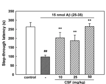
4. Effect of CSF on Aβ (25-35)-induced memory impairment
In the initial acquisition trial of the passive avoidance task, the step-through latency did not differ among the four groups (data not shown). The step-through latency of the Aβ (25-35)-treated group in the retention trial significantly decreased to 97.4± 9.6 s,
compared with 263.2± 24.1 s in the control group, indicating that Aβ (25-35) impaired memory in mice. Chronically administered CSF markedly protected against the memory impairment produced by Aβ (25-35). The step-through latency in groups administered CSF was 202.5 ± 29.8, 187.0 ± 30.4 and 265.3± 15.8 s at doses of 10, 25 and 50 ㎎/㎏, respectively (Fig.
5). We failed to find the dose-dependency of CSF.
Fig. 3. Inhibitory effect of CSF on Aβ (25-35)-induced [Ca2+]i elevation in cultured cortical neurons. [Ca2+]i was monitored using Fluo-4 AM dye and a confocal laser scanning microscope. All images were processed to analyze changes in [Ca2+]i at the single cell level. Results are expressed as the relative fluorescence intensity (RFI).
Each trace shows a single cell that is representative of at least 3 independent experiments.
Fig. 4. Inhibitory effect of CSF on Aβ (25-35)-induced ROS generation in cultured cortical neurons. ROS was monitored using H2DCF-DA dye and a confocal laser scanning microscope. Results are expressed as mean± S.E.M. of RFI obtained from 3 independent experiments.
##P < 0.01 vs control (Student’s t-test); **P < 0.01 vs 10 µM Aβ (25-35) (Tukey’s test).
Fig. 5. Protective effect of CSF on Aβ (25-35)-induced memory impairment in mice. The learning and memory performance was assessed by the passive avoidance test.
Values are expressed as mean± S.E.M. of step-through latency (n = 8-12). ##P < 0.01 vs sham-operated control (Student’s t-test); **P < 0.01 vs 15 nmol Aβ (25-35) (Tukey’s test).
To determine the effect of CSF on cholinergic function, brain cholinesterase activity was measured. No significant increase of cholinesterase activity was shown in brains exposed to 15 nmol Aβ (25-35). However, chronic administration of 50 ㎎/㎏ CSF resulted in a significant reduction of cholinesterase activity showing 545.5± 44.7 mol/h/g brain compared with 710.5 ± 30.2 mol/h/g brain in Aβ (25-35)-treated brains (Table 1).
DISCUSSION
Aβ (25-35) forms a β-sheet structure and induces neuronal cell death, neuritic atrophy, synaptic loss, and memory impairment, as an active partial fragment of Aβ, although it is not found in the AD brain (Maurice et al., 1996; Tohda et al., 2004). Previously, we have reported that Aβ (25-35) caused neuronal cell death, which was blocked by treatment with (5R,10S)-(+)-5-methyl- 10,11-dihydro-5H-dibenzo[a,d]cyclohepten-5,10-imine (MK-801), an NMDA antagonist, verapamil, a L-type Ca2+ channel blocker, and NG-nitro-L-arginine methyl ester (L-NAME), a nitric oxide synthase (NOS) inhibitor (Ban et al., 2006; Lee et al., 2005).
This result implies the involvement of NMDA-glutamate receptor activation, an increase of Ca2+ influx and generation of ROS in Aβ (25-35)-induced neurotoxicity in cultured cortical neurons, as evidenced in other studies (Ekinci et al., 2000; Gray and Patel, 1995; Ueda et al., 1997). The primary event following Aβ (25-35) treatment of cultured neurons has been suggested to be Ca2+ influx, apparently via L-type voltage-dependent Ca2+
channel (L-VDCC), since blockage of this channel and/or Ca2+
chelation prevents all other consequences (Ekinci et al., 2000;
Ueda et al., 1997). It has been reported that vitamin-E, an antioxidant, blocked the Aβ-induced generation of ROS, but not Ca2+ influx, and reduction of extracellular Ca2+ inhibited the Aβ- induced increase in intracellular Ca2+ as well as generation of ROS, indicating that ROS generation is a consequence of Ca2+
accumulation (Ekinci et al., 2000). In the present study, Aβ (25- 35) elicited gradual and significant [Ca2+]i increase, ROS generation, and neuronal cell death in cultured cortical neurons, which were blocked by CSF. These results indicate that CSF might prevent Aβ (25-35)-induced Ca2+ entry through VDCC- and/or NMDA-receptor-coupled channels to inhibit ROS generation and then neuronal death. Many experiments, however, have demonstrated that free radicals are responsible for the increase of [Ca2+]i. The ROS-induced membrane damage causes further Ca2+ influx and resultantly accentuated Ca2+ influx in turn will induce the generation of further ROS (Cotman et al., 1992).
CSF has been proven to be rich in antioxidant constituents such as flavonoids and vitamin C (Hamauz et al., 2005) Therefore, it also can be suggested that the CSF could inhibit the delayed [Ca2+]i increase through suppression of ROS generation, and resultantly ameliorated Aβ (25-35)-induced neuronal death.
Many researchers have demonstrated that Aβ triggers apoptotic degeneration in in vitro neuronal experiments (Ekinci et al., 1999; Yan et al., 1999). In the present work, cultured cortical neurons exposed to Aβ (25-35) for 36 h showed increased chromatin condensation, a typical feature of apoptotic cell death, which was reduced by CSF. Activation of caspases after increased [Ca2+]i signaling and ROS generation, or inflammatory responses in Aβ-stimulated neurons have been proposed to play pivotal roles in apoptosis (Costa et al., 2010;
Gasparini et al., 2004). We have also demonstrated that an increase of caspase-3 activity in Aβ (25-35)-treated cultured cortical neurons is correlated with the increase of [Ca2+]i, ROS generation and neuronal apoptotic death (Ban et al., 2006; Lee et al., 2005). In the present study, CSF might inhibit caspase activity to reduce Aβ (25-35)-induced neuronal apoptosis. The molecular mechanism for the prevention of neuronal apoptosis by CSF should be further clarified.
I.c.v. injection of Aβ (25-35) into experimental rodents induces memory impairment in different behavioral paradigms, including spontaneous alternation, the water maze, and passive avoidance (Maurice et al., 1996; Um et al., 2006). Memory impairment in the passive avoidance test was also confirmed in mice 7 days after the i.c.v. injection of Aβ (25-35) in the present work.
Chronic treatment with CSF effectively protected the mice against Aβ (25-35)-induced memory deficit. This result was consistent with its protective effect on Aβ (25-35)-induced neurotoxicity in vitro. Aβ accumulation associated with cognitive impairment in AD is accompanied by an increase in cholinesterase activity (Maurice et al., 1996). To slow the Table 1. Effect of CSF on brain cholinesterase activity in mice.
Group Dose cholinesterase activity*
(µmol/h g brain−1)
Control − 672.7 ± 68.4
Aβ (25-35) 15 nmol/animal 710.5 ± 30.2
+ CSF 10 ㎎/㎏ 652.6 ± 33.1
+ CSF 25 ㎎/㎏ 616.9 ± 40.3
+ CSF 50 ㎎/㎏ 545.5 ± 44.7*
*Results are expressed as mean ± S.E.M. of cholinesterase activity in brain (n = 6-8 mice/group). **P < 0.01 vs Aβ (25-35) alone (Tukey’s test).
progression of AD and improve cognitive function, it was proposed to restore the cholinergic balance through inhibition of acetylcholine breakdown by cholinesterase (Francis et al., 1999;
van Marum, 2008). Cholinesterase inhibitors such as tacrine and donepezil have been developed for the treatment of cognitive loss in AD based on the cholinergic deficits hypothesis (van Marum, 2008). Although a significant increase of cholinesterase activity was not produced by Aβ (25-35), it was inhibited by CSF (50㎎/㎏) in Aβ (25-35)-treated mice brain in the present study. This result suggests that CSF may increase cholinergic activity and be able to treat memory impairment in AD.
On the other hand, elevated levels of Aβ induce oxidative stress, increasing the appearance of ROS such as superoxide and NO and subsequently producing ONOO− by a rapid interaction, could mediate the damage seen in AD (Kontush, 2001; Smith et al., 1997). A scavenger of ONOO− and antioxidants such as α- tocopherol protect against learning and memory deficits induced by Aβ (Alkam et al., 2007; Yamada et al., 1999). In the present study, 10µM Aβ (25-35) significantly increased the ROS level in cultured neurons, and this was inhibited by CSF. In addition, CSF contains flavonoids as one of the main phytochemical constituents, which are effective in preventing free radical-caused oxidative damage (Zhou et al., 2007; Zhang et al., 2009).
Therefore, it is possible that the favorable effect of CSF on Aβ (25-35)-induced cognitive deficits can be attributed to the inhibition of ROS generation. It has been reported that aqueous extract of CSF inhibited memory deficit induced by the intrahippocampal injection of Aβ (1-40) in mice through inhibition of cytokine expression and ROS generation (Jung and Lee, 2004). In their experiments, delayed memory impairment was produced through 10 weeks by injection of very low level of Aβ (1-40) (10 µM, 0.5 ㎕), and 284 ㎎/㎏ of CSF, which was more than 5 times higher than the highest dosage of 50㎎/㎏
used in the present study, was daily administered for 8 weeks. It was not until 4 weeks that the water extract of CSF revealed the inhibition of Aβ (1-40)-induced memory deficit in mice. We used an ethanol extract of CSF for 1 week to produce antidementia. These results may explain that there are considerable differences of active components between water and ethanol extract of CSF. The present results demonstrated a novel pharmacological activity, a protective effect on Aβ-induced neurotoxicity in cultured neurons, of an ethanol extract of CSF.
In conclusion, this study demonstrated that an ethanol extract of CSF protected against Aβ (25-35)-induced neuronal damage in cultured rat cortical neurons and memory impairment in mice.
These results may explain the inhibitory action of CSF on the progression of AD. Further studies should determine the specific components of CSF that are responsible for preventing the neuronal damage.
ACKNOWLEDGEMENTS
This work was supported by the research grant of the Chungbuk National University in 2011.
LITERATURE CITED
Alkam T, Nitta A, Mizoguchi H, Itoh A and Nabeshima T.
(2007). A natural scavenger of peroxynitrites, rosmarinic acid, protects against impairment of memory induced by A beta (25- 35). Behavioral Brain Research. 180:139-145.
Ban JY, Cho SO, Koh SB, Song KS, Bae K and Seong YH.
(2006). Protection of amyloid β protein (25-35)-induced neurotoxicity by methanol extract of Smilacis chinae rhizome in cultured rat cortical neurons. Journal of Ethnopharmacology.
106:230-237.
Ban JY, Cho SO, Kwon SH, Kim JB, Seong NS, Bae KW, Song KS and Seong YH. (2005). Protection of amyloid β protein (25-35)-induced neuronal cell damage by methanol extract of new stem of Phyllostachys nigra Munro var. henonis Stapf in cultured rat cortical neuron. Korean Journal of Medicinal Crop Science. 13: 95-102.
Butterfield DA and Lauderback CM. (2002). Lipid peroxidation and protein oxidation in Alzheimer's disease brain: potential causes and consequences involving amyloid beta-peptide- associated free radical oxidative stress. Free Radical Biology and Medicine. 32:1050-1060.
Butterfield DA, Reed T, Newman SF and Sultana R. (2007).
Roles of amyloid beta-peptide-associated oxidative stress and brain protein modifications in the pathogenesis of Alzheimer's disease and mild cognitive impairment. Free Radical Biology and Medicine. 43:658-677.
Cho SO, Ban JY, Kim JY, Jeong HY, Lee IS, Song KS, Bae K and Seong YH. (2009). Aralia cordata protects against amyloid β protein (25-35)-induced neurotoxicity in cultured neurons and has antidementia activities in mice. Journal of Pharmacological Sciences. 111: 22-32.
Costa RO, Ferreiro E, Cardoso SM, Oliveira CR and Pereira CM. (2010). ER stress-mediated apoptotic pathway induced by A beta peptide requires the presence of functional mitochondria.
Journal of Alzheimers Diseases. 20:625-636.
Cotman CW, Pike CJ and Copani A. (1992). β-amyloid neurotoxicity: A discussion of in vitro findings. Neurobiology of Aging. 13:587-590.
Demuro A, Parker I and Stutzmann GE. (2010). Calcium signaling and amyloid toxicity in Alzheimer disease. Journal of Biological Chemistry. 285:12463-12468.
Ekinci FJ, Linsley MD and Shea TB. (2000). Beta-amyloid- induced calcium influx induces apoptosis in culture by
oxidative stress rather than tau phosphorylation. Molecular Brain Research. 76:389-395.
Ekinci FJ, Malik KU and Shea TB. (1999). Activation of the L voltage-sensitive calcium channel by mitogen-activated protein (MAP) kinase following exposure of neuronal cells to beta- amyloid. MAP kinase mediates beta-amyloid-induced neurodegeneration. Journal of Biological Chemistry. 274:30322- 30327.
Francis PT, Palmer AM, Snape M and Wilcock GK. (1999).
The cholinergic hypothesis of Alzheimer’s disease: a review of progress. Journal of Neurology Neurosurgery and Psychiatry.
66:137-147.
Gasparini L, Ongini E, and Wenk G. (2004). Non-steroidal anti- inflammatory drugs (NSAIDs) in Alzheimer's disease: old and new mechanisms of action. Journal of Neurochemistry. 91:521- 536.
Gray CW and Patel AJ. (1995). Neurodegeneration mediated by glutamate and beta-amyloid peptide: a comparison and possible interaction. Brain Research. 691:169-179.
Hamauzu Y, Yasui H, Inno T, Kume C and Omanyuda M.
(2005). Phenolic profile, antioxidant property, and anti-influenza viral activity of Chinese quince (Pseudocydonia sinensis Schneid.), quince (Cydonia oblonga Mill.), and apple (Malus domestica Mill.) fruits. Journal of Agricultural and Food Chemistry. 53:928-934.
Hamauzu Y, Irie M, Kondo M and Fujita T. (2008). Anti- ulcerative properties of crude polyphenols and juice of apple, and Chinese quince extracts. Food Chemistry. 108:488-495.
Hestrin S. (1949). The reaction of acetylcholine and other carboxylic acid derivatives with hydroxylamine, and its analytical application. Journal of Biological Chemistry. 180:249- 261.
Hu HP, Han YL and Zhang F. (2008). Preliminary study on antioxidant effects of Chaenomeles sinensis fruit extracts. (in Chinese). Food Science. 29:645-648.
Ivins KJ, Ivins JK, Sharp JP and Cotman CW. (1999). Multiple pathways of apoptosis in PC12 cells. CrmA inhibits apoptosis induced by beta-amyloid. Journal of Biological Chemistry.
274:2107-2112.
Jung IC and Lee SR. (2004). Effects of Chaenomeles fructus extract on the Alzheimer’s disease mice model induced by βA.
Korean Journal of Oriental Physiology & Pathology. 18:1795- 1804.
Kim JY, Jeong HY, Ban JY, Yoo JK, Bae K and Seong YH.
(2009). Ethanol extract of three plants of curcumalongae radix, Phellinus linteus, and Scutellariae radix inhibits amyloid β protein (25-35)-induced neurotoxicity in cultured neurons and memory impairment in mice. Korean Journal of Medicinal Crop Science. 17: 388-396.
Kontush A. (2001). Amyloid-beta: an antioxidant that becomes a pro-oxidant and critically contributes to Alzheimer's disease.
Free Radical Biology and Medicine. 31:1120-1131.
Lee BY, Ban JY and Seong YH. (2005). Chronic stimulation of GABAA receptor with muscimol reduces amyloid β protein (25-35)-induced neurotoxicity in cultured rat cortical cells.
Neuroscience Research. 52:347-356.
Lee MH, Son YK and Han YN. (2002). Tissue factor inhibitory
flavonoids from the fruits of Chaenomeles sinensis. Archives of Pharmacl Research. 25:842-850.
Maurice T, Lockhart BP and Privat A. (1996). Amnesia induced in mice by centrally administered beta-amyloid peptides involves cholinergic dysfunction. Brain Research. 706:181-193.
Oku H, Ueda Y and Ishiguro K. (2003). Antipruritic effects of the fruits of Chaenomeles sinensis. Biological & Pharmaceutical Bulletin. 26:1031-1034.
Osawa K, Arakawa T, Shimura S, Takeya K. (2001). New quinic acid derivatives from the fruits of Chaenomeles sinensis (Chinese quince). Natural Medicines. 55:255-257.
Osawa K, Miyazaki K, Imai H, Arakawa T, Yasuda H and Takeya K. (1999). Inhibitory effects of Chinese quince (Chaenomeles sinensis) on hyaluronidase and histamine release from rat mast cells. Natural Medicines (in Japanese). 53:188- 193.
Osawa K, Yasuda H, Morita H, Takeya K and Itokawa H.
(1997). Antibacterial and antihemolytic activity of triterpenes and β-sitosterol isolated from Chinese quince (Chaenomeles sinensis). Natural Medicines (in Japanese). 51:365-367.
Ros JM, Laencina J, Hellín P, Jordán MJ, Vila R and Rumpunen K. (2004). Characterization of juice in fruits of different Chaenomeles species. LWT-Food Science and Technology. 37:301-307.
Ruan CJ, Si JY, Zhang L, Chen DH, Du GH and Su L. (2009).
Protective effect of stilbenes containing extract-fraction from Cajanus cajan L. on A beta (25-35)-induced cognitive deficits in mice. Neuroscience Letters. 467:159-163.
Sawai R, Kuroda K, Shibata T, Gomyou R, Osawa K and Shimizu K. (2008). Anti-influenza virus activity of Chaenomeles sinensis. Journal of Ethnopharmacology. 118:108- 112.
Smith MA, Richey Harris PL, Sayre LM, Beckman JS and Perry G. (1997). Widespread peroxynitrite-mediated damage in Alzheimer's disease. Journal of Neuroscience. 17:2653-2657.
Thomas M, Crépeau MJ, Rumpunen K and Thibault JF.
(2000). Dietary fibre and cell wall polysaccharides in the fruits of Japanese quince (Chaenomeles japonica). LWT-Food Science and Technology. 33:124-131.
Tohda C, Matsumoto N, Zou K, Meselhy MR and Komatsu K.
(2004). A beta (25-35)-induced memory impairment, axonal atrophy, and synaptic loss are ameliorated by M1, A metabolite of protopanaxadiol-type saponins. Neuropsychopharmacology.
29:860-868.
Ueda K, Shinohara S, Yagami T, Asakura K and Kawasaki K.
(1997). Amyloid beta protein potentiates Ca2+ influx through L- type voltage-sensitive Ca2+ channels: a possible involvement of free radicals. Journal of Neurochemistry. 68:265-271.
Um MY, Choi WH, Aan JY, Kim SR and Ha TY. (2006).
Protective effect of Polygonum multiflorum Thunb on amyloid beta-peptide 25-35 induced cognitive deficits in mice. Journal of Ethnopharmacology. 104:144-148.
Van Dam D and De Deyn PP. (2006). Drug discovery in dementia: the role of rodent models. Nature Reviews Drug Discovery. 5:956-970.
Van Marum RJ. (2008). Current and future therapy in Alzheimer's disease. Fundamental & Clinical Pharmacology.
22:265-274.
Yamada K, Tanaka T, Han D, Senzaki K, Kameyama T and Nabeshima T. (1999). Protective effects of idebenone and alpha-tocopherol on beta-amyloid-(1-42)-induced learning and memory deficits in rats: implication of oxidative stress in beta- amyloid-induced neurotoxicity in vivo. European Journal of Neuroscience. 11:83-90.
Yan XZ, Qiao JT, Dou Y and Qiao ZD. (1999). Beta-amyloid peptide fragment 31-35 induces apoptosis in cultured cortical neurons. Neuroscience. 92:177-184.
Zhang LH, Xu HD and Li SF. (2009). Effects of micronization
on properties of Chaenomeles sinensis (Thouin) Koehne fruit powder. Innovative Food Science & Emerging Technologies.
10; 633-637.
Zhang T, Mi MT, Tang Y and Zhao J. (2007). The extraction of polyphenol contents of Chaenomeles sinensis and its effect on scavenging DPPH radical. (in Chinese). Acta Nutrimenta Sinica. 29:485-489.
Zhou XL, Shao Z, Li TT, Zhang YB, Teng LR and Lv JH.
(2007). Extraction for flavonoids from Carica papaya and its anti-oxidation effect in vitro. (in Chinese). Science and Technology of Food Industry. 28:170-172.