과산화수소수로 유도된 배양 뇌신경세포손상에 대한 왕머루 잎과 줄기 추출물의 보호효과
김주연*·주현수*·반주연**·송경식***·배기환****·성연희*†
*충북대학교 수의과대학, **경희대학교 의과대학, ***경북대학교 농업생명과학대학, ****충남대학교 약학대학
Protective Effect of Vitis amurensis Stems and Leaves Extract on Hydrogen Peroxide-induced Oxidative Neuronal Cell Damage in Cultured Neurons
Joo Youn Kim
*, Hyun Soo Ju
*, Ju Yeon Ban
**, Kyung-Sik Song
***, KiHwan Bae
****, and Yeon Hee Seong
*†*
College of Veterinary Medicine, Chungbuk National University, Cheongju, Chungbuk 361-763, Korea.
**Department of Pharmacology, College of Medicine, Kyung Hee University, Seoul 130-701, Korea.
***College of Agriculture and Life-Sciences, Kyungpook National University, Daegu 702-701, Korea.
****College of Pharmacy, Chungnam National University, Daejon, 305-764 Korea.
ABSTRACT : Vitis amurensis (VA; Vitaceae) has long been used in oriental herbal medicine. It has been reported that roots and seeds of VA have anti-inflammatory and antioxidant effects. In the present study, the protective effect of ethanol extract from stems and leaves of VA on hydrogen peroxide (H
2O
2) (100 µ M)-induced neuronal cell damage was examined in primary cultured rat cortical neurons. VA (10-100 µ g/ml) concentration-dependently inhibited H
2O
2-induced apop- totic neuronal cell death measured by 3-[4,5-Dimethylthiazol-2-yl]-2,5-diphenyl-tetrazolium bromide (MTT) assay and Hoechst 33342 staining. VA inhibited H
2O
2-induced elevation of intracellular Ca
2+concentration ([Ca
2+]
i) and generation of reactive oxygen species (ROS), which were measured by fluorescent dyes. Pretreatment of VA also prevented glutamate release into medium induced by 100 µ M H
2O
2, which was measured by HPLC. These results suggest that VA showed a neuroprotective effect on H
2O
2-induced neuronal cell death by interfering with H
2O
2-induced elevation of [Ca
2+]
i, glutamate release, and ROS generation. This has a significant meaning of finding a new pharmacological activity of stems and leaves of VA in the CNS.
Key Words : Vitis amurensis , Hydrogen Peroxide, Neurotoxicity, Neuroprotection, Anti-oxidant
INTRODUCTION
Oxidative stress occurs when the balance between free radical production and cellular defense mechanisms are disturbed and is implicated in the neuronal cell death that is associated with many neurodegenerative disorders such as Alzheimer’s disease, Parkinson disease and amyotrophic lateral sclerosis (Alexi et al ., 2000; Behl, 1999; Behl et al ., 1994; Chen et al ., 2008; Jellinger, 2000). It has been also indicated that oxidative stress is considered one of the primary risk factors that exacerbate the damage by cerebral ischemia (Chan, 2001). Several components of reactive oxygen species (ROS) generated after ischemia/reperfusion
injury, including superoxide anions (O
2−), hydroxyl radical (OH
−), hydrogen peroxide (H
2O
2), and peroxynitrite radical (ONOO
−), are known to promote DNA damage, peroxida- tion of lipids, proteins and carbohydrates, blood brain-barrier break-down, and microglial infiltration in the ischemic territory (Camacho and Massieu, 2006; Crack et al ., 2005).
Therefore, in vitro H
2O
2toxicity has become a well- established model for studying the neuropathology of oxidative stress in CNS disorders. Many researches have demonstrated the involvement of glutamate in H
2O
2-induced neurotoxicity in cultured neurons (Gardner et al ., 1997).
H
2O
2and O
2−inhibit uptake of glutamate and enhance the release of glutamate, resulting in NMDA receptor oversti-
†
Corresponding author: (Phone) +82-43-261-2968 (E-mail) [email protected]
Received January 20, 2009 / Revised February 5, 2009 / Accepted February 10, 2009
mulation (Mailly et al ., 1999; Volterra et al ., 1994). There are some reports on H
2O
2-induced intracellular Ca
2+concen- tration ([Ca
2+]
i) increase (Das et al ., 2008; Numakawa et al ., 2007). NMDA receptor is a ligand-gated/voltage-sensitive cation channel, especially highly permeable to Ca
2+. Calcium influx through NMDA receptor-coupled Ca
2+channel appears to be a critical role in the H
2O
2-induced neurotoxicity (Mailly et al . 1999). Moreover, Ca
2+signals activate enzymes, which lead to further ROS generation (Ekinci et al ., 2000; Butterfield et al ., 2001). Conversely, ROS generation can facilitate [Ca
2+]
iincreases by damaging the [Ca
2+]
iregulatory mechanism and activating Ca
2+release from intracellular Ca
2+stores (Duffy and Macviar, 1996).
Vitis species, the family Vitaceae, is widely distributed over the world and has been used as foods and medicinal herbs (Nassiri-Asl and Hooeinzadeh, 2009). Grape seeds extract from Vitis vinifera has been reported to have excellent free radical scavenging and cardioprotective pro- perties such as inhibition of cardiac ischemia-reperfusion injury, myocardiac infarction, H
2O
2-induced oxidant injury in heart and reactive oxygen intermediates release (Bagchi et al ., 2000; Sato et al ., 1999; Shao et al ., 2003; Vitseva et al ., 2005). Furthermore, Vitis vinifera and its active components including polyphenols and flavanols exhibited neuroprotection against stroke and memory impairment in both in vitro cultured neurons and animal models (Anderson et al ., 2003; Angelo et al ., 2008; Chun et al ., 2008;
Vauzour et al ., 2007; Wang et al ., 2005; Wang et al ., 2006; Wang et al ., 2008). In terms of Vitis amurensis (VA), the roots extract and resveratrols isolated from it showed inhibition of cholinesterase, β -amyloid aggregation and β - amyloid-induced neuronal cell damage indicating its beneficial effect on Alzheimer disease (Jang et al ., 2007; Jang et al ., 2008) and showed anti-tumor and anti-inflammatory activities (Huang et al ., 2001; Lee et al ., 2006). The fruits extract of VA and polyphenols from it showed anti-allergic and anti- oxidant effects (Kim et al ., 2008; Wang et al ., 2000). Inter- estingly, however, there is no report to prove phar- macological activity of leaves and stems of VA. In the present study, the protective effect of ethanol extract from leaves and stems of VA against H
2O
2-induced neuronal cell damage was investigated in primary cultured rat cortical neurons to evaluate its therapeutic possibility in oxidative stress-induced brain damage.
MATERIALS AND METHODS
1. Plant material and extraction
The leaves and stems of VA were collected at Keryong Mountain in Daejeon, Korea, in July 2007. Botanical identification was performed by Professor KiHwan Bae and the voucher specimen (CNU-1552) was deposited at the herbarium of the College of Pharmacy, Chungnam National University, Korea. The dried leaves and stems (500 g) of VA were extracted with 50% ethanol in water at 90 ℃ in the water bath, three times. The combined ethanol extract was filtered and concentrated until the constant weight to yield an ethanol extract (98.5 g).
2. Experimental animals
Pregnant Sprague-Dawley (SD) rats were purchased from Daehan Biolink Co. Ltd. (Chungbuk, Korea) and housed singly in an environmentally controlled room at 22 ± 2 ℃ , with a relative humidity of 55 ± 5%, a 12-h light/dark cycle, and food and water ad libitum. The procedures involving experimental animals complied with the regulations for the care and use of laboratory animals of the animal ethics committee of Chungbuk National University.
3. Induction of neurotoxicity in primary cultures of rat cerebral cortical neurons
Primary cortical neuronal cultures were prepared using embryonic day 15 to 16 SD rat fetuses as described previously (Ban et al ., 2005; Lee et al ., 2007a). Isolated cells were plated onto poly-L-lysine coated 12 well-plates at a density of 4 × 10
5cells/well in DMEM supplemented with 10% fetal bovine serum. Neurotoxicity experiments were performed on neurons after 5-7 days in culture. For every experiment, H
2O
2was diluted freshly in a HEPES buffer (pH 7.4) containing 8.6 mM HEPES, 154 mM NaCl, 5.6 mM KCl and 2.3 mM CaCl
2. For the measurement of cell viability, neurons were treated with 100 µ M H
2O
2in HEPES buffer at 37 ℃ for 15 min, followed by incubation for 12 h in H
2O
2- and serum-free DMEM medium. Neurons were pretreated with VA (10, 50 and 100 ㎍ / ㎖ ) for 15 min prior to H
2O
2treatment, and then again during the H
2O
2exposure and post-exposure period. VA was dissolved in ethanol (100 ㎎ / ㎖ ) and then diluted further in experimental buffers. The final concentration of ethanol was
≤ 0.1%, which did not affect cell viability.
4. Measurements of H
2O
2-induced neuronal death and intracellular biochemical changes
A 3-[4,5-dimethylthiazol-2-yl]-2,5-diphenyl-tetrazolium bromide (MTT; Sigma Chemical Co.) assay and Hoechst 33342 (Molecular Probes Inc., Eugene, OR, USA) staining were performed to measure neuronal death at the end of the post-exposure period, as described previously (Ban et al ., 2005). The change of [Ca
2+]
iwas measured with Fluo-4 AM (Molecular Probes), a Ca
2+-sensitive fluorescent dye, using a laser scanning confocal microscope (LSM 510, Carl Zeiss Oberkochen, Germany) with a 488-nm excitation argon laser and 515- ㎚ longpass emission filters (Ban et al ., 2005;
Lee et al ., 2007). Glutamate secreted into the medium after treatment with 100 µ M H
2O
2in HEPES buffer at 37 ℃ for 15 min, followed by post-incubation for 3 h in H
2O
2- and serum-free DMEM medium was quantified by HPLC with an electrochemical detector (MF series, BAS, IN, USA) (Lee et al ., 2007). The microfluorescence assay of 2'7'- dichlorofluorescein, the fluorescent product of 2'7'-dichloro- dihydrofluorescein diacetate (H
2DCF-DA; Molecular probes), and laser scanning confocal microscope (MRC1024ES, Bio- Rad, Maylands, UK) with 488 ㎚ excitation and 510 ㎚ emission filters were used to monitor the generation of ROS in neurons treated with 100 µ M H
2O
2for 20 min (Ban et al ., 2005).
5. Statistical analysis
Data are expressed as mean ± S.E.M. and statistical significance was assessed by one-way analysis of variance (ANOVA) and Tukey's test. P<0.05 was considered significant.
RESULTS
1. Protective effect of VA on H
2O
2-induced neuronal cell death
In a previous report, we demonstrated that H
2O
2over the concentration range of 50-200 µ M produced a concentration- dependent reduction of cell viability in cultured cortical neurons (Park et al ., 2006). For the present experiments, 100 µ M of H
2O
2was used. Following exposure to 100 µ M H
2O
2, the viability (MTT reduction rate) of cortical neurons decreased to 48.0 ± 5.0% relative to untreated controls. We observed that this decrease in MTT reduction could be ameliorated in a VA concentration-dependent manner, to 80.3
± 1.9 and 91.9 ± 2.5% with 50 and 100 ㎍ / ㎖ VA, respec-
tively (Fig. 1).
Chromatin condensation and nuclear fragmentation were observed in neurons treated with 100 µ M H
2O
2, whereas the control culture contained the round blue nuclei of viable neurons (Lee et al . 2007b). As shown in Fig. 2, apoptosis was observed in 35.8 ± 1.7% of cultured cortical neurons treated with 100 µ M H
2O
2, as compared with 13.2 ± 2.4%
of neurons in control cultures. On the other hand, the addition of VA (100 ㎍ / ㎖ ) significantly decreased H
2O
2- induced apoptotic neuronal death to 22.4 ± 2.1%.
Fig. 1.
Inhibitory effect of VA on H
2O
2-induced neuronal death in cultured cerebral cortical neurons. Neuronal cell death was measured using the MTT assay. The MTT absorbance from untreated cells was normalized to 100%. Values represent mean ± S.E.M. of data obtained from 4 independent experiments.
##P<0.01 vs. control;
**P<0.01 vs. 100
µM H
2O
2.
Fig. 2.
Inhibitory effect of VA on H
2O
2-induced apoptosis of cultured cerebral cortical neurons. Apoptotic cells measured by Hoechst 33342 staining were counted from 5 to 6 fields per well. Values represent mean ± S.E.M. of data obtained from 3 independent experiments.
##P<
0.01 vs. control;
**P< 0.01 vs. 100
µM H
2O
2.
2. Inhibitory effect of VA on H
2O
2-induced elevation of [Ca
2+]
iMany studies have shown that an increase in [Ca
2+]
iis associated with H
2O
2-induced cell death (Duffy and MacVicar, 1996). In cultured cortical neurons, treatment with 100 µ M H
2O
2produced a slow but gradual increase in [Ca
2+]
i, with the maximum fluorescence intensity (ca. 195, compared to a basal level of 110) observed about 10 min after H
2O
2application. In contrast, pretreatment with VA (100 ㎍ / ㎖ ) significantly inhibited this increase in [Ca
2+]
ithroughout the measurement period (Fig. 3). VA did not affect basal [Ca
2+]
i.
3. Inhibitory effect of VA on H
2O
2-induced glutamate release
Glutamate release into the extracellular medium for 3 h was quantified 15 min after the incubation of cells with 100
µ M H
2O
2. H
2O
2elevated basal glutamate level to 3.86 ± 0.40 µ M from 0.44 ± 0.16 µ M in control cultures. VA (100
㎍ / ㎖ ) significantly blocked H
2O
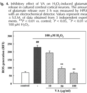
2-induced elevation of glutamate release showing 1.14 ± 0.19 µ M (Fig. 4).
4. Inhibitory effect of VA on H
2O
2-induced elevation of ROS
In H
2DCF-DA-loaded cortical neurons, H
2O
2increased the fluorescence intensity, indicating generation of ROS. A ca.
3-fold increase in fluorescence intensity was observed in
neurons treated for 20 min with 100 µ M H
2O
2, compared to control neurons (228.9 ± 12.8 and 69.6 ± 6.7, respectively).
However, the H
2O
2-induced increase in ROS generation was inhibited significantly by VA (50 and 100 ㎍ / ㎖ ) (Fig. 5).
DISCUSSION
H
2O
2causes neuronal cell death by inducing a delayed accumulation of extracellular glutamate and NMDA receptor
Fig. 3.
Inhibitory effect of VA on H
2O
2-induced [Ca
2+]
iincrease in cultured cerebral cortical neurons. [Ca
2+]
iwas monitored using Fluo-4 AM dye and a confocal laser scanning microscope. Results are expressed as the relative fluorescence intensity (RFI). Each trace shows a single cell that is representative of at least 3 independent experi- ments.
Fig. 4.
Inhibitory effect of VA on H
2O
2-induced glutamate release in cultured cerebral cortical neurons. The amount of glutamate release over 3 h was measured by HPLC with an electrochemical detector. Values represent mean
± S.E.M. of data obtained from 3 independent experi- ments.
##P< 0.01 vs. control;
*P< 0.05,
**P< 0.01 vs.
100
µM H
2O
2.
Fig. 5