Curcuma longae
Radix,Phellinus linteus
및Scutellariae
Radix 혼합추출물의 Aβ (25-35) 유도 배양신경세포독성 및 마우스기억손상 억제효과김주연*·정하연*·반주연**·유재국***·배기환***·성연희*†
*충북대학교수의과대학, **단국대학교치과대학, ***한국신약, ***충남대학교약학대학
Ethanol Extract of Three Plants of Curcuma longae Radix, Phellinus linteus , and Scutellariae Radix Inhibits Amyloid
βProtein (25-35)-Induced Neurotoxicity
in Cultured Neurons and Memory Impairment in Mice
Joo Youn Kim*, Ha Yeon Jeong*, Ju Yeon Ban**, Jae Kuk Yoo***, KiHwan Bae****, and Yeon Hee Seong*†
*
College of Veterinary Medicine, Chungbuk National University, Cheongju, Chungbuk 361-763, Korea.**
School of Dentistry, Dankook University, Cheonan, Chungnam 330-714, Korea.***
Han Kook Shin Yak, Nonsan, Chungnam 320-854, Korea.****
College of Pharmacy, Chungnam National University, Taejon 305-764, Korea.ABSTRACT : The present study investigated an ethanol extract (HS0608) of a mixture of three medicinal plants of Curcuma longae radix, Phellinus linteus,and Scutellariae radix for possible neuroprotective effects on neurotoxicity induced by amyloid β protein (Aβ) (25-35) in cultured rat cortical neurons and antidementia activity in mice. Exposure of cultured cortical neurons to 10µM Aβ (25-35) for 36 h induced neuronal apoptotic death. At 1-50㎍/㎖, HS0608 inhibited neuronal death, elevation of intra- cellular calcium concentration ([Ca2+]i), and generation of reactive oxygen species (ROS) induced by Aβ (25-35) in primary cul- tures of rat cortical neurons. Memory loss induced by intracerebroventricular injection of ICR mice with 15 nmol Aβ (25-35) was inhibited by chronic treatment with HS0608 (25, 50 and 100㎎/㎏, p.o. for 7 days) as measured by a passive avoidance test. From these results, we suggest that the antidementia effect of HS0608 is due to its neuroprotective effect against Aβ (25-35)-induced neurotoxicity and that HS0608 may have a therapeutic role in preventing the progression of Alzheimer’s disease.
Key Words:Curcuma longae Radix, Phellinus linteus, Scutellariae Radix, Neuroprotection, Amyloid β protein, Cultured Neurons, Memory Impairment
INTRODUCTION
Curcuma longae radix, the root of Curcuma longa, has been extensively studied for its biological activities, such as anti-inflammatory (Guo et al., 2008), anti-platelet (Srivastava et al., 1995), hypoglycemic (Sharma et al., 2006), anti- oxidant (Adaramoye, 2002) and neuroprotective effects (Rajakrishnan et al., 1999). Phytochemical screenings have shown that the main constituents of Curcuma longae radix are curcumins, curcuminoids, zingiberine, phelandreen, and essential oils (Kapoor, 1990; Srinivasan, 1953). Phellinus linteus has been used for its anti-cancer, anti-diabetes and anti-oxidant activities (Ajith and Janardhanan, 2002; Sliva et
al., 2008). Recently, it was defined that Phellinus linteus and its active component, hispolon, shows anti-inflammation and analgesic effects via inhibition of nitric oxide (NO) and prostaglandin (PG) E2 production and antioxidative activity (Chang et al., 2009; Kim et al., 2007). Scutellariae radix from Scutellaria baicalensis Gergi (Labiatae) has long been used as a medicinal herb in Asia due to its antipyretic, antibacterial, and anti-inflammatory properties (Bensky, 1992;
Huang, 1999). Several flavonoids such as baicalin, baicalein and wogonin have been demonstrated as active components of Scutellariae radix (Huang, 1999). Furthermore, Scutellariae radix and its flavonoids have shown neuroprotection against ischemic brain damage, 6-hydroxydopamine-induced parkin-
†Corresponding author: (Phone) +82-43-261-2968 (E-mail) [email protected] Received 2009 August 11 / Revised 2009 October 20 / Accepted 2009 December 7
sonism, and Aβ (25-35)-induced amnesia (Mu et al., 2009;
Wang et al., 2004; Zhang et al., 2006).
Alzheimer’s disease (AD) is characterized by neuronal loss and extracellular senile plaque, whose major constituent is amyloid β protein (Aβ), a 39-43 amino acid peptide derived from amyloid precursor protein (Ivins et al., 1999). Both in vitro and in vivo studies have reported the toxic effects of Aβ or Aβ peptide fragments suggesting an important role of Aβ in the pathogenesis of AD. In cultures, Aβ can directly induce neuronal cell death (Ueda et al., 1997) and can render neurons vulnerable to excitotoxicity (Koh et al., 1990) and oxidative insults (Chen et al., 2007). The mechanisms underlying Aβ-neurotoxicity are complex but may involve N-methyl-D-aspartate (NMDA) receptor, a glutamate receptor subtype, modulation induced by glutamate release, sustained elevations of intracellular Ca2+ concen- tration ([Ca2+]i), and oxidative stresses (Ekinci et al., 2000;
Gray and Patel, 1995; Ueda et al., 1997).
An experimental model that mimics the progression of AD was developed using an intracerebroventricular (i.c.v.) injection of Aβ in mice (Van Dam and De Deyn, 2006).
A continuous infusion of full-length Aβ, Aβ (1-40), into the cerebral ventricle in rats resulted in learning and memory deficits (Nitta et al., 1997). The deposition of Aβ in the pathogenesis of AD is invariably associated with oxidative stress and inflammatory responses. (Butterfield and Lauderback, 2002; Butterfield et al., 2007). Aβ has been reported to produce hydrogen peroxide and lipid peroxide in neurons and superoxide and proinflammatory cytokines in astrocytes and microglial cells (Gitter et al., 1995;
McDonald et al., 1997). Antioxidants such as α-tocopherol protect against cytotoxicity induced by Aβ in vitro as well as against learning and memory deficits in vivo (Yamada et al., 1999). Also, α-tocopherol and anti-inflammatory agents such as indomethacin reportedly slow the progression of AD (Rogers et al., 1993; Sano et al., 1997). Aβ (25-35), the core toxic fragment of Aβ (1-40), produces similar neurodegenerative properties as Aβ (1-40), including oxidative damage, inflammatory responses, and memory impairment (Pike et al., 1995; Richardson et al., 1996).
Since reactive oxygen species (ROS) and inflammation are part of the complex series of pathophysiological events that contribute to neurodegenerative diseases such as AD and stroke (Hoehn et al., 2005; Pitchumoni and Doraiswamy, 1998), free radical scavengers and anti-inflammatory agents
have attracted considerable attention as potential neuropro- tective agents. We hypothesized that the combination of three plants of Curcuma longae radix, Phellinus linteus, and Scutellariae radix might protect neurons against neurode- generative diseases such as AD and ischemia on the basis of their antioxidant, anti-inflammatory, and neuroprotective activities. The aim of the present study was to determine whether an ethanol extract of a mixture of three herbs of Curcuma longae radix, Phellinus linteus, and Scutellariae radix, which was named as HS0608, has a protective effect against Aβ (25-35)-induced memory deficits in mice and Aβ (25-35)-induced neuronal damage in cultured rat cortical neurons.
MATERIALS AND METHODS
1. Plant materials and extraction
The dried Curcuma longae radix and Scutellariae radix were purchased at Kyungdong Folk Medicine market, Seoul, Korea, and fruiting bodies of cultured Phellinus linteus were supplied from Han Kook Shin Yak, Nonsan, Chungnam.
These plants were identified by one of the authors, Dr.
KiHwan Bae, Chungnam National University. Each 100 g of the three plants was mixed, refluxed in 3 L ethanol at room temperature for 3 h, filtered, and lyophilized to yield an ethanol extract (HS0608, 64 g), which was then stored at
−20℃ until required.
2. Experimental animals
Pregnant Sprague-Dawley rats for primary neuronal culture and male ICR mice for the passive avoidance test were supplied by Daehan BioLink Co., Ltd. (Chungbuk, Korea) and housed in an environmentally controlled room at 22±2
℃, with a relative humidity of 55±5%, a 12-h light/dark cycle, and food and water ad libitum. The procedures involving experimental animals complied with the animal care guidelines of the National Institutes of Health and the animal ethics committee of Chungbuk National University.
3. Induction of neurotoxicity in primary cultures of rat cerebral cortical neurons
Primary cortical neuron cultures were prepared using embryonic day 15 to 16 Sprague-Dawley rat fetuses, as described previously (Ban et al., 2006). Neurotoxicity experiments were performed on neurons after 3-4 days in
culture. Cultured neurons were treated with 10µM Aβ (25- 35) (Bachem, Bubendorf, Switzerland) in serum-free Dulbecco’s modified Eagle’s medium (DMEM) (Sigma) at 37℃ for 36 h (unless otherwise indicated) to produce neurotoxicity. An Aβ (25-35) stock solution of 2 mM was prepared in sterile distilled water, stored at −20℃, and incubated for more than 2 days at 37℃ to aggregate before use. HS0608 was dissolved in dimethylsulfoxide (DMSO) at concentrations of 50㎎/㎖ and further diluted in experimental buffers. The final concentration of DMSO was
≤0.1%, which did not affect cell viability. For each experiment, HS0608 was applied 15 min prior to treatment with 10µM Aβ (25-35). It was also present in the medium during Aβ (25-35) incubation.
4. Measurements of Aβ (25-35)-induced neuronal death and intracellular biochemical changes
A 3-(4,5-dimethylthiazol-2-yl)-2,5-diphenyl-tetrazolium bromide (MTT; Sigma) assay and Hoechst 33342 (Molecular Probes, Eugene, OR, USA) staining were performed to measure neuronal death 36 h after exposure of cultured neurons to 10µM Aβ (25-35), as described previously (Ban et al., 2006). Changes in [Ca2+]i were measured with Fluo-4 AM (Molecular Probes), a calcium-sensitive fluorescent dye, using a laser scanning confocal microscope (TCS SP2 AOBS;
Leica, Heidelberg GmbH, Germany) with a 488-㎚
excitation argon laser and 515-㎚ longpass emission filters (Ban et al., 2006; Lee et al., 2007). The microfluorescence of 2',7'-dichlorofluorescein, the fluorescent product of 2',7'- dichlorodihydrofluorescein diacetate (H2DCF-DA; Molecular Probes), and a laser scanning confocal microscope (TCS SP2 AOBS; Leica, Heidelberg GmbH, Germany) with 488-㎚ excitation and 510-㎚ emission filters were used to monitor the generation of ROS in neurons treated with 10
µM Aβ (25-35) for 36 h (Ban et al., 2006; Kim et al., 2008; Lee et al., 2007).
5. Examination of learning and memory; Passive avoidance task
On day 1, HS 0608 was administered to 5-week-old ICR mice and 30 min later, the aggregated form of Aβ (25-35) was administered i.c.v. using a microsyringe with a 27- gauge stainless-steel needle (Hamilton), the tip of which had been adjusted to a depth of 3㎜. The needle was inserted unilaterally 2㎜ to the right of the midline point equidistant
from each eye, at an equal distance between the eyes and the ears, and perpendicular to the plane of the skull (Maurice et al., 1996)). The injection volume of Aβ (25-35) was 15㎕ (15 n㏖). Fifteen ㎕ of saline was injected i.c.v.
in sham-operated mice. HS0608 (25, 50 and 100㎎/㎏, p.o.) suspended in distilled water was administered daily for 7 days. Sham-operated and Aβ (25-35)-injected control groups were administered distilled water as a vehicle. Thirty min after administration of HS0608 on day 7, mice were trained on a one-trail step-through passive avoidance task.
The passive avoidance box was divided into two chambers separated by a guillotine door and equipped with a grid floor and a shock generator (Gemini Avoidance System, San Diego, CA, USA). During the acquisition trial, each mouse was placed into the start chamber and kept in the dark until it was illuminated. After 50 s, the start chamber was illuminated and the door was opened for mouse to move into the dark chamber freely. Immediately after the mouse entered the dark chamber, the door was closed and the mouse received an inescapable electric shock (0.3 mA, 2 s).
In the retention trial given 24 h after the acquisition trial, the mouse was again placed in the chamber and illuminated 50 s later, and the time it took until it re-entered the dark chamber was measured (the step-through latency; maximum testing limit was 300 s). To test for any possible effect of Aβ (25-35) on motor function, the locomotor activity of the animals was measured using a photobeam monitoring system (AM1051, Benwick Electronics, Benwick, UK), and motor coordination was measured using a rota-rod apparatus (Daejong Inc., Seoul, Korea). Each mouse was placed in the center of the activity cage and the total number of beam interruptions was registered for 5 min. Then the mouse was put on the rota-rod for 2 min and number of falls from the rod was noted.
6. Statistical analysis
Data are expressed as mean±SEM and statistical significance was assessed by one-way analysis of variance (ANOVA) and Tukey’s tests. P< 0.05 was considered significant.
RESULTS
1. HS0608 inhibits Aβ (25-35)–induced neuronal cell death
The concentration of 10µM of Aβ (25-35) was used for
determining Aβ (25-35)-induced neuronal cell damage in the present experiments based on our previous result (Lee et al., 2005). When cortical neurons were exposed to 10µM Aβ (25-35) for 36 h, absorbance in the MTT assay was 70.6±2.2% of that of the untreated controls (Fig. 1), indicating that Aβ (25-35) caused neuronal cell death.
Pretreatment of cortical neurons with 10 and 50㎍/㎖
HS0608 reduced the neuronal death induced by 10µM Aβ
(25-35) (absorbance, 83.7±2.4% and 95.1±4.0% of control, respectively; Fig. 1).
An additional experiment was performed with Hoechst 33342 staining to detect condensed or fragmented DNA, which is indicative of Aβ (25-35)-induced neuronal apoptotic death. Treatment of neurons with 10µM Aβ (25-35) produced apoptosis of 39.2±2.0% of the total population of cultured cortical neurons, as compared with 14.8±1.1% of apoptotic neurons in control cultures. The addition of HS0608 (1, 10 and 50㎍/㎖) significantly decreased the Aβ
(25-35)-induced apoptotic cell death, showing 22.9±1.6, 20.3
±1.1 and 13.4±0.6%, respectively (Fig. 2).
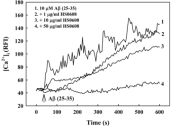
2. HS0608 inhibits Aβ (25-35)–induced [Ca2+]i elevation Increases in [Ca2+]i have been associated with Aβ-induced cell death. In our cell cultures, [Ca2+]i gradually increased in response to treatment with 10µM Aβ (25-35) with intermittent fluctuations over 10 min (Fig. 3). In contrast, pretreatment with HS0608 (1, 10 and 50㎍/㎖) showed inhibition of the increase of [Ca2+]i induced by 10µM Aβ
(25-35) in a concentration-dependent manner. HS0608 did not affect basal [Ca2+]i.
3. HS0608 inhibits Aβ (25-35)–induced ROS generation To clarify the involvement of oxidative stress in Aβ Fig. 1. Inhibitory effect of HS0608 on Aβ (25-35)-induced
neuronal cell death in cultured cortical neurons. Neuronal cell death was measured using the MTT assay. The MTT absorbance from untreated cells was normalized to 100%. Results are expressed as mean ± S.E.M. of data obtained from 5 independent experiments. ##P< 0.01 vs control; *P< 0.05, **P< 0.01 vs 10µM Aβ (25-35).
Fig. 2. Inhibitory effect of HS0608 on Aβ (25-35)-induced apoptosis of cultured cortical neurons. Apoptotic cells measured by Hoechst 33342 staining were counted in 5 to 6 fields per well. The values represent the apoptotic cells as a percentage of the total number of cells expressed as mean ± S.E.M. of data obtained from 3 independent experiments. ##P< 0.01 vs control, **P<
0.01 vs 10µM Aβ (25-35).
Fig. 3. Inhibitory effect of HS0608 on Aβ (25-35)-induced [Ca2+]i elevation in cultured cortical neurons. [Ca2+]i was monitored using Fluo-4 AM dye and a confocal laser scanning microscope. All images were processed to analyze changes in [Ca2+]i at the single cell level. Results are expressed as the relative fluorescence intensity (RFI).
Each trace shows a single cell that is representative of at least 3 independent experiments.
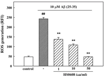
neurotoxicity, we measured the accumulation of ROS after the exposure of the cells to Aβ (25-35) for 36 h. In H2DCF-DA-loaded cerebral cortical neurons, 10µM Aβ (25- 35) increased the fluorescence intensity, indicating that ROS were generated. In neurons treated with 10µM Aβ (25-35), the relative fluorescence increased approximately 2.5-fold to 242.8±7.3 compared with the value in control neurons (47.63±4.2; Fig. 4). The Aβ (25-35)-induced increase in ROS generation was significantly inhibited by HS0608 (1, 10 and 50㎍/㎖).
4. HS0608 inhibits Aβ (25-35)–induced memory impairment
In the initial acquisition trial of the passive avoidance task, the step-through latency did not differ among the 5 groups (control, 15 n㏖ Aβ (25-35), Aβ (25-35) + 25㎎/㎏
HS0608, Aβ (25-35) + 50㎎/㎏ HS0608, and Aβ (25-35) + 100㎎/㎏ HS0608; data not shown). The step-through latency of the Aβ (25-35)-treated group in the retention trial significantly decreased to 62.2±16.0 s, compared with 274.3
±23.1 s in the control group, indicating that Aβ (25-35) impaired memory in mice. Chronically administered HS0608 markedly protected against the memory impairment produced by Aβ (25-35). The step-through latency in groups administered HS0608 was 165.4±25.3, 222.0±24.2 and 279.0±14.8 s at doses of 25, 50 and 100㎎/㎏, respectively (Fig. 5). To elucidate whether HS0608 affects general motor functions, we measured spontaneous locomotor activity and motor coordination in the mice. Neither HS0608 nor Aβ (25-35) significantly affected locomotor and rota-rod activity (data not shown), indicating that the observed improvement of memory by HS0608 was not due to immobility.
DISCUSSION
The present study provides evidence that Aβ (25-35)- induced injury to rat cortical neurons can be prevented by HS0608, an ethanol extract of a mixture of Curcuma longae radix, Phellinus linteus, and Scutellariae radix.
HS0608 was able to reduce the Aβ (25-35)-induced [Ca2+]i
increase, ROS generation, and, in result, attenuate neuronal apoptotic death in primarily cultured rat cortical neurons.
Furthermore, HS0608 prevented memory loss induced by i.c.v. injection of Aβ (25-35) in mice.
The involvement of the disruption of [Ca2+]i homeostasis and ROS generation in Aβ-induced neurotoxicity has been demonstrated in various studies. Our previous studies confirmed that Aβ (25-35) caused neuronal cell death, which was blocked by treatment with MK-801, verapamil, an L-type Ca2+ channel blocker, and NG-nitro-L-arginine methyl ester (L-NAME), a NO synthase (NOS) inhibitor (Ban and Seong, 2005; Lee et al., 2005). This result implies the involvement of NMDA-glutamate receptor activation, an increase of Ca2+ influx and generation of ROS in Aβ (25-35)-induced neurotoxicity in cultured cortical
Fig. 4. Inhibitory effect of HS0608 on Aβ (25-35)-induced ROS generation in cultured cortical neurons. ROS was monitored using H2DCF-DA dye and a confocal laser scanning microscope. Results are expressed as mean ± S.E.M. of RFI obtained from 4 independent experiments.
##P< 0.01 vs control; **P< 0.01 vs 10µM Aβ (25-35).
Fig. 5. Protective effect of HS0608 on Aβ (25-35)-induced memory impairment in mice. The learning and memory performance was assessed by the passive avoidance test.
Values are expressed as mean ± S.E.M. of step-through latency (n = 8-12). ##P< 0.01 vs sham-operated control;
**P< 0.01 vs 10µM Aβ (25-35).
neurons, as evidenced in other studies (Ekinci et al., 2000;
Gray and Patel, 1995; Ueda et al., 1997). The primary event following Aβ (25-35) treatment of cultured neurons has been suggested to be Ca2+ influx, apparently via L-type voltage-dependent Ca2+ channel (L-VDCC), since blockage of this channel and/or Ca2+ chelation prevents all other consequences (Ekinci et al., 1999; Ueda et al., 1997). In the present study, Aβ (25-35) elicited gradual and significant [Ca2+]c increase, which was blocked by HS0608. Many reports demonstrated the involvement of ROS formation in Aβ-induced neurotoxicity (Miranda et al., 2000; Morais Cardoso et al., 2002). It has been reported that vitamin-E, an antioxidant, blocked the Aβ-induced generation of ROS, but not Ca2+ influx, and reduction of extracellular Ca2+
inhibited the Aβ-induced increase in intracellular Ca2+ as well as generation of ROS, indicating that ROS generation is a consequence of Ca2+ accumulation (Ekinci et al., 2000).
HS0608 also decreased the Aβ (25-35)-induced increase of ROS generation. These results indicate that HS0608 might prevent Aβ (25-35)–induced Ca2+ entry through VDCC- and/
or NMDA-receptor–coupled channels to inhibit ROS generation and then neuronal death, although the mechanism by which HS0608 blocks the channels is not clear.
Curcuma longae radix, Phellinus linteus, and Scutellariae radix, the constituents of HS0608, have been reported to possess antioxidant principles, curcumin, hispolon, and baicalein, baicalin and wogonin (Chang et al., 2009; Guo et al., 2008; Su et al., 2008; Zhang et al., 2006), repectively, suggesting that inhibition of Aβ (25-35)–induced neuronal death by HS0608 might be due to their ROS scavenging activity. Further study to elucidate the precise mechanism should be performed.
Many researchers have demonstrated that Aβ triggers apoptotic degeneration in in vitro neuronal experiments (Ekinci et al., 2000; Yan et al., 1999). In the present work, cultured cortical neurons exposed to Aβ (25-35) for 36 h showed increased chromatin condensation, a typical feature of apoptotic cell death, which was reduced by HS0608. Aβ- induced apoptosis was also associated with COX-2 upregulation, and COX has been suggested to be an important source of ROS in the pathologic brain (Chan, 2001; Jang and Surh, 2005). Curcuma longae radix, Phellinus linteus, and Scutellariae radix have been reported to inhibit expression of inflammatory mediators including NFκB, COX-2 and iNOS (Chang et al., 2009; Guo et al.,
2008; Kim et al., 2007; Yune et al., 2009). Therefore, the protective effect of HS0608 on the Aβ-induced neurotoxicity might result from the inhibition of the inflammatory process.
The molecular mechanism for the prevention of neuronal apoptosis by HS0608 should be further clarified.
I.c.v. injection of Aβ (25-35) into experimental rodents induces memory impairment in different behavioral paradigms, including spontaneous alternation, the water maze, and passive avoidance (Maurice et al., 1996; Um et al., 2006). Aβ (25-35) preferentially impairs spatial and non- spatial short-term memory, and these effects remain evident up to 6 months after even a single i.c.v. injection of the peptide (Stepanichev et al., 2003). Memory impairment in the passive avoidance test was also confirmed in mice 7 days after the i.c.v. injection of Aβ (25-35) in the present work. Chronic treatment with HS0608 effectively protected the mice against Aβ (25-35)-induced memory deficit. This result was consistent with its protective effect on Aβ (25- 35)-induced neurotoxicity in vitro. Aβ accumulation associated with cognitive impairment in AD is accompanied by an increase in cholinesterase activity (Atack et al., 1983). Therefore, it is suggested that HS0608 may increase cholinergic activity.
On the other hand, other studies have indicated that oxidative stress is responsible for the onset of the cognitive dysfunction as well as the progression of AD (Butterfield et al., 2001; Kontush, 2001). Elevated levels of Aβ induce oxidative stress, increasing the appearance of ROS such as superoxide and NO and subsequently producing ONOO− by a rapid interaction, could mediate the damage seen in AD (Kontush, 2001; Smith et al., 1997). A scavenger of ONOO− protects against Aβ (25-35)-induced memory impairment (Alkam et al., 2007), and antioxidants such as
α-tocopherol protect against cytotoxicity in vitro as well as against learning and memory deficits induced by Aβ
(Yamada et al., 1999). In the present study, 10µM Aβ (25- 35) significantly increased the ROS level in cultured neurons, and this was inhibited by HS0608. In addition, the constituents of HS0608 contain antioxidant components (Chang et al., 2009; Guo et al., 2008; Su et al., 2008;
Zhang et al., 2006). Therefore, it is possible that the favorable effect of HS0608 on Aβ (25-35)-induced cognitive deficits can be attributed to the inhibition of ROS generation. Meanwhile, the attenuation of memory impair- ment by HS0608 is likely to be a corollary of its inhibition
of Aβ (25-35)-induced [Ca2+]i, because memantine was demonstrated to have therapeutic benefits on AD due to its affinity for the NMDA receptor Ca2+ channel (Wenk, 2006)). Moreover, the possible contribution of the blocking effect of donepezil, an acetylcholinesterase inhibitor, on the voltage-gated Ca2+ channels to the neuroprotective effect in AD was reported (Solntseva et al., 2007). In support of this hypothesis, verapamil, an L-type Ca2+ channel blocker, inhibited Aβ (25-35)-induced memory impairment in a previous study (Cho et al., 2009).
The three constituent plants of HS0608 and their active principles have been reported for neuroprotective activities.
Curcuma longae radix and curcumins have been shown to protect against Aβ-induced cognitive deficits, cerebral ischemia and heavy metals-induced neurotoxicity and have anti-depressant activity (Dairam et al., 2007; Frautschy et al., 2001; Shukla et al., 2008; Wang et al., 2005; Yu et al., 2002). Recently, Phellinus linteus has been demonstrated to reduce infarction of ischemic rats (Suzuki et al., 2009).
Neuroprotective effects of Scutellariae radix and its active components, wogonin, baicalein and baicalin, have been widely studied (Heo et al., 2009; Mu et al., 2009; Wang et al., 2004; Zhang et al., 2006). Therefore, the preparation of HS0608 might reveal synergistic effect of these three plants in protection of Aβ-induced memory impairment.
In conclusion, the protection against Aβ (25-35)-induced neuronal cell damage in culture and Aβ (25-35)-induced memory deficit in vivo may explain the inhibitory action of HS0608 on the progression of AD. Further studies should determine the specific components in three plants of HS0608 that are responsible for preventing the memory impairment.
ACKNOWLEDGEMENTS
This work was supported by the research grant of the Chungbuk National University in 2009.
LITERATURE CITED
Adaramoye OA, Adesanoye OA, Olusola A and Akinloye O.
(2002). Antioxidant activity of turmeric extracts (Curcuma longa L.) and its effect on iron/ascorbate induced lipid peroxidation. Biokemistri. 12:127-135.
Ajith TA and Janardhanan KK. (2002). Antioxidant and antihepatotoxic activities of Phellinus rimosus (Berk) Pilat.
Journal of Ethnopharmacology. 81:387-391.
Alkam T, Nitta A, Mizoguchi H, Itoh A and Nabeshima T. (2007). A natural scavenger of peroxynitrites, rosmarinic acid, protects against impairment of memory induced by Abeta (25- 35). Behavioral Brain Research. 180:139-145.
Atack JR, Perry EK, Bonham JR, Perry RH, Tomlinson BE, Blessed G and Fairbairn A. (1983). Molecular forms of acetylcholinesterase in senile dementia of Alzheimer type:
selective loss of the intermediate (10S) form. Neuroscience Letters. 40:199-204.
Ban JY, Jeon SY, Bae K, Song KS and Seong YH. (2006).
Catechin and epicatechin from Smilacis chinae rhizome protect cultured rat cortical neurons against amyloid beta protein (25- 35)-induced neurotoxicity through inhibition of cytosolic calcium elevation. Life Sciences. 79:2251-2259.
Ban JY and Seong YH. (2005). Blockade of 5-HT3 receptor with MDL72222 and Y25130 reduces β-amyloid protein (25-35)- induced neurotoxicity in cultured rat cortical neurons. European Journal of Pharmacology. 520:12-21.
Bensky D, Gamble A and Kaptchuk T. (1992). Chinese Herbal Medicine Materia Medica. Eastland Press, Seattle, WA. pp.107- Butterfield DA, Drake J, Pocernich C and Castegna A.109. (2001).
Evidence of oxidative damage in Alzheimer's disease brain:
central role for amyloid beta-peptide. Trends in Molecular Medicine. 7:548-554.
Butterfield DA and Lauderback CM. (2002). Lipid peroxidation and protein oxidation in Alzheimer's disease brain: potential causes and consequences involving amyloid beta-peptide- associated free radical oxidative stress. Free Radical Biology and Medicine. 32:1050-1060.
Butterfield DA, Reed T, Newman SF and Sultana R. (2007).
Roles of amyloid beta-peptide-associated oxidative stress and brain protein modifications in the pathogenesis of Alzheimer's disease and mild cognitive impairment. Free Radical Biology and Medicine. 43:658-677.
Chan PH. (2001). Reactive oxygen radicals in signaling and damage in the ischemic brain. Journal of Cerebral Blood Flow and Metabolism. 21:2-14.
Chang HY, Sheu MJ, Yang CH, Lu TC, Chang YS, Peng WH, Huang SS and Huang GJ. Analgesic effects and the mechanisms of anti-inflammation of hispolon in mice.
Evidence-Based Complementary and Alternative Medicine.
eCAM Advance Access published April 6, 2009. 1-8.
Chen CY, Jang JH, Park MH, Hwang SJ, Surh YJ and Park OJ. (2007). Attenuation of Abeta-induced apoptosis of plant extract (Saengshik) mediated by the inhibition of mitochondrial dysfunction and antioxidative effect. Annals of the New York Academy of Sciences.1095:399-411.
Cho SO, Ban JY, Kim JY, Jeong HY, LiS, Song KS, Bae K and Seong YH. (2009). Aralia cordata protects against amyloid
β protein (25-35)-induced neurotoxicity in cultured neurons and has antidementia activities in mice. Journal of Pharmacological Sciences. 111:22-33.
Dairam A, Limson JL, Watkins GM, Antunes E and Daya S.
(2007). Curcuminoids, curcumin, and demethoxycurcumin reduce lead-induced memory deficits in male Wistar rats.
Journal of Agricultural Food Chemistry. 55:1039-1044.
Ekinci FJ, Linsley MD and Shea TB. (2000). Beta-amyloid- induced calcium influx induces apoptosis in culture by oxidative stress rather than tau phosphorylation. Molecular Brain Research. 76:389-395.
Ekinci FJ, Malik KU and Shea TB. (1999). Activation of the L voltage-sensitive calcium channel by mitogen-activated protein (MAP) kinase following exposure of neuronal cells to beta- amyloid. MAP kinase mediates beta-amyloid-induced neurodegeneration. Journal of Biological Chemistry. 274:30322- 30327.
Frautschy SA, Hu W, Kim P, Miller SA, Chu T, Harris-White ME and Cole GM. (2001). Phenolic anti-inflammatory antioxidant reversal of Abeta-induced cognitive deficits and neuropathology. Neurobiology of Aging. 22:993-1005.
Gitter BD, Cox LM, Rydel RE and May PC. (1995). Amyloid beta peptide potentiates cytokine secretion by interleukin-1 beta-activated human astrocytoma cells. Proceedings of the National Academy of Sciences U S A. 92:10738-10741.
Gray CW and Patel AJ. (1995). Neurodegeneration mediated by glutamate and beta-amyloid peptide: a comparison and possible interaction. Brain Research. 691:169-179.
Guo LY, Cai XF, Lee JJ, Kang SS, Shin EM, Zhou HY, Jung JW and Kim YS. (2008). Comparison of suppressive effects of demethoxycurcumin and bisdemethoxycurcumin on expressions of inflammatory mediators in vitro and in vivo. Archives Pharmacal Research. 31:490-496.
Heo H, Shin Y, Cho W, Choi Y, Kim H and Kwon YK. (2009).
Memory improvement in ibotenic acid induced model rats by extracts of Scutellaria baicalensis. Journal of Ethnopharmacology.
122:20-27.
Hoehn BD, Palmer TD and Steinberg GK. (2005). Neurogenesis in rats after focal cerebral ischemia is enhanced by indomethacin. Stroke. 36:2718-2724.
Huang KC. (1999). The Pharmacology of Chinese Herbs. CRC press, Boca Raton, FL. pp.385
Ivins KJ, Ivins JK, Sharp JP and Cotman CW. (1999). Multiple pathways of apoptosis in PC12 cells. CrmA inhibits apoptosis induced by beta-amyloid. Journal of Biological Chemistry.
274:2107-2112.
Jang JH and Surh YJ. (2005). Beta-amyloid-induced apoptosis is associated with cyclooxygenase-2 up-regulation via the mitogen-activated protein kinase-NF-kappaB signaling pathway.
Free Radical Biology and Medicine 38:1604-1613.
Kapoor LD. (1990). Handbook of Ayurvedic Medicinal plants.
CRC Press, Boca Raton. pp.149-150.
Kim JY, Ju HS, Ban JY, Song KS and Seong YH. (2008).
Moutan cortex extract inhibits amyloid β protein (25-35)- induced neurotoxicity in cultured rat cortical neurons. Korean Journal of Medicinal Crop Sciences. 16:409-415.
Kim HG, Yoon DH, Lee WH, Han SK, Shrestha B, Kim CH, Lim MH, Chang W, Lim S, Choi S, Song WO, Sung JM, Hwang KC and Kim TW. (2007). Phellinus linteus inhibits inflammatory mediators by suppressing redox-based NF-kappaB and MAPKs activation in lipopolysaccharide-induced RAW 264.7 macrophage. Journal of Ethnopharmacology. 114:307-315.
Koh JY, Yang LL and Cotman CW. (1990). Beta-amyloid
protein increases the vulnerability of cultured cortical neurons to excitotoxic damage. Brain Research. 533:315-320.
Kontush A. (2001). Amyloid-beta: an antioxidant that becomes a pro-oxidant and critically contributes to Alzheimer's disease.
Free Radical Biology and Medicine. 31:1120-1131.
Lee BY, Ban JY and Seong YH. (2005). Chronic stimulation of GABAA receptor with muscimol reduces amyloid beta protein (25-35)-induced neurotoxicity in cultured rat cortical cells.
Neuroscience Research. 52:347-356.
Lee SB, Kim JY, Cho SO, Ban JY, Ju HS, Bae K and Seong YH. (2007). Extract of Cedrela sinensis leaves protects neuronal cell damage induced by hydrogen peroxide in cultured rat neurons. Korean Journal of Medicinal Crop Scidence.
15:444-450.
Maurice T, Lockhart BP and Privat A. (1996). Amnesia induced in mice by centrally administered beta-amyloid peptides involves cholinergic dysfunction. Brain Research. 706:181-193.
McDonald DR, Brunden KR and Landreth GE. (1997).
Amyloid fibrils activate tyrosine kinase-dependent signaling and superoxide production in microglia. Journal of Neuroscience.
17:2284-2294.
Miranda S, Opazo C, Larrondo LF, Munoz FJ, Ruiz F, Leighton F and Inestrosa NC. (2000). The role of oxidative stress in the toxicity induced by amyloid beta-peptide in Alzheimer's disease. Progress in Neurobiology. 62:633-648.
Morais Cardoso S, Swerdlow RH and Oliveira CR. (2002).
Induction of cytochrome c-mediated apoptosis by amyloid beta 25-35 requires functional mitochondria. Brain Research.
931:117-125.
Mu X, He G, Cheng Y, Li X, Xu B and Du G. (2009). Baicalein exerts neuroprotective effects in 6-hydroxydopamine-induced experimental parkinsonism in vivo and in vitro. Pharmacology, Biochemistry and Behavior. 92:642-648.
Nitta A, Fukuta T, Hasegawa T and Nabeshima T. (1997).
Continuous infusion of beta-amyloid protein into the rat cerebral ventricle induces learning impairment and neuronal and morphological degeneration. Japanese Journal of Pharmacology.
73:51-57.
Pike CJ, Walencewicz-Wasserman AJ, Kosmoski J, Cribbs DH, Glabe CG and Cotman CW. (1995). Structure-activity analyses of beta-amyloid peptides: contributions of the beta 25- 35 region to aggregation and neurotoxicity. Journal of Neurochemistry. 64:253-265.
Pitchumoni SS and Doraiswamy PM. (1998). Current status of antioxidant therapy for Alzheimer's Disease. Journal of the American Geriatrics Society. 46:1566-1572.
Rajakrishnan V, Viswanathan P, Rajasekharan KN and Menon VP. (1999). Neuroprotective role of curcumin from curcuma longa on ethanol-induced brain damage. Phytotherapy Research.
13:571-574.
Richardson JS, Zhou Y and Kumar U. (1996). Free radicals in the neurotoxic actions of beta-amyloid. Annals of the New York Academy of Sciences. 777:362-367.
Rogers J, Kirby LC, Hempelman SR, Berry DL, McGeer PL, Kaszniak AW, Zalinski J, Cofield M, Mansukhani L, Willson P et al. (1993). Clinical trial of indomethacin in Alzheimer's disease. Neurology. 43:1609-1611.
Sano M, Ernesto C, Thomas RG, Klauber MR, Schafer K, Grundman M, Woodbury P, Growdon J, Cotman CW, Pfeiffer E, Schneider LS and Thal LJ. (1997) A controlled trial of selegiline, alpha-tocopherol, or both as treatment for Alzheimer's disease. The Alzheimer's Disease Cooperative Study. New England Journal of Medicine. 336:1216-1222.
Sharma S, Kulkarni SK and Chopra K. (2006). Curcumin, the active principle of turmeric (Curcuma longa), ameliorates diabetic nephropathy in rats. Clinical and Experimental Pharmacology and Physiology. 33:940-945.
Shukla PK, Khanna VK, Ali MM, Khan MY and Srimal RC.
(2008). Anti-ischemic effect of curcumin in rat brain.
Neurochemical Research. 33:1036-1043.
Sliva D, Jedinak A, Kawasaki J, Harvey K and Slivova V.
(2008). Phellinus linteus suppresses growth, angiogenesis and invasive behaviour of breast cancer cells through the inhibition of AKT signalling. British Journal of Cancer. 98:1348-1356.
Smith MA, Richey Harris PL, Sayre LM, Beckman JS and Perry G. (1997). Widespread peroxynitrite-mediated damage in Alzheimer's disease. Journal of Neuroscience. 17:2653-2657.
Solntseva EI, Bukanova JV, Marchenko E and Skrebitsky VG.
(2007). Donepezil is a strong antagonist of voltage-gated calcium and potassium channels in molluscan neurons.
Comparative Biochemistry and Physiology. 144:319-326.
Srinivasan KR. (1953) A chromatographic study of the curcuminoids in Curcuma longa, L. Journal of Pharmacy and Pharmacology. 5:448-457.
Srivastava KC, Bordia A and Verma SK. (1995). Curcumin, a major component of food spice turmeric (Curcuma longa) inhibits aggregation and alters eicosanoid metabolism in human blood platelets. Prostaglandins Leukotriens and Essential Fatty Acids. 52:223-227.
Stepanichev MY, Moiseeva YV, Lazareva NA, Onufriev MV and Gulyaeva NV. (2003). Single intracerebroventricular administration of amyloid-beta (25-35) peptide induces impairment in short-term rather than long-term memory in rats. Brain Research Bulletin. 61:197-205.
Su S, He CM, Li LC, Chen JK and Zhou TS. (2008). Genetic characterization and phytochemical analysis of wild and cultivated populations of Scutellaria baicalensis. Chemistry and Biodiversity. 5:1353-1363.
Suzuki S, Kawamata T, Okada Y, Kobayashi T, Nakamura T and Hori T. Filtrate of Phellinus linteus Broth Culture Reduces Infarct Size Significantly in a Rat Model of Permanent Focal Cerebral Ischemia. Evidence-Based Complementary and
Alternative Medicine. eCAM Advance Access published Janu- ary 20, 2009. 1-7.
Ueda K, Shinohara S, Yagami T, Asakura K and Kawasaki K.
(1997). Amyloid beta protein potentiates Ca2+ influx through L- type voltage-sensitive Ca2+ channels: a possible involvement of free radicals. Journal of Neurochemistry. 68:265-271.
Um MY, Choi WH, Aan JY, Kim SR and Ha TY. (2006).
Protective effect of Polygonum multiflorum Thunb on amyloid beta-peptide 25-35 induced cognitive deficits in mice. Journal of Ethnopharmacology. 104:144-148.
Van Dam D and De Deyn PP. (2006). Drug discovery in dementia: the role of rodent models. Nature Reviews Drug Discovery. 5:956-970.
Wang Q, Sun AY, Simonyi A, Jensen MD, Shelat PB, Rottinghaus GE, MacDonald RS, Miller DK, Lubahn DE, Weisman GA and Sun GY. (2005). Neuroprotective mechanisms of curcumin against cerebral ischemia-induced neuronal apoptosis and behavioral deficits. Journal of Neuroscience Research. 82:138-148.
Wang SY, Wang HH, Chi CW, Chen CF and Liao JF. (2004).
Effects of baicalein on beta-amyloid peptide-(25-35)-induced amnesia in mice. European Journal of Pharmacology. 506:55- Wenk GL.61. (2006). Neuropathologic changes in Alzheimer's disease: potential targets for treatment. Journal of Clinical Psychiatry. 67 Suppl 3:3-7; quiz 23.
Yamada K, Tanaka T, Han D, Senzaki K, Kameyama T and Nabeshima T. (1999). Protective effects of idebenone and alpha-tocopherol on beta-amyloid-(1-42)-induced learning and memory deficits in rats: implication of oxidative stress in beta- amyloid-induced neurotoxicity in vivo. European Journal of Neuroscience. 11:83-90.
Yan XZ, Qiao JT, Dou Y and Qiao ZD. (1999). Beta-amyloid peptide fragment 31-35 induces apoptosis in cultured cortical neurons. Neuroscience. 92:177-184.
Yu ZF, Kong LD and Chen Y. (2002). Antidepressant activity of aqueous extracts of Curcuma longa in mice. Journal of Ethnopharmacology. 83:161-165.
Yune TY, Lee JY, Cui CM, Kim HC and Oh TH. (2009).
Neuroprotective effect of Scutellaria baicalensis on spinal cord injury in rats. Journal of Neurochemistry. 110:1276-1287.
Zhang Y, Wang X, Xu Z, Liu Z, Ni Q, Chu X, Qiu M, Zhao A and Jia W. (2006). Protective effect of flavonoids from Scutellaria baicalensis Georgi on cerebral ischemia injury.
Journal of Ethnopharmacology. 108:355-360.