R E S E A R C H Open Access
4-Hexylresorcinol induced angiogenesis potential in human endothelial cells
Min-Keun Kim
1, Seong-Gon Kim
1*and Suk Keun Lee
2*Abstract
Background: 4-Hexylresorcinol (4HR) is able to increase angiogenesis. However, its molecular mechanism in the human endothelial cells has not been clarified.
Methods: As endothelial cells are important in angiogenesis, we treated the human umbilical vein endothelial cells (HUVECs) with 4HR and investigated protein expressional changes by immunoprecipitation high-performance liquid chromatography (IP-HPLC) using 96 antisera.
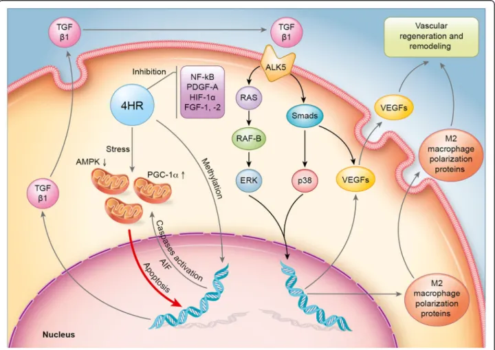
Results: Here, we found that 4HR upregulated transforming growth factor- β (TGF-β)/SMAD/vascular endothelial growth factor (VEGF) signaling, RAF-B/ERK and p38 signaling, and M2 macrophage polarization pathways. 4HR also increased expression of caspases and subsequent cellular apoptosis. Mechanistically, 4HR increased TGF- β1 production and subsequent activation of SMADs/VEGFs, RAF-B/ERK and p38 signaling, and M2 macrophage polarization.
Conclusion: Collectively, 4HR activates TGF- β/SMAD/VEGF signaling in endothelial cells and induced vascular regeneration and remodeling for wound healing.
Keywords: 4HR, HUVEC, IP-HPLC, TGF- β1, Angiogenesis
Background
4-Hexylresorcinol (4HR) is a substituted phenol that is synthesized from resorcinol and caproic acid [1]. It is used as an antimicrobial in tooth pastes and skin lotions [2] and as a preservative for fresh fruits and vegetables [3]. It has bactericidal [ 4], anthelmintic [5], and potential antineoplastic activities [6], and thus, it is also used as an antiseptic in mouthwashes and skin wound cleansers [7]. 4HR may also inhibit oxidative DNA damage by en- hancing the activities of antioxidant enzymes, including glutathione peroxidase and glutathione reductase, which facilitate the scavenging reactive oxygen species by gluta- thione [8], and thus, it is also used to prevent the en- zymatic browning of shrimps and different fruits [9].
A recent study demonstrated that 4HR increases the expression level of vascular endothelial growth factor (VEGF) in RAW264.7 cells and angiogenesis in the ani- mal model [10]. 4HR increases M2 markers, and broad- spectrum matrix metalloproteinase (MMP) inhibitor (PD166793) can reduce 4HR-induced VEGF expression.
However, MMPs are also highly expressed in the inflam- matory phase, and the expression of MMPs is mostly regulated by hypoxic stress [11]. Interestingly, the action of PD166793 is mediated by chelating zinc ion [12]. Ac- cordingly, zinc-dependent protein like transforming growth factor- β1 (TGF-β1) may be regulated by 4HR and induce VEGF and angiogenesis.
Immunoprecipitation high-performance liquid chro- matography (IP-HPLC) had been used previously by sev- eral authors to detect organic compounds quantitatively, including peptides, but the technique used was compli- cated and of limited applicability [13, 14]. Recently, a new IP-HPLC protocol was developed to determine pro- tein expression levels in different biological fluids, such
© The Author(s). 2020 Open Access This article is licensed under a Creative Commons Attribution 4.0 International License, which permits use, sharing, adaptation, distribution and reproduction in any medium or format, as long as you give appropriate credit to the original author(s) and the source, provide a link to the Creative Commons licence, and indicate if changes were made. The images or other third party material in this article are included in the article's Creative Commons licence, unless indicated otherwise in a credit line to the material. If material is not included in the article's Creative Commons licence and your intended use is not permitted by statutory regulation or exceeds the permitted use, you will need to obtain permission directly from the copyright holder. To view a copy of this licence, visithttp://creativecommons.org/licenses/by/4.0/.
* Correspondence:
kimsg@gwnu.ac.kr;sukkeunlee@hanmail.net1
Department of Oral and Maxillofacial Surgery, College of Dentistry, Gangneung-Wonju National University, and Institute of Oral Science, 123 Chibyun-dong, Gangneung 210-702, Republic of Korea
2
Department of Oral Pathology, College of Dentistry, Gangneung-Wonju
National University, and Institute of Oral Science, 123 Chibyun-dong,
Gangneung 210-702, Republic of Korea
as blood serum, urine, saliva [15], inflammatory exudates [16–18], and different protein extracts from cells [19–
21], liver [22], and cancer tissues [21]. Recent IP-HPLC results demonstrate that 4HR administration increases the expression of TGF-β1 in the osteoblast-like cells [23]. IP-HPLC is comparable to enzyme-linked immuno- sorbent assay (ELISA), but the former uses protein A/G agarose beads in buffer solution and ultraviolet spectros- copy to determine protein concentrations, whereas the latter uses fluorescence-conjugated antibodies fixed in plastic wells and fluoroscopy. Furthermore, multiple tri- als have shown that IP-HPLC can be used to rapidly de- termine multiple protein levels accurately (± 5%
standard deviation) and reproducibly.
In this study, differentially expressed proteins by 4HR were screened by IP-HPLC in a human endothelial cell line (human umbilical vein endothelial cells [HUVECs]) using our antibody library. IP-HPLC results demonstrated that TGF-β1 played a key role in 4HR-induced activation of angiogenesis-associated signal pathway in HUVEC cells.
To confirm this hypothesis, additional western blotting was done with TGF-β1 and its signal blocker.
Methods
HUVEC culture in the presence of 4HR
HUVECs (Lonza, Walkersville, MD, USA) were pur- chased and cultured in an endothelial basal medium supplemented with 1 μg/mL hydrocortisone, 12 μg/mL bovine brain extract, 50 μg/mL gentamicin, 50 ng/mL amphotericin-B, 10 ng/mL epidermal growth factor (EGF), VEGF, FGF-2, heparin, ascorbic acid, and 10%
fetal calf serum (EGM
TM-2, Clonetics®, Lonza, Walkers- ville, MD, USA) in 5% CO
2at 37.5 °C. Cells were tested for mycoplasma on a regular basis to ensure that only mycoplasma-free cells were assayed.
About 70% confluent HUVECs grown on Petri dish surfaces were treated with 10 μg/mL 4HR (with a single dose given safely given in dog; 100–300 mg/kg, WHO food additives Series 35, 835) for 8, 16, or 24 h; control cells were treated with 1 mL of normal saline. Cultured cells were harvested with protein lysis buffer (PRO- PREP
TM, iNtRON Biotechnology INC, Korea) and im- mediately preserved at − 70 °C until required.
Immunoprecipitation high-performance liquid chromatography (IP-HPLC)
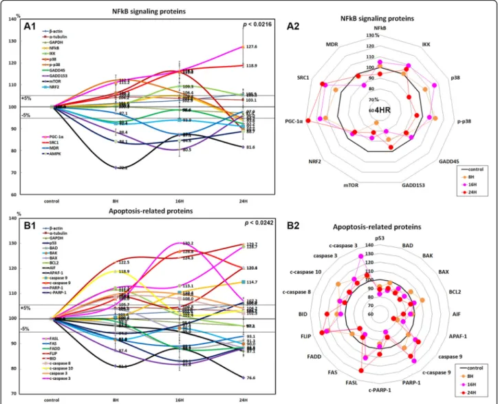
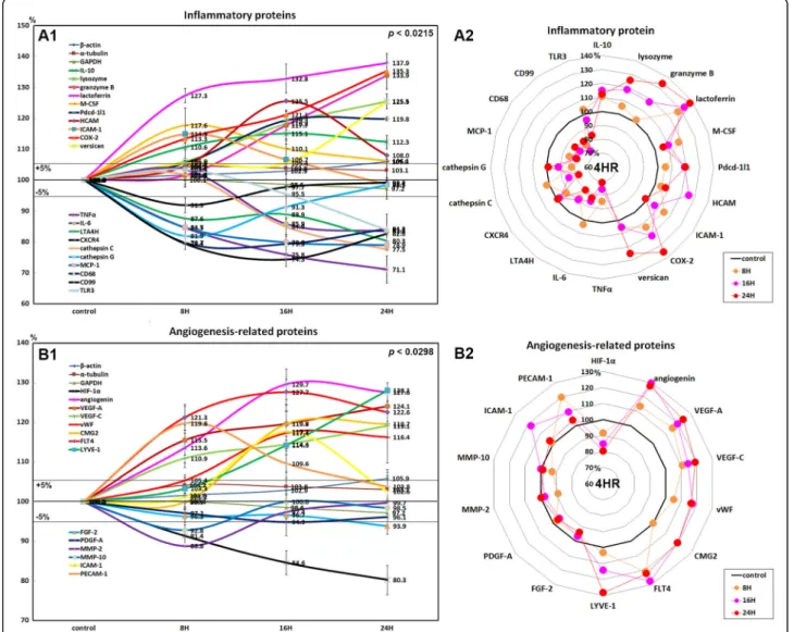
Protein extracts (100 μg) were subjected to immunopre- cipitation using a protein A/G agarose column (Amico- gen, Korea). Protein A/G agarose columns were separately pre-incubated with 1 μg of 96 different anti- sera for growth factor-related proteins (n = 10), RAS sig- naling proteins (n = 22), NFkB signaling proteins (n = 12 [2]), apoptosis-related proteins (n = 20), inflammatory proteins (n = 20), angiogenesis-related proteins (n = 14
[3]), and control housekeeping proteins (n = 3) (num- bers in brackets indicate the number of overlapping anti- bodies; Table 1).
Briefly, protein samples were mixed with 5 mL of bind- ing buffer (150 mM NaCl, 10 mM Tris pH 7.4, 1 mM EDTA, 1 mM EGTA, 0.2 mM sodium vanadate, 0.2 mM PMSF, and 0.5% NP-40) and incubated in protein A/G agarose (Amicogen, Korea) columns on a rotating stirrer for 1 h at 4 °C. After washing columns with PBS (phos- phate-buffered saline solution), target proteins were eluted using 150 μL of IgG elution buffer (Pierce, USA).
Immunoprecipitated proteins were analyzed using an HPLC unit (1100 series, Agilent, USA) equipped with a reverse phase column and a micro-analytical detector system (SG Highteco, Korea). Elution was performed using 0.15M NaCl/20% acetonitrile solution at 0.4 mL/
min for 30 min, and proteins were detected using an ultraviolet spectrometer at 280 nm. Control and experi- mental samples were run sequentially to allow compari- sons. For IP-HPLC, whole protein peak areas (mAU*s) were calculated after subtracting negative control anti- body peak areas, and square roots of protein peak areas were calculated to normalize concentrations. Protein percentages in total proteins in experimental and control groups were plotted. Results were analyzed using the chi-squared test [19–21].
The housekeeping proteins β-actin, α-tubulin, and glyceraldehyde 3-phosphate dehydrogenase (GAPDH) were used as internal controls. Expressional changes of housekeeping proteins were adjusted to < ± 5% using a proportional basal line algorithm. Protein expressional changes of ≤ ± 5%, ± 5–10%, ± 10–20%, and ≥ ± 20%
change were defined as minimal, slight, meaningful, or marked, respectively.
Statistical analysis
Proportional data (%) of the experimental and control groups were plotted into line graphs and star plots, and analyses were repeated two to six times until standard deviations were ≤ ± 5%. Line graphs revealed the similar- ities of the expression pattern between the relevant pro- teins, and star plots revealed the differences in the expression levels of the whole objective proteins. Results were analyzed using the chi-squared test. The expres- sions of control housekeeping proteins, that is, β-actin, α-tubulin, and GAPDH, were nonresponsive (≤ 5%) to 12, 24, or 48 h of 4HR treatment.
Results
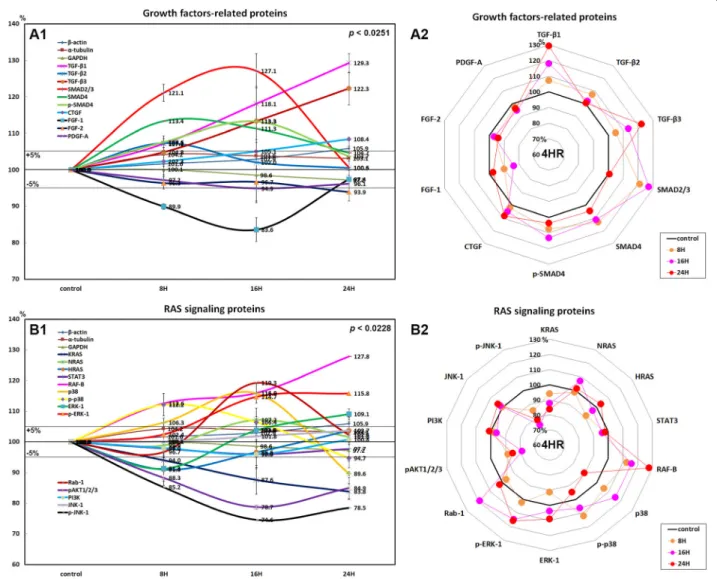
Effects of 4HR on the expressions of growth factor- related proteins in HUVECs
HUVECs treated with 4HR showed marked increases in
the expressions of TGF-β1 (29.3% at 24 h), TGF-β2
(7.3% at 8 h), TGF-β3 (22.3% at 24 h), SMAD2/3 (27.1%
at 16 h), SMAD4 (13.4% at 8 h), and p-SMAD4 (13.3% at 16 h) and a slight increase in the expression of connect- ive tissue growth factor (8.4% at 24 h) as compared with nontreated control, but a decrease in the expression of fibroblast growth factor-1 (FGF-1; 16.4% at 16 h), FGF-2 (6.1% at 24 h), and platelet-derived growth factor-A (PDGF-A; 5.1% at 16 h; Fig. 1A1, A2).
These results indicate 4HR increased the expressions of growth factors associated with TGF-β/SMAD pathways in HUVECs but slightly decreased the expressions of FGF-1, FGF-2, and PDGF-A. Therefore, we considered that 4HR provided dominant TGF-β-dependent angiogenesis in HUVECs despite downregulation of matrix angiogenetic factors (e.g., FGF-1, FGF-2, and PDGF-A).
Effects of 4HR on the expressions of RAS signaling proteins in HUVECs
The expressions of RAS signaling proteins were variable in HUVECs treated with 4HR for 24 h. K-RAS expression grad- ually decreased by 16.2% at 24 h, H-RAS expression de- creased by 9% at 8 h but increased by 3.7% at 24 h versus
nontreated control, while N-RAS increased by 2% at 16 h and by 1.6% at 24 h. Downstream signal proteins SOS1/2 and STAT3 tended to be decreased by 11.3% and 5% at 16 h, respectively.
4HR upregulated RAF-B, a growth signal transduction pro- tein kinase, by 27.8% at 2 h in HUVECs and subsequently upregulated mitogen-activated protein kinase 3, also known as extracellular signal-regulated kinase (ERK-1) and p-ERK-1 (Thr 202/Tyr 204) by 9.1% and 15.8% at 24 h, respectively.
4HR also upregulated p38 mitogen-activated protein kinase (p38, 15.8% at 16 h) and phosphorylated p38 (p-p38, 12.2%
at 8 h). The critical mediator of growth factor-induced signals pAKT1/2/3 (Thr 308) was consistently downregulated by 21.3% at 24 h and by 15.1% at 8 h, and phosphorylated c-Jun N-terminal kinase-1 (p-JNK-1, 89; Thr 183/Tyr 185), which is responsible for stress stimuli, such as cytokinesultraviolet irradiation, heat shock, and osmotic shock, was also down- regulated by 24.5% at 16 h and by 21.5% at 24 h, although the expression of non-phosphorylated JNK-1 was slightly in- creased by 3.7% at 24 h (Fig. 1)B1, B2). On the other hand, 4HR-treated HUVECs showed decreases in the Table 1 Antibodies used in the study
Protein No. Antibodies
Growth factor-related protein
10 FGF-1*, FGF-2*, CTGF, TGF- β1
#, TGF- β2*, TGF-β3*, SMAD4*, SMAD2/3, p-SMAD4, PDGF-A*
RAS signaling proteins 22 NRAS
$, KRAS
$, HRAS, PI3K, pAKT1/2/3, RAF-B*, JNK-1*, p-JNK-1, ERK-1*, p-ERK-1
$, Rab 1*, STAT3, p38*, p-p38*
NFkB signaling proteins 12 (2)
NFkB*, IKK*, GADD45*, GADD153*, mTOR
@, NRF-2*, PGC-1 α, SRC-1*, MDR, AMPK (p38*, p-p38*)
Inflammatory proteins 20 IL-10*, lysozyme*, granzyme, lactoferrin, M-CSF, Pdcd-1/1, HCAM, ICAM-1, COX2*, versican, TNF α
@, IL-6*, LTA4H
&, CXCR4, cathepsin C, cathepsin G*, MCP-1, CD68, CD99, TLR3
Apoptosis-related proteins
20 p53*, BAD*, BAK*, BAX, BCL2, AIF*, APAF-1, caspase 9*, c-caspase 9*, PARP-1*, c-PARP-1*, FASL*, FAS*, FADD*, FLIP*, BID, c-caspase 8*, c-caspase-10, caspase 3*, c-caspase 3*
Angiogenesis-related proteins
14 (3)
HIF-1 α
&, angiogenin
$, VEGF-A*, VEGF-C*, vWF
$, CMG2
$, FLT-4
$, LYVE-1*, MMP-2, MMP-10, PECAM-1 (FGF-2, PDGF-A, ICAM-1)
Control housekeeping proteins
3 α-Tubulin*, β-actin*, GAPDH*
Total 101
(5)
The number of antibodies that overlapped is indicated in parentheses
Abbreviations: AIF apoptosis inducing factor, AMPK AMP-activated protein kinase, pAKT v-akt murine thymoma viral oncogene homolog, p-Akt1/2/3 phosphorylated (p-Akt, Thr 308), BAD BCL2-associated death promoter, BAK BCL2 antagonist/killer, BAX BCL2-associated X, CMG2 capillary morphogenesis protein 2, COX-2 cyclooxygenase-2, CTGF connective tissue growth factor, CXCR4 C-X-C chemokine receptor type 4, FADD FAS-associated via death domain, FAS CD95/Apo1, FASL FAS ligand, FGF-1 fibroblast growth factor-1, FLIP FLICE-like inhibitory protein, FLT-4 Fms-related tyrosine kinase 4, GADD45 growth arrest and DNA damage- inducible 45, GAPDH glyceraldehyde 3-phosphate dehydrogenase, HCAM (CD44) homing cell adhesion molecule, HDAC-10 histone deacetylase 10, HIF-1α hypoxia- inducible factor-1α, HRAS GTPase HRas, HSP-70 heat shock protein-70, ICAM (CD54) intercellular adhesion molecule 1, IKK ikappaB kinase, IL-1 interleukin-1, JNK-1 Jun N-terminal protein kinase, KRAS V-Ki-ras2 Kirsten rat sarcoma viral oncogene homolog, LTA4H leukotriene A4 hydrolase, LYVE-1 lymphatic vessel endothelial hyaluronan receptor 1, MCP-1 monocyte chemotactic protein 1, M-CSF macrophage colony-stimulating factor, MDR multiple drug resistance, MMP-2 matrix metalloprotease-2, mTOR mammalian target of rapamycin, NCAM (CD56) neural cell adhesion molecule 1, NF-1 neurofibromin 1, NFkB nuclear factor kappa-light- chain-enhancer of activated B cells, NRAS neuroblastoma RAS viral oncogene homolog, NRF2 nuclear factor (erythroid-derived)-like 2, PARP-1 poly-ADP ribose polymerase 1, c-PARP-1 cleaved PARP-1, Pdcd-1/1 (CD279) programmed cell death protein 1, PDGF-A platelet-derived growth factor-A, PECAM-1 (CD31) platelet endothelial cell adhesion molecule-1, PGC-1α peroxisome proliferator-activated receptor gamma coactivator 1-α, PI3K phosphatidylinositol-3-kinase, PTEN phosphatase and tensin homolog, Rab 1 Rab GTPases, RAF-B v-Raf murine sarcoma viral oncogene homolog B, SMAD4 mothers against decapentaplegic, drosophila homolog 4, SRC1 steroid receptor coactivator-1, STAT3 signal transducer and activator of transcription-3, TGF-β1 transforming growth factor-β1, TNFα tumor necrosis factor-α, VEGF-A vascular endothelial growth factor A, vWF von Willebrand factor
*Santa Cruz Biotechnology, CA, USA
#DAKO, Denmark
$Neomarkers, CA, USA
@ZYMED, CA, USA
&
Abcam, Cambridge, UK