• 검색 결과가 없습니다.

Unilateral Diaphragmatic Paralysis after Thyroid Surgery

N/A
N/A
Protected

Academic year: 2021

Share "Unilateral Diaphragmatic Paralysis after Thyroid Surgery"

Copied!
5
0
0

로드 중.... (전체 텍스트 보기)

전체 글

(1)

Introduction

In Korea, the incidence of thyroid cancer has increased continuously since 1996, becoming one of the most common types of cancer in 2010.

1)

As such, the number of operations to treat thyroid cancer is increasing annually.

1)

Accordingly,

there has been a gradual increase in the need to evaluate com- plications and side-effects associated with thyroid cancer surgery. The typical complications of thyroid surgery are la- ryngeal nerve paralysis and hypoparathyroidism, with occur- rences of 2-5% and 1-4%, respectively.

2)

Approximately 0.4% of patients with isobilateral recurrent laryngeal nerve paralysis also experience acute respiratory distress due to adduction of the vocal cords.

3)

However, an ad- ditional report assessing clinical precautions for the occur- rence of dyspnea-related damage of the phrenic nerve during thyroid surgery reported that the occurrence of unilateral phrenic nerve paralysis was 5.5%.

4)

Phrenic nerve paralysis is one of the reasons for dyspnea that is commonly disregard-

대한 두경부 종양 학회지

제 30 권 제 2 호 2014

Unilateral Diaphragmatic Paralysis after Thyroid Surgery

Jong Kyu Byun, MD

1*

, Sang Youl Rhee, MD

1*

, Yu Jin Kim, MD

1

, Yu Jin Um, MD

1

, Seul Ki Kim, MD

1

, Jung Il Son, MD

1

, Sang Ouk Chin, MD

1

, Suk Chon, MD

1

, Woo-Shik Kim, MD

2

, Joo Young Kim, MD

3

,

Byoung Wook Lee, MD

3

, Jeong-taek Woo, MD

1

, Young Seol Kim, MD

1

Department of Endocrinology and Metabolism

1

and Cardiology,

2

Kyung Hee University School of Medicine, Seoul, Korea Department of Internal Medicine,

3

Dongsuwon General Hospital, Suwon, Korea

갑상선 수술 후 발생한 편측 횡격막 마비 1예

경희대학교 의과대학 내분비대사내과학교실,

1

심장내과학교실,

2

동수원병원 내과

3

변종규1*·이상열1*·김유진1·엄유진1·김슬기1·손정일1·진상욱1 전 숙1·김우식2·김주영3·이병욱3·우정택1·김영설1

= 국 문 초 록 =

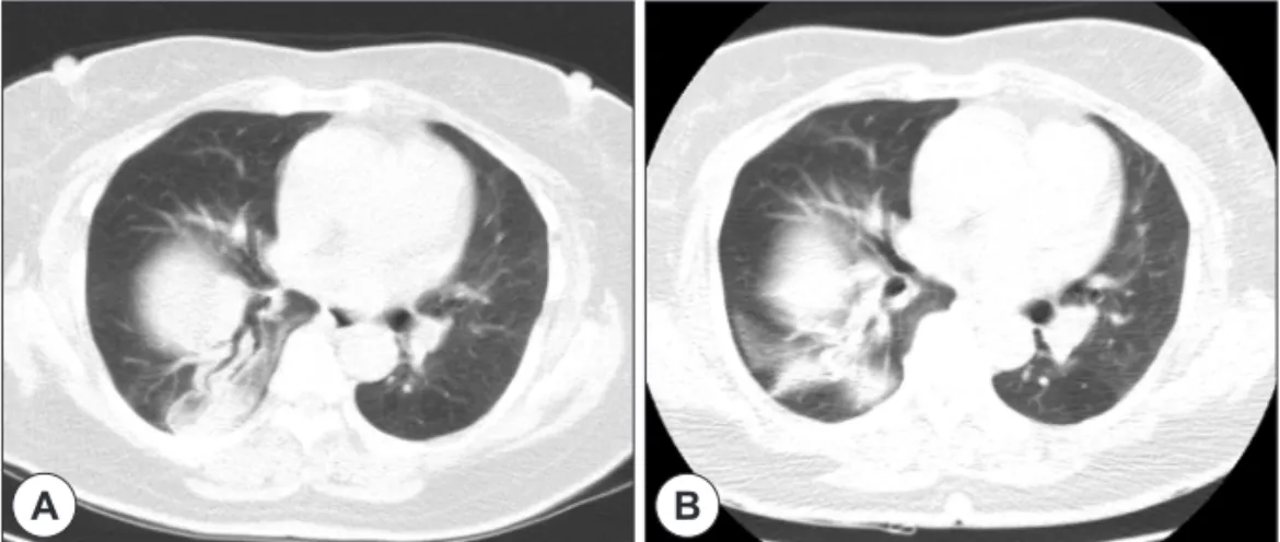
갑상선 암 진단과 치료기술이 발전하면서 최근 그 수술 건 수가 급격하게 증가하고 있다. 하지만이와 관련된 합병 증과 부작용을 면밀하게 평가해야 할 필요 역시 점차 늘어나고 있다. 갑상선 암 수술 후 발생할 수 있는 드문 합병증 의 하나로 횡격막 신경마비(phrenic nerve paralysis)가 있다. 이러한 횡격막신경마비는 대부분 증상이 경미하고 쉽게 호전되어 임상적으로 크게 중요하게 다루어지지 않았다. 하지만, 갑상선 수술 후 갑작스런 호흡곤란이 발생한다면 횡 격막 신경마비에 의한 횡격막 마비(diaphragmatic paralysis)와 관련되었을 가능성을 놓치지 말아야 한다. 저자들은 최 근 갑상선암 수술 후 발생한 호흡곤란으로 2년 동안 심각한 호흡곤란을 호소하던 73세 여자환자에서 투시촬영(fluo- roscopy) 상 편측으로 상승되고 운동성이 저하된 횡격막을 확인하여 일측성 횡격막신경마비(Unilateral phrenic nerve paralysis)를 확진 하였다. 갑상선수술 후 발생하는 일측 횡경막 신경마비는 임상에서 드물게 관찰되는 수술 합병증이 기에 환자는 상당기간 이에 대한 감별이 제대로 이루어지지 않았다. 우리는 횡격막 마비의 조기 진단과 적극적인 치 료를 통하여 심한 호흡곤란을 호소하는 환자의 증상 및 병의 경과를 호전 시킬 수 있었다.

중심 단어

:횡격막 마비ㆍ횡격신경ㆍ갑상선 유두암ㆍ갑상선 수술.

Received : July 31, 2014 / Revised : September 12, 2014 Accepted : September 15, 2014

교신저자 : 우정택, 130-872 서울 동대문구 경희대로 23 경희대학교 의과대학 내분비대사내과학교실

전화 : (02) 958-8200 · 전송 : (02) 968-1848 E-mail : [email protected]

*Contributed equally to this study as first authors.

online©MLComm

(2)

ed ; therefore, it is necessary to determine whether phrenic nerve paralysis and inexplicable dyspnea are present.

In this study, we confirmed the diagnosis of unilateral phrenic nerve paralysis by identifying the unilateral increase and reduced mobility of the diaphragm, measured with flu- oroscopy, in a 73-year-old female patient who had complained of severe difficulty breathing for 2 years following thyroid cancer surgery.

1. Patient

A 73-year-old female patient visited our hospital with a chief complaint of dyspnea. She had been diagnosed with thyroid papillary cancer(right thyroid, upper pole, 1 cm sized) with right lymph node metastasis in right level III(T3N1M0), 2 years previously and had been treated by thyroidectomy with right modified radical neck dissection (total thyroidec- tomy with both central neck dissection with lymph node dis- section level II III IV) ; she was also taking 150 μg/day levo- thyroxine. After surgery, the patient had to sleep sitting upright due to exacerbated dyspnea when in the supine position. She was prescribed alprazolam 0.25 mg twice daily due to con- tinuous anxiety syndrome caused by consistent sleep distur- bance. There was no medical history of significant chronic illness, such as high blood pressure, diabetes, or tuberculosis, and no significant family medical history.

At presentation, the results of the physical examination of the patient were as follows : height 150 cm, weight 66 kg, blood pressure 130/80 mmHg, pulse 70 beats per minute, body tem- perature 36.5℃, respiration rate 20 times per minute, and 98% oxygen saturation, as measured by pulse oximetry. Ex- amination by stethoscopy revealed that the pulmonary sound of the lower part of the right lung was decreased, yet not sig- nificantly. No other specific abnormalities were observed. The laboratory results were as follows : white blood cell count 5840×103/μL(seg 45.8%), hemoglobin(Hb) 12.3 g/dL, as-

partate aminotransferase/alanine aminotransferase(AST/

ALT) 28/23 U/L, blood urea nitrogen/serum creatinine(BUN/

Cr) 14/0.5 mg/dL, C-reactive peptide(CRP) <0.3 mg/dL, thy- roid stimulating hormone(TSH) 0.06 μU/mL, and free-T4 2.05 ng/dL.

A pulmonary function test was then performed to identify the reason for dyspnea. Forced vital capacity(FVC) was 1.64 L(73% of the normal predictive value) and forced expiratory volume in one second(FEV1) was 1.39 L(90% of the normal predictive value). The ratio between FVC and FEV1 was 72%, and no other abnormalities were observed other than a minor restrictive disorder. The results of a DLCO(carbon monoxide diffusing capacity) test were normal. Her 6 minute gait test result was 418 m, and Borg Scale was 4 points. No specific abnormalities were observed on gastroscopy, echocardiogra- phy, coronary angiogram CT scan, or chest CT scan, which were all conducted to rule out any other reasons for the dys- pnea.

The symptom transition signs pre- and post-operation were identified in detail to determine the cause of dyspnea in the patient. Immediately after surgery, she showed symptoms of dyspnea, which remained constant without further deteriora- tion or improvement. Therefore, we determined that dyspnea could be related to a surgical complication. As such, all of the previous radiology results associated with the chest were ver- ified sequentially. Two weeks after the operation, a right dia- phragmatic line was elevated significantly on a chest X-ray (Fig. 1B) compared with the pre-operative chest X-ray(Fig.

1A). Subsequent images obtained 1 year after surgery dem- onstrated the continual increase of the right diaphragmatic line(Fig. 1C). Chest CT scans performed 2 and 9 months af- ter the operation also identified segmental atelectasis on the right middle lobe, which is located next to the right diaphragm and right lower lobe(Fig. 2). Therefore, we suspected that the patient had clinically significant diaphragmatic dysfunction

A B C

Fig. 1. A : Pre-operative Chest radiograph. B : Chest radiograph at 2 weeks. C : 1year after the surgery. Chest X-ray at 2weeks after

operation shows an elevated hemidiaphragm compared with preoperative picture, and still elevated diaphragm is noted 1 year af-

ter the surgery.

(3)

based on the imaging studies. Imaging studies have previous- ly suggested a close relationship between dyspnea and phren- ic nerve dysfunction. A sniff test using fluoroscopic study was performed, and this confirmed unilateral phrenic nerve pa- ralysis(Fig. 3).

Conservative treatment was performed in the patient based on life expectancy and physical status. We were able to iden- tify phrenic nerve paralysis, but the abnormality of the dia- phragm had not changed significantly.

Discussion

The diaphragm is a respiration-related muscle that plays a critical role in determining the inspiratory phase time, and it is responsible for 60-70% of the total tidal volume at rest.

5)

Phrenic nerve paralysis can be caused by problems with sev- eral factors, such as the brain, spinal cord, phrenic nerve, neu- romuscular junction, and the muscle itself.

Diaphragmatic dysfunction can cause dyspnea, hypokine- sia, sleep disorders, reduced quality of life, atelectasis, and

respiratory failure. The true prevalence of unilateral phrenic nerve paralysis is unclear, since it is frequently discovered in- cidentally by chest X-ray and is present without symptoms ; yet, it is more common than bilateral phrenic nerve paraly- sis. Many symptoms, including difficulty breathing during exercise, labored respiration, and asymmetry of chest exer- cises, occur during diaphragmatic paralysis due to impair- ment of the phrenic nerve. Depending on the posture change of the patient, respiratory distresses including dyspnea, wheez- ing, and sleep disorders can develop occasionally. These con- ditions can result in the misdiagnosis of phrenic nerve paral- ysis as heart failure, pulmonary artery embolism, chronic ob- structive pulmonary disease, or interstitial lung disease. There- fore, unnecessary invasive test procedures are often performed on the patient.

Patients with phrenic nerve paralysis exhibit diaphragmat- ic elevation on chest X-rays, as well as paradoxical movement of the diaphragm and mediastinum on fluoroscopy. The di- agnosis of phrenic nerve paralysis can be confirmed based on these observations. Pulmonary function tests can also be

A B

Fig. 2. A : Computerized tomography of the chest at 2 months after surgery. B : 9 months after surgery. Segmental atelectasis was identified on the right middle lobe, which was located next to the right diaphragm and right lower lobe.

A B

Fig. 3. Fig. 3 shows fluoroscopic “sniff” test, in which diaphragmatic movement is observed fluoroscopically while the patient sniffs

forcefully. The sniff test showed paradoxically elevation of the paralyzed hemidiaphragm with inspiration compared with the rapid

descent of the normal hemidiaphragm. A : Inspiration. B : Expiration.

(4)

performed to evaluate the deterioration of diaphragm func- tion. As a result, the FVC of our patient was 70-80% of the expected level, which was reduced due to bilateral phrenic nerve paralysis.

During bilateral phrenic nerve paralysis, FVC measured in the supine position is commonly reduced by 15-25% com- pared with in the sitting position.

6)

Phrenic nerves branch off from C3, C4, and C5 vertebrae, and the anterior portions of the nerves protrude between the scalene muscle and its cov- ering fascia, which originates from the anterior neck. The nerves then spread on the same side as the diaphragm by lo- calizing to the back of the subclavian vein and entering the thoracic cavity from the neck. They also spread to the pericar- dium, pneumomediastinum, pleura, and peritoneum. Impair- ment of the hemidiaphragm is caused mainly by the pulling and cooling of the phrenic nerve during cardiac surgery, cer- vical spondylosis, or nerve compression on the cervical spine due to cancer, injury, surgery, and pneumonia.

7)

There are also many reports of nerve damage due to nerve compression caused by iatrogenic impairment, which can occur during sur- gery, or it can develop on the trachea or mediastinum.

8)

There are no official statistics analyzing the development of phrenic nerve paralysis after thyroid surgery. However, when we performed follow-up monitoring of 82 patients that had been diagnosed and treated for thyroid cancer at a tertia- ry hospital from 1996-2000 for 1-10 years, nine patients ex- hibited surgical complications, and one developed permanent impairment of the phrenic nerve.

9)

In addition, two cases of unilateral phrenic nerve paralysis after thyroid surgery were reported in male patients, aged 70 and 54 years, who had suf- fered cervico-mediastinal goiters.

10)

A previous study reported that -40-60% of patients diag- nosed with thyroid papillary carcinoma develop cervical lymphadenopathy.

11)

In this study, modified or selective radi- cal neck dissection were performed. Modified radical neck dissection removes each lymph node from levels 1-5, simi- lar to radical dissection, but one or more of the accessory ner- ves, internal jugular veins, and sternocleidomastoid muscles are left intact.

The phrenic nerve can be dissected from the outside to the inside, taking special care to avoid damage while perform- ing radical neck dissection from levels 2-4. This process can damage the phrenic nerve, for example if the nerve is dis- sected together with fibroid adipose tissue, the prevertebral fascia region is detached too deeply, or a cut is too close when cervical plexus dissection is performed. To reduce these po-

tential damages, it is recommended to dissect at least 1 cm from the lateral part of the nerve.

Phrenic nerve paralysis also occurs rarely in benign thyroid disease, and some cases are due to compression of the phren- ic nerve when a thyroid goiter invades into the pleural cavity.

An additional case report described the disappearance of pa- ralysis symptoms following partial thyroidectomy.

12)

Howev- er, the occurrence of phrenic nerve paralysis due to benign thyroid disease was recently reduced because most surgical dissections are now performed prior to enlargement of thy- roid goiters.

Phrenic nerve damage can also occur during surgery for head and neck tumors or during cardiothoracic surgery. Dia- phragm nerve damage was reported in 16% of patients who underwent extensive lymphadenectomy and in 1.9% of pa- tients with esophageal cancer.

13)

Phrenic nerve damage was also reported in 14(8%) out of 176 patients with head and neck tumors that underwent modified radical neck dissection in 1991,

14)

as well as in two out of 26 patients with cervical lymph- adenopathy.

15)

It can also occur during relatively non-invasive procedures ; for example, it occurred in 10(1.3%) out of 814 patients with radiofrequency ablation for pulmonary tumors

16)

and in up to 61% of patients during cervical plexus-blocking anesthesia for carotid endarterectomy.

17)

Unilateral diaphragmatic paralysis typically appears tem- porarily and then disappears without any complications ; yet, severe respiratory distress was reported in a case of bilateral diaphragmatic paralysis due to bilateral phrenic nerve inju- ry.

18)

However, many cases are not reported due to lack of rec- ognition of minor symptoms, which are easy for a clinician to miss if a patient complains of nonspecific symptoms. Ac- cording to one study describing phrenic nerve paralysis, it can take several weeks to 15 years for diagnosis.

19)

The treatment of phrenic nerve paralysis is determined by the severity of the paralysis. If the paralysis is minor, it is rec- ommended to use conservative and non-invasive treatments, such as artificial respiration or treatment of lung secretion. In contrast, diaphragmatic plication is required for patients with recurrent pneumonia and/or atelectasis, and those in need of long term mechanical ventilation. In general, phrenic nerve paralysis can be recovered 1-3 months after conservative or surgical treatment. Although the function of the diaphragm was not completely recovered, some report that diaphragmat- ic placation may help to diminish symptoms and improve pul- monary function.

20)

The prognosis of phrenic nerve damage varies according

(5)

to the mechanism that caused the injury, and it can be worse when the nerve is pulled or cut compared with cold injuries.

21)

The mechanism of cold injury is considered to be axonal de- generation and demyelination of the nerve, which will regen- erate in -3 years. In contrast, there is a high probability of permanent nerve damage if the nerve has been cut ; thus, sur- gical treatment should be considered at an early stage.

This case study presented a patient who developed unilat- eral diaphragmatic paralysis due to phrenic nerve injury, which occurred after thyroidectomy and modified radical neck dis- section. This patient differed from other cases due to the pres- ence of severe respiratory distress, which developed over 2 years. The importance of diagnosing phrenic nerve paralysis in this patient cannot be overemphasized, since the prolonged dyspnea of unknown cause improved with conservative treat- ment alone. If dyspnea occurs after thyroid surgery, the pos- sibility that it is related to phrenic nerve damage should not be overlooked.

References

1) Kweon SS, Shin MH, Chung IJ, Kim YJ, Choi JS. Thyroid can- cer is the most common cancer in women, based on the data from population-based cancer registries, South Korea. Jpn J Clin On- col. 2013;43:1039-1046.

2) Roher HD, Goretzki PE, Hellmann P, Witte J. [Complications in thyroid surgery. Incidence and therapy]. Chirurg. 1999;70:999- 1010.

3) Rosato L, Avenia N, Bernante P, De Palma M, Gulino G, Nasi PG, et al. Complications of thyroid surgery: analysis of a multi- centric study on 14,934 patients operated on in Italy over 5 years.

World J Surg. 2004;28:271-276.

4) Pan YF, Zhang XH, Ye GH, Lin BR, Chen XM, Jia XJ, et al. [One stage thyroidectomy and bilateral neck dissection for well-dif- ferentiated thyroid carcinoma]. Zhonghua Zhong Liu Za Zhi. 2006;

28:389-392.

5) Fell SC. Surgical anatomy of the diaphragm and the phrenic nerve.

Chest Surg Clin N Am. 1998;8:281-294.

6) Mier-Jedrzejowicz A, Brophy C, Moxham J, Green M. Assess- ment of diaphragm weakness. Am Rev Respir Dis. 1988;137:877- 883.

7) McCaul JA, Hislop WS. Transient hemi-diaphragmatic paraly- sis following neck surgery: report of a case and review of the literature. J R Coll Surg Edinb. 2001;46:186-188.

8) Salati M, Cardillo G, Carbone L, Rea F, Marulli G, Brunelli A,

et al. Iatrogenic phrenic nerve injury during thymectomy: the extent of the problem. J Thorac Cardiovasc Surg. 2010;139:e77- e78

9) Spear SA, Theler J, Sorensen DM. Complications after the sur- gical treatment of malignant thyroid disease. Mil Med. 2008;

173:399-402

10) Rosato L, Nasi PG, Porcellana V, Varvello G, Mondini G, Ber- tone P. Unilateral phrenic nerve paralysis: a rare complication after total thyroidectomy for a large cervico-mediastinal goitre.

G Chir. 2007;28:149-152.

11) Hundahl SA, Fleming ID, Fremgen AM, Menck HR. A Nation- al Cancer Data Base report on 53,856 cases of thyroid carcino- ma treated in the U.S., 1985-1995 [see commetns]. Cancer. 1998;

83:2638-2648.

12) van Doorn LG, Kranendonk SE. Partial unilateral phrenic nerve paralysis caused by a large intrathoracic goitre. Neth J Med. 1996;48:216-219.

13) Sayama J, Shineha R, Hirayama K, Nishihira T, Mori S. [Four cases of phrenic nerve paralysis following surgery of thoracic esophageal carcinoma]. Nihon Kyobu Geka Gakkai Zasshi. 1994;

42:562-567.

14) de Jong AA, Manni JJ. Phrenic nerve paralysis following neck dissection. Eur Arch Otorhinolaryngol. 1991;248:132-134.

15) Zhang S, Wang L, Li S. [Surgery for mass at root of the neck-- with 26 cases report]. Lin Chung Er Bi Yan Hou Tou Jing Wai Ke Za Zhi. 2011;25:529-530.

16) Matsui Y, Hiraki T, Gobara H, Uka M, Masaoka Y, Tada A, et al. Phrenic nerve injury after radiofrequency ablation of lung tumors: retrospective evaluation of the incidence and risk fac- tors. J Vasc Interv Radiol. 2012;23:780-785.

17) Castresana MR, Masters RD, Castresana EJ, Stefansson S, Shaker IJ, Newman WH. Incidence and clinical significance of hemidiaphragmatic paresis in patients undergoing carotid end- arterectomy during cervical plexus block anesthesia. J Neuro- surg Anesthesiol. 1994;6:21-23.

18) Yaddanapudi S, Shah SC. Bilateral phrenic nerve injury after neck dissection: an uncommon cause of respiratory failure. J Laryngol Otol. 1996;110:281-283.

19) Ben-Dov I, Kaminski N, Reichert N, Rosenman J, Shulimzon T.

Diaphragmatic paralysis: a clinical imitator of cardiorespira- tory diseases. Isr Med Assoc J. 2008;10:579-583.

20) Celik S, Celik M, Aydemir B, Tunckaya C, Okay T, Dogusoy I.

Long-term results of diaphragmatic plication in adults with uni- lateral diaphragm paralysis. J Cardiothorac Surg. 2010;5:111.

21) Ostrowska M, de Carvalho M. Prognosis of phrenic nerve inju- ry following thoracic interventions: four new cases and a review.

Clin Neurol Neurosurg. 2012;114:199-204.

수치

Fig. 1. A : Pre-operative Chest radiograph. B : Chest radiograph at 2 weeks. C : 1year after the surgery
Fig. 2. A : Computerized tomography of the chest at 2 months after surgery. B : 9 months after surgery

참조

관련 문서

Head-up tilt test is an effective diagnostic test in noncardiac syncope and EEG is a valuable test in neuropychiatric syncope, especially in seizure.. Key words

수평각측정시 에 필요한 조정 전부, 횡선의 조정, 망원경수준기의 조정, 연직분도원

영반과 유엔개발계획(UNDP)의 지원으로 평양시 보통강구역에 설립된

삼각수준측량- 측각기(트랜싯 또는 데오도라이트)에 의하여 연직각 및 거 리를 측정하여 삼각법에 의한 계산을 하여 높이차를

④ 한쌍의 정준나사를 서로 반대방향으로 같은 양만큼 돌리면 반수준기의 기포는 좌 무지(left thumb)의 방향과 같은 방향으로 움직이므로 반수준기의 수포가 중앙에

※ Accessory spleen의 유무 확인 또는 Diaphragmatic rupture를 동반한 trauma후에 chest를 보기 위 한 검사일

 Combine with seismic data to define plate boundary geometry, measure and interpret physical processes of strain accumulation and release.  Improve understanding of

Second, at the test of affective domain between two groups, before test controlled group showed higher points at physical strength & health, mind