식물병연구
Research Article Open Access
Res. Plant Dis. 20(1) : 21-24(2014)
http://dx.doi.org/10.5423/RPD.2014.20.1.021
Research in Plant Disease
The Korean Society of Plant Pathology pISSN 1598-2262, eISSN 2233-9191
배추 뿌리혹병균 Plasmodiophora brassicae의 종 특이적 프라이머 개발
Development of Species-Specific Primers for Plasmodiophora brassicae, Clubroot Pathogen of Kimchi Cabbage
최진수 · 양슬기 · 송정영 · 김홍기*
충남대학교 응용생물학과
Jin Su Choi, Seul Gi Yang, Jeong Young Song and Hong Gi Kim*
Department of Applied Biology, Chungnam National University, Daejeon 305-764, Korea
Clubroot caused by the obligate biotrophic protist Plasmodiophora brassicae Woronin is one of the most dam- aging diseases of Brassicaceae family. In this study, we developed species-specific primer sets for rapid and accurate detection of P. brassicae. The primer sets developed amplified a specific fragment only from P. bras- sicae DNA while they did not amplify a band from 10 other soilborne pathogens or from Kimchi cabbage. In sensitivity test, the species-specific primer set ITS1-1/ITS1-2 could work for approximately 10 spores/ml of genomic DNA showing more sensitivity and accuracy than previous methods. With quantitative real-time PCR test, the primer set detected less spores of P. brassicae than before, confirming that the species-specific primer set could be useful for rapid and accurate detection of P. brassicae.
Keywords : Clubroot pathogen, Detection, Plasmodiophora brassicae, Quantitative real-time PCR, Species- specific primer
서 론
뿌리혹병은 십자화과 채소에 가장 피해가 큰 병해로서 배 추, 무, 양배추, 순무, 케일, 갓, 브로콜리 이외에 애기장대와 냉 이뿐 아니라 비트, 한련화, 파파야 등에도 그 발생이 보고되었 으며(Ludwig-Müller, 1999) 국내의 경우 배추에서 피해규모가 가장 크다(Cho 등, 2003). 병원균인 Plasmodiophora brassicae (Woron.)는 소련의 Woronin(1878)에 의해 최초로 발견되었고 한국에서는 1990년대 초부터 경기도 북부와 강원도의 전통적 배추재배지역을 위시하여 전국적으로 급격히 확산, 큰 피해를 주고 있다.
이 병원균은 기주 세포 내에서 다핵성 변형체를 형성하여 기 주 조직의 이상비대를 초래, 뿌리로부터 혹을 형성하게 된다 (Fähling 등, 2004). 조직 내에 증식된 다핵성 변형체로 인해 뿌
리의 유관속부가 정상적으로 분화하지 못하고 양분과 수분 흡 수가 저해되어, 지상부는 생육이 부진하고 점점 시드는 증상 이 심화, 결국에는 감염식물의 고사를 초래한다(Voorrips와 Kanne, 1997).
뿌리혹병균의 검출은 형광현미경을 이용한 형태 관찰법 이 사용되고 있으나 검출에 시간이 많이 소용되고 고가의 장비가 필요하다. 뿌리혹병균의 검출을 위해 Ito 등(1999), Wallenhammar 등(2012)에 의해 종 특이적 프라이머가 제작되 었고 국내 병원균 검출을 위한 프라이머가 Soh 등(2013)에 의 해 개발되었다. 본 연구에서는 뿌리혹병균 검출 및 병원균의 양 을 정량할 수 있는 보다 개선된 프라이머를 개발하고자 하였다.
재료 및 방법
공시 균주. 사용한 배추뿌리혹병균(P. brassicae)은 강원도 평창, 인제, 홍천, 대관령, 강릉, 원주와 경기도 안성, 충남 홍성, 전라남도 해남, 진도 등 10여 곳의 배추 재배지역 포장으로부터
*Corresponding author Tel : +82-42-821-5768 Fax: +82-42-823-8679 E-mail: [email protected]
Received November 29, 2012 Revised February 14, 2014 Accepted February 21, 2014
21-24 507 김홍기(o).indd 21 2014-03-26 오후 6:49:38
Research in Plant Disease Vol. 20 No. 1
22
500여개의 뿌리혹을 채집하여 휴면포자를 분리한 다음 실험에 사용하였다.
휴면포자의 분리. 채집된 뿌리혹을 흐르는 물로 수 차례 씻 어내어 토양입자와 불순물을 최대한 제거해 낸 후, 멸균된 메 스와 가위를 이용하여 잘게 잘라낸 다음, 굵은 시험관(Pyrex, Ø30 mm)에 넣고 Homogenizer(IKA, 25 μm)로 잘게 마쇄하여 뿌리조직 내 휴면포자를 나출시켰다. 나출된 포자를 식물조직 및 토양 잔재물로부터 분리하기 위해 8겹의 거즈를 통해 걸러 낸 후, 13,000 rpm에서 15분간 원심분리하여 수거한 pellet에 멸균수를 첨가하고 원심분리기에서 15분간 씻어내는 과정을 2 차례 반복하였다. 최종적으로 수거한 pellet을 다음 실험 때까 지 -20oC에 보관하였다.
Genomic DNA의 분리. DNA는 Jang(2006)의 방법으로 분 리 및 정제하였다. 수거된 휴면포자로부터 DNA를 추출하기 위한 동결건조 전처리 과정으로써 pellet에 50 unit/ml 농도 의 DNase I (Sigma, D-4263) 50 μl, Streptomycin 300 ppm과 Rifampicin 100 ppm을 처리하여 37oC에서 1시간 동안 반응시 켰다. DNase I의 불활성화는 5 μl Proteinase K(25 μg/μl)를 37oC 에서 1시간 동안 처리한 후, 5 mM EDTA 300 μl를 처리하고, 최 종적으로 80oC hot block에서 5 분간 고열처리 하였다. DNase I 이 불활성화된 포자현탁액은 다시 4,000 rpm에서 15분간 원심 분리하여 상등액을 제거한 다음 동결건조하여 DNA 추출에 사 용하였다. 전처리 후 동결건조된 pellet은 미세한 휴면포자의
세포벽을 효과적으로 파쇄하기 위해 Glass bead(0.09-0.15 mm)를 첨가하여 Pellet Pestile (Sigma)로 마쇄한 다음, 400 μl의 extrac- tion buffer[200 mM Tris-Cl(pH 8.0), 200 mM EDTA, 1.4 M NaCl, 1% PVP를 첨가하고 600 μl의 chloroform으로 추출하였다.
DNA는 column purification(Intron, Korea) 과정을 통해 정제하 고 100 μl의 distilled water에 녹인 다음, 이후 실험에 사용하 였다.
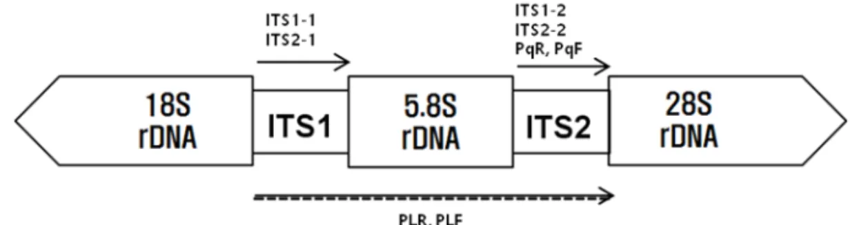
종 특이적 프라이머의 개발. 종 특이적 프라이머의 선발 을 위해 Genbank(http://www.ncbi.nlm.nih.gov)와 EMBL-EBI (http://www.ebi.ac.uk)로부터 얻어진 P. brassicae의 ITS 영역 염 기서열 정보를 토대로 참고하여 clustralW와 Oligonucleotide Properties Calculator(http://www.basic.north western.edu)를 활용하여 ITS1-1/ITS1-2와 ITS2-1/ITS2-2, PLF/PLR, PqF/PqR 등 총 4개의 프라이머 세트를 설계하였다(Fig. 1). 프라이머의 합성 은 Genotech(Korea)사에 의뢰하였다(Table 1).
프라이머의 특이성 및 민감도 검정. 프라이머의 종 특이성 을 조사하고자 10종의 주요 토양전염성 병원균과 배추의 DNA 에 대해 앞서 개발한 프라이머 세트를 이용해 PCR 검정을 실시 하였다. 또한 민감도를 알아보기 위해 P. brassicae의 포자를 105 spore/ml부터 10배씩 연속적으로 희석한 후 DNA를 추출 하여 PCR 증폭을 실시하였다. PCR 반응 조건은 initial denatur- ation을 94oC에서 5분 간 실시한 후 denaturation 94oC/30초, annealing 65oC/20초, extension은 72oC/20초로 35 cycles을 수
Table 1. List of primer sets used in this study
Primer set Primer Nucleotide sequence (5’-3’) Product size
Primer 1 set ITS1-1
ITS1-2
CTA GCG CTG CAT CCC ATA TC TGT TTC GGC TAG GAT GGT TC
129 bp
Primer 2 set ITS2-1
ITS2-2
CGG CAT AGC TTG AAC GAA G GTG TGT GTC GAT CTG CGA TT
167 bp
Primer 3 set PLF
PLR
TCC TCC GCT TAT TGA TAT GCT T GAA CCT GCG GAA GGA TCA
548 bp
Primer 4 set PqF
PqR
GCA AGA CAA TGA GCT TTG CTG TGT GTG TGT CGA TCT GCG ATT
131 bp Fig. 1. Diagram of the ribosomal DNA repeat of Plasmodiophora brassicae showing the approximate locations of primers.
21-24 507 김홍기(o).indd 22 2014-03-26 오후 6:49:38
Research in Plant Disease Vol. 20 No. 1 23
행하고 final extension을 72oC/5분 실시하였다.
Quantitative real-time PCR 분석은 증폭산물의 크기가 129 bp 로 real-time PCR에 적용 가능한 ITS1-1과 ITS1-2 프라이머를 이 용하였다. PCR 반응액의 총량은 20 μl로 하고 주형 DNA는 1 μl를 사용하였고, 2 μM의 primer를 사용하였으며 iQTM SYBR Green Supermix(BIO-RAD)를 첨가하였다. PCR 조건은 95oC에 서 5 분간 initial denaturation시켰고, 95oC/30초, 65oC/20초, 72oC/20초로 40 cycles 반응시킨 다음 72oC에서 5분간 final extension시킨 후, melt curve 값은 65oC에서 95oC까지 증가시 켜 산출하였다.
결과 및 고찰
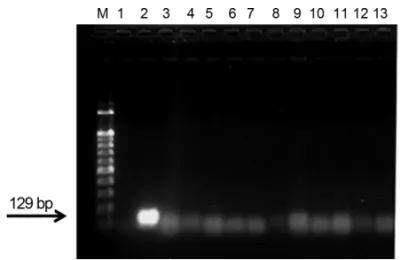
프라이머 ITS1-1/ITS1-2와 ITS2-1/ITS2-2, PLF/PLR, PqF/PqR를 이용하여 3개의 이병 배추뿌리혹으로부터 분리한 휴면포자의 DNA를 PCR 증폭한 결과, 4개의 primer set 모두 P. brassicae의 DNA를 증폭하였다. 증폭산물의 크기가 129 bp로 quantitative real-time PCR 사용에 가장 적합했던 ITS1-1/ITS1-2를 이용하여 특이성 검정을 실시한 결과, 토양 내에 존재하는 주요 병원균 인 Alternaria sojae, Fusarium oxysporum, Didymella bryoniae, Phytophthora infestans, Pythium ultimum, Colletotrichum acuta- tum, Botrytis cinerea, Cylindrocarpon destructans, Rhizoctonia solani, Verticillium dahliae 등 다른 병원균 및 식물체의 DNA는 증폭이 이루어지지 않고 뿌리혹병균의 DNA만 증폭된 것을 확 인할 수 있었다(Fig. 2).
민감도 검정을 위해 뿌리혹병균의 포자현탁액을 105 spore/
ml로 조정한 후 10배씩 희석한 다음 DNA를 분리하였다. 분리 한 DNA를 앞서 개발한 primer ITS1-1/ITS1-2를 이용하여 1차
PCR 증폭을 수행한 결과 포자 수 10개까지 검출이 가능하였다 (Fig. 3). 이전 연구에서 사용되었던 Faggian 등(1999)에 의해 개 발된 특이적 primer PbITS set는 검출 민감도가 first round PCR 에서는 포자 수 100개까지, nested PCR에서는 10개의 포자까 지 증폭이 가능하여 ITS1-1/1-2의 민감도와 같지만 nested PCR 은 극히 민감한 PCR로 실험방법도 더 까다로워 실제적으로는 first round PCR만을 수행하는 경우가 많기 때문에 본 연구에서 개발된 ITS1-1/1-2를 사용한 PCR 검정이 병원균 검출에 보다 유 용할 것으로 판단된다. 또한 quantitative real-time PCR 검정을
Fig. 2. Specificity of primer set ITS1-1/ITS1-2 for Plasmodiophora brassicae using conventional PCR. M: 100 bp marker, lane 1: negative control (not containing DNA), lane 2: P. brassicae DNA, lanes 3-12 (other soilborne fungi): Alternaria sojae (3), Fusarium oxysporum (4), Didymella bryoniae (5), Phytophthora infestans, (6), Pythium ultimum (7), Colletotrichum acutatum (8), Botrytis cinerea (9), Cylindrocarpon destructans (10), Rhizoctonia solani (11), Verticillium dahliae (12), lane 13: Kimchi cabbage DNA.
Fig. 3. PCR sensitivity of primer set ITS1-1/ITS1-2 for Plasmodiophora brassicae DNAs with different concentration. M: 100 bp marker, lanes 1-7: serially diluted spores (from 105 spores/ml), N.C: negative control (not containing DNA).
Fig. 4. Sensitivity of primer set ITS1-1/ITS1-2 for Plasmodiophora brassicae on quantitative real-time PCR using SYBR Green. A Real-time amplification curves of different concentrations of DNAs (a) and standard curves (b), numbers 1-5: 10-1 spores/ml (1), 1 spore/ml (2),10 spores/ml (3), 102 spores/ml (4), 103 spores/ml (5).
21-24 507 김홍기(o).indd 23 2014-03-26 오후 6:49:39
Research in Plant Disease Vol. 20 No. 1
24
위해 포자현탁액을 103 spores/ml로 조정한 후 DNA를 분리한 것을 10배씩 희석하고 ITS1-1/ITS1-2를 이용하여 quantitative real-time PCR을 수행했을 때 10-1까지 검출이 가능하였다(Fig.
4). 이는 실험방법과 샘플에 다소 차이가 있으나 민감도에서 기 존의 Wallenhammar 등(2012)에 의해 개발된 real-time PCR용 프라이머의 민감도 500 resting spores/g-1 soil 보다 높은 민감 도를 보였으며, 또한 희석된 DNA의 threshold cycle 값에 의해 작성된 standard curve 값(Fig. 4a)을 통해 여러 샘플 간의 DNA 농도에 대한 상대적인 정량이 가능하였다. 본 결과를 토대로 Real-time PCR에서 SYBR Green을 활용한 균주 특이적인 quan- titative real- time PCR 검정이 가능할 것으로 사료된다.
요 약
Plasmodiophora brassicae는 십자화과 작물에 뿌리혹병을 일 으키는 주요 병원균이다. 본 연구에서는 뿌리혹병균의 신속 정 확한 검출을 위해서 뿌리혹병균에 대한 새로운 종 특이적 프라 이머를 개발하고자 하였다. 새롭게 개발된 프라이머들은 10종의 주요 토양병원균을 비롯하여 기주인 배추 DNA와는 반 응하지 않고 P. brassicae와만 반응하는 특이성을 갖고 있었다.
그 가운데 Primer ITS1-1/1-2는 민감도 검정 결과, 10 spores/ml 의 DNA까지 검출이 가능함으로써, first round PCR용임에도 불 구하고 이전의 검출법 보다 감도가 높고 정확한 결과를 얻었다.
Quantitative real-time PCR로 분석할 경우에는 더 적은 수의 포 자까지 안정적으로 검출해 낼 수 있어 새로운 P. brassicae 종 특 이적 프라이머로서의 유용성을 확인할 수 있었다.
Acknowledgement
This study was supported by research fund of Chungnam National University, Korea.
References
Cho, W. D., Kim, W. G. and Takahashi, K. 2003. Occurrence of club- root in cruciferous vegetable crops and races of the pathogen in Korea. Plant Pathology J. 19: 64-68.
Faggian, R., Bulman, S. R., Lawrie, A. C. and Porter, I. J. 1999. Specific polymerase chain reaction primers for the detection of Plasmo- diophora brassicae in soil and water. Phytopathology 89: 392- 397.
Fähling, M., Graf, H. and Siemens, J. 2004. Characterization of single- spore isolate population of Plasmodiophora brassicae resulting from a single club. J. Phytopathology 152: 438-444.
Ito, S., Maehara, T., Maruno, E., Tanaka, S., Kameya-Iwaki, M. and Kishi, F. 1999. Development of a PCR-based assay for the detec- tion of Plasmodiophora brassicae in soil. J. Phytopathology 147:
83-88.
Jang, S. J. 2006. Characteristics of infection and novel single-spore isolation method through two-step inoculation of clubroot pathogen, Plasmodiophora brassicae. Master’s thesis, Chun- gnam National University, Korea. 67 pp.
Ludwig-Müller, J., Bennett, R. N., Kiddle, G., Ihmig, S., Ruppel, M. and Hilgenberg, W. 1999. The host range of Plasmodiophora bras- sicae and relationship to endogenous glucosinolate content.
New Phytol. 141: 443-458.
Soh, J. W., Han, K. S., Lee, S. C. and Lee, J. S. 2013. Contamination of Chinese cabbage soil with Plasmodiophora brassicae. Res. Plant Dis. 19: 201-207. (In Korean)
Voorrips, R. E. and Kanne, H. J. 1997. Genetic analysis of resistance to clubroot (Plasmodiophora brassicae) in Brassica oleracea. II.
Quantitative analysis of root symptom measurements. Euphytica 93: 41-48.
Wallenhammar, A.-C., Almquist, C., Söderström M. and Jonsson, A.
2012. In-field distribution of Plasmodiophora brassicae mea- sured using quantitative real-time PCR. Plant Pathol. 61: 16-28.
Woronin, M. 1878. Plasmodiophora brassicae, Urheber de Kohlpflan- zen-Hernie. Jb. Wiss. Bot. 11: 548.
21-24 507 김홍기(o).indd 24 2014-03-26 오후 6:49:39