Articles published in Obstet Gynecol Sci are open-access, distributed under the terms of the Creative Commons Attribution Non-Commercial License (http://creativecommons.
org/licenses/by-nc/3.0/) which permits unrestricted non-commercial use, distribution, and reproduction in any medium, provided the original work is properly cited.
Copyright © 2014 Korean Society of Obstetrics and Gynecology http://dx.doi.org/10.5468/ogs.2014.57.6.534
pISSN 2287-8572 · eISSN 2287-8580
Introduction
Ovarian steroid cell tumors are tumors derived from the stro- mal component of the ovary. Also known as sex-cord stromal tumors, these tumors constitute <0.1% of all ovarian tumors.
The majority of these tumors are unilateral, benign, and char- acterized by the proliferation of steroid cells [1-3]. Steroid cell tumor, not otherwise specified (NOS) constitutes approximate- ly 60% of all steroid cell tumors, with approximately one-third of NOS tumors reported as malignant [4].
Approximately half of all steroid cell tumors, NOS exhibit symptoms from excessive androgen secretion. Hirsutism and virilization are the most common primary symptoms, followed by anovulation, clitoromegaly, temporal hair loss, obesity, hy- pertension, impaired glucose tolerance, abdominal striae, and polycythemia. In approximately 8% of all cases, the increase in female hormones leads to symptoms such as abnormal uterine bleeding and sexual precocity among prepubescent females.
Other symptoms exhibited due to this increase in female hor- mones include abdominal distension, abdominal pain, and on rare occasions, ascites [4,5].
When steroid cell tumors are accompanied by higher stage, large size, gross necrosis, and/or hemorrhage, the tumor gen- erally exhibits higher malignant potential and worse prognosis.
However, recurrence or metastasis rarely occurs, and antican- cer treatment for these tumors is generally not required [4].
Here, we report on a rare case involving a patient who had undergone a total abdominal hysterectomy with bilateral salpingo-oophorectomy 5 years ago due to an ovarian steroid cell tumor, NOS. Five years later, the patient exhibited multiple peritoneal and hepatic recurrence, and debulking surgery and radiofrequency ablation of the liver metastases were per- formed to induce an optimal cytoreduction condition. Subse- quently, adjuvant chemotherapy consisting of the bleomycin, etoposide, and cisplatin (BEP) regimen was administered to elicit a complete response.
Case report
A 51-year-old obese female patient with fatty liver underwent abdominal ultrasonography at her local clinic, and was admit-
Received: 2014.4.30. Revised: 2014.6.4. Accepted: 2014.6.18.
Corresponding author: Byoung Ryun Kim
Department of Obstetrics and Gynecology, Wonkwang University School of Medicine, 460 Iksan-daero, Iksan 570-974, Korea Tel: +82-63-859-1520 Fax: +82-63-852-7520
E-mail: [email protected]
Recurrent ovarian steroid cell tumor, not otherwise specified managed with debulking surgery,
radiofrequency ablation, and adjuvant chemotherapy
Jin Suk Kim, Seong Nam Park, Byoung Ryun Kim
Department of Obstetrics and Gynecology, Wonkwang University School of Medicine, Iksan, Korea
Steroid cell tumors, not otherwise specified, are infrequently encountered ovarian neoplasms, which constitute <0.1%
of all ovarian tumors. Most of these tumors are unilateral, and almost one-third of all cases are reportedly malignant.
However, because most of these tumors are diagnosed in the early stage, and do not recur or metastasize, little is known about their response to therapies such as chemotherapy or radiation. Here, we present a rare case of recurrent steroid cell tumor, not otherwise specified that showed a complete response after debulking surgery, radiofrequency ablation, and adjuvant chemotherapy.
Keywords: Adjuvant chemotherapy; Debulking surgery; Radiofrequency ablation; Recurrent steroid cell tumor
ted to our hospital due to liver masses, 25 and 15 mm in size.
The patient had been on medication for diabetes and hyper- tension for 10 years. She had been admitted to the hospital 5 years ago with chief complaints of ascites and a 77-mm solid mass in the left ovary, which were confirmed by abdominopel- vic computed tomography (CT). Under the clinical impression of ovarian cancer, total abdominal hysterectomy with bilateral salpingo-oophorectomy, omentectomy, and selective lymph- adenectomy were performed. Postoperatively, the lesion was diagnosed as an ovarian steroid cell tumor, NOS.
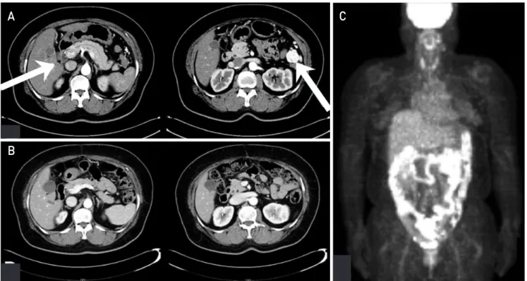
Physical examinations did not yield any remarkable findings and the patient exhibited normal vital signs. Laboratory tests yielded normal findings of hemoglobin (13.3 g/dL), white blood cells (9,250/µL), CA-125 (3.73 IU/mL), and testosterone levels (0.074 ng/mL). Abdominopelvic CT revealed 24- and 10- mm arterial well-enhancing nodules at segment 5 and the left lateral segment of the liver, respectively. Moreover, approxi- mately 10 to 30 mm multiple enlarged round masses were observed at the perigastric, gastrohepatic, and hepatoduo- denal mesentery, and at the site of ligamentum teres fissure (Fig. 1A). In order to differentiate hepatocellular carcinoma and recurrent ovarian steroid cell tumor, NOS, we performed
a sonography-guided liver biopsy. Immunohistochemically, the same findings were observed as in the specimen from 5 years ago, and accordingly, the specimen was diagnosed as a meta- static lesion.
We performed an exploratory laparotomy and noted ap- proximately 30 metastatic masses, ranging from 5 to 30 mm in size, at the omentum, lesser sac, gastrosplenic ligament, and mesentery. In addition, numerous metastatic nodules, 1 to 2 mm in size, were observed. Subsequently, we performed multiple washing cytology and omentectomy and metastasec- tomy to induce an optimal cytoreduction state without resid- ual lesions >5 mm in size. With respect to the liver metastasis, difficulties in resectability resulted in radiofrequency ablation being performed 5 days postsurgery. The patient recovered without any notable postoperative complications and was dis- charged from the hospital 9 days postoperation.
Postoperative histopathological analyses revealed tumor cells with abundant eosinophilic to clear cytoplasm. Moreover, immunohistochemical analyses revealed positive findings of inhibin-a and calretinin, confirming the diagnosis of recurrent ovarian steroid cell tumor, NOS (Fig. 2).
The patient was subjected to adjuvant chemotherapy 3
Fig. 1. (A) Preoperative abdominopelvic computed tomography shows 24-mm arterial well-enhancing mass at segment 5 of liver and 30-mm well-en- hancing omental mass at paracolic gutter space. (B,C) Abdominopelvic computed tomography and positron emission tomography/computed tomography after the completion of treatment shows no definitive noticeable nodules and discrete fluorodeoxyglucose uptake noted at the peritoneal cavity and liver.
A C
B
weeks postsurgery. The chemotherapy regimen administered to the patient consisted of bleomycin (20 units/m² every 3 weeks×four cycles), etoposide (75 mg/m² on days 1–5, every 3 weeks×four cycles), and cisplatin (20 mg/m² on days 1–5, ev- ery 3 weeks×four cycles). The patient successfully completed the chemotherapy regimen without any notable complica- tions, except grade III neutropenia.
The patient was examined every 3 months after the comple- tion of chemotherapy. Upon abdominopelvic CT and positron emission tomography/CT performed at 9 months, the patient exhibited a complete response to the treatment, with no radiological findings of definitive noticeable nodules and dis- crete fluorodeoxyglucose uptake noted at the peritoneal cavity and liver (Fig. 1B, C). Currently , the patient is under follow-up without notable problems.
Discussion
Although ovarian steroid cell tumors were formerly referred to as lipid cell tumors, Hayes and Scully [4] proposed that the tumors be termed steroid cell tumors, as 25% of the tumors do not contain intracellular lipids. Depending on the originating cell, the steroid cell tumors are categorized into three subtypes:
stromal luteoma, Leydig cell tumor, and steroid cell tumor, NOS.
Steroid cell tumors, NOS constitute approximately 60% of all steroid cell tumors. Steroid tumor cells typically occur in patients in their 40s and 50s, with the average age reported as 43 years.
However, in rare occurrences, postmenopausal and prepubes- cent females are also diagnosed with steroid cell tumors. The sizes of the reported tumors range from 1 to 45 cm, with an average reported size of 8.4 cm [1,6-8]. In general, ovarian ste-
roid cell tumors are benign and exhibit low differentiation. In- terestingly, in cases where the tumors are pathologically benign but clinically malignant, 20% of patients present with intraperi- toneal metastatic lesions, but rarely exhibit distant metastasis [9]. According to one report, 94% of all steroid cell tumors are unilateral and 28.6% of the tumors are malignant [4].
Steroid cell tumor, NOS typically exhibits clear boundaries with solid formation, and occasionally exhibits bands; they are morphologically polygonal and divided into eosinophilic and liquid cytoplasm. According to a report by Hayes and Scully [4], pathologic findings that suggest malignancy are observed in 92% of cases where ≥2 differentiations are observed under a high-powered microscope, 86% of cases where the tumor is accompanied by necrosis, 78% of cases where the size of the tumor is >7 cm, 77% of cases where the tumor is accompa- nied by hemorrhaging, and in 64% of cases where the tumor exhibits stage 2 to 3 dysplasia. However, most of the steroid cell tumors are screened in early stages and reports of recurrences or metastasis are rare.
In the diagnosis of steroid cell tumor, NOS, immunohistochem- ical staining of inhibin-a is generally useful. Inhibin-a not only exists in supporting and stromal cells, but also in granulosa cells and luteal cells of the ovary. Particularly, among the pituitary gonadotropins, it is inhibited by follicle stimulating hormone.
Moreover, calretinin has recently been reported to be expressed by steroid cell tumors, NOS. Calretinin, a calcium binding pro- tein, is originally found in nerve tissues, but also exists in the ovaries and testicles. According to a report by Deavers et al.
[10], positive calretinin and inhibin-a expressions are observed in 60% to 90% and in 5% to 90% of all steroid cell tumors, NOS, respectively, and accordingly, in the present case, both inhibin-a and calretinin were positive upon immunohistochemistry.
Fig. 2. (A) Steroid cell tumor, not otherwise specified composed of cells with abundant eosinophilic to clear cytoplasm. The cells have an appearance similar to adrenal cortical cells (H&E, ×200). (B) Diffuse nuclear and cytoplasmic staining for inhibin-a in steroid cell tumor (×200). (C) Diffuse nuclear and cytoplasmic staining for calretinin in steroid cell tumor (×200).