Conceptual changes in small-for-size graft and small-for-size syndrome in living donor liver transplantation
9
0
0
전체 글
(2) Korean J TransplantㆍDecember 2019ㆍVolume 33ㆍIssue 4. 35% of the lower limit of GV/SLV, corresponding to a. HIGHLIGHTS. GRWR of 0.7% under aggressive graft inflow modulation. Small-for-size graft expected graft recipient weight ratio (GRWR) cutoff of 0.8. Volume-related factors may significantly affect graft survival in living donor liver transplantation (LDLT). Early allograft dysfunction characterized by prolonged cholestasis and coagulopathy on day 7 after LDLT may fit the clinical presentations of post-LDLT graft dysfunction better than small-for-size synd-. [12]. In 2001, the Tokyo group reported prolonged cholestasis and elongation of prothrombin time for GV/SLV <40%, resulting in poor graft survival (80%); thus, a GV/SLV of 40% has been used as the lower limit for graft selection since [13]. A literature review showed that the major LDLT transplant centers in Eastern and Western countries apply a GRWR of 0.8 or. rome. Graft selection to maintain an expected GRWR >0.8, and full venous drainage and inflow modulation using. GV/SLV of 40% of the expected graft size as the safe. splenic artery ligation may become the standard.. of 0.8 or GV/SLV of 40%, although small-for-size syn-. limit for successful LDLT without inflow modulation [14-18]. Currently, SFSG can be defined as a GRWR drome (SFSS) after an LDLT graft could be attributed. tion as a case with 23% of the expected total liver size.. to multiple factors including not only graft size but also. They reported that an SFS graft <50% of the expected. donor age, graft steatosis, recipient condition, and por-. total liver size showed characteristic pathological pre-. tal hypertension [19-23].. sentations including ischemia, cholestasis, and regeneration after LDLT. Under such circumstances, the Hong Kong group [7] in turn reported that right lobe. DIFFERENCES IN EXPECTED AND ACTUAL GRAFT VOLUMES. grafts with the middle hepatic vein should be used in adult LDLT to prevent SFS-related graft dysfunction, demon-. When debating the graft volume (GV), we should consid-. strating the safety of removing the extended right lobe. er the differences between expected and actual GVs.. in donors.. Urata et al. [24] showed that the conversion ratio for 3 liver weight (g) and liver volume (cm ) was not 1.0 and. DEFINING SFSG. should instead be 1.12 g/mL, with a possible error of 12%. In 2001, Marcos et al. [25] described differences. In 1997, the Kyoto group also showed negative out-. between hepatic venous-oriented radiological anatomy. comes of left lobe grafts with graft recipient weight ratio. and portal-oriented surgical anatomy regarding the. (GRWR) <1.0, especially in older donors [8] and re-. boundaries between the right and left lobes, possibly re-. vised their strategy in 2000 to use mainly right lobe. sulting in volume errors of 100 g with a 1-cm deviation. grafts without the middle hepatic vein in adult LDLT. in the cutting line. However, three-dimensional (3D). with GRWR ≥0.8 [9]. Recently, the group decreased. volumetry software with possible portal-oriented 3D re-. the lower limit of graft weight to a GRWR of 0.7 for un-. construction of LDLT grafts and portal demarcation. der extensive inflow modulation using splenectomy. line-oriented donor hepatectomy technique, which was. [10]. The Kyushu group initially started the LDT pro-. recently reported by Suh et al. [15], might address this. gram with a lower limit of GV/SLV of 30% with suc-. bias. Hiroshige et al. [26] performed an experimental. cessful outcomes in the initial series [11]. However, the. study in small animals to evaluate the changes in pre-. initial 50 cases performed with this strategy resulted in. served graft weights, reporting that procured grafts lost. prolonged cholestasis and ascites output in 20% of. 4% of their weight in 15 minutes during preservation due. cases. Therefore, to decrease the incidence of graft. to the high osmolarity of the preservation solution.. dysfunction and maximize the success rate, the Kyushu. Radtke et al. [27] evaluated changes in liver volume by. group instead used more right lobe grafts, not less than. infusing contrast medium during computed tomography. 66.
(3) Ikegami T, et al. SFSG, SFSS, and EAD in LDLT. (CT) scans, reporting enlargement by as much as 7%. total bilirubin >5 mg/dL and daily ascites output ex-. during the venous phase from the plain phase and reduc-. ceeding 1 L at 14 days after LDLT. They revised the. tion of as much as 11% during procurement. Because. definition in 2005 as prolonged functional cholestasis,. 3D-CT volumetry is usually performed using the venous. with total bilirubin >10 mg/dL and daily ascites pro-. CT phase, the differences between expected and actual. duction >1 L at postoperative day 14 with a graft sur-. volumes could be 18%.. vival rate of 90% [12]. In contrast, Dahm et al. [32]. Because of these innate errors between the expected. defined SFSS as GRWR <0.8 and the presence of two. and actual GV, Yoneyama et al. [28] established co-. of the following for 3 consecutive days during the first. efficient factors of 0.84 and 0.86 for the right and left. postoperative week: total bilirubin >5 mg/dL, interna-. lobe grafts, respectively, to determine the expected and. tional normalized ratio (INR) >2 and encephalopathy. actual GVs. They also reported no difference in actual. grade 3 or 4, excluding technical, immunological and. and expected cirrhotic liver volume for a coefficient fac-. infectious factors. Their definition focused on primary. tor of 1.01, suggesting that a soft and normal liver graft. non-function after deceased donor liver transplantation. with good elasticity might have larger differences in. [33] with a prompt presentation of non-functional. predicted. volume.. liver. It also had problems including the exclusion of. Kayashima et al. [29] found higher graft shrinkage. GRWR ≥0.8 and focusing only on the very early period. (14%) during preservation in LDLT grafts procured. after LDLT without prolonged cholestasis. Thereafter,. from younger donors compared to those from older do-. Hill et al. [34] defined SFSS in 2009 as the presence. nors (4.4%). Thus, most surgical papers might be de-. of significant cholestasis with serum bilirubin >10. scribing actual GV, as highlighting good graft function. mg/dL after postoperative day 7, coagulopathy with an. even with small graft size. However, strategic descrip-. INR >1.5, and daily ascites output >2 L in the absence. tions of the graft selection may refer to the expected. of technical problems. They used grafts with an ex-. volume; the actual GV may be 15%-20% smaller. A. pected GRWR ≥0.8 and compared graft outcomes be-. transplant surgeon might have a chance to have an. tween the cases with actual GRWR ≥0.8 and those. LDLT graft with enough predicted GV with a largely re-. <0.8. They concluded that graft size was not the only. duced GV on a back-table. Thus, the definition of an. determinant of outcome after LDLT and that inflow. SFS graft could be 0.8 for the “expected” GRWR or 40%. modification may help to prevent certain problems as-. for the expected GV/SLV rather than for the actual. sociated with LDLT. Since the early 2000s, LDLT. GRWR or GV/SLV.. centers have adopted their own institutional policies re-. and. actual. graft. weight. and. garding the lower cutoff for graft selection as 0.8 for. DEFINING SFSS. GRWR and 40% of GV/SLV; thus, there is significant selection bias in terms of GV in the reports since that. Ben-Haim et al. [30] first used the term “small-for-size. time. In the 2010s, factors including prolonged choles-. syndrome (SFSS)” to describe an LDLT graft with. tasis and coagulopathy, in addition to GV, become the. cholestasis and coagulopathy in disparity with normal-. focus in describing graft dysfunction in LDLT caused. ization of transaminase levels together with graft blood. by multiple factors besides GV.. flow, although no objective definitions were described. They mentioned that recipient factors could contribute to SFSS and that GRWR as low as 0.6% can be used. EARLY ALLOGRAFT DYSFUNCTION RATHER THAN SFSS. without SFSS in patients within Child A level, whereas GRWR ≥0.85% was necessary to avoid SFSS in those. In 2012, Ikegami et al. [35] advocated a conceptual. with Child-Pugh class B or C. Soejima et al. [31] pro-. shift from SFSS to primary graft dysfunction after. posed the first objective definition of SFSS in 2003 as. LDLT. Their review of adult LDLT cases showed a. 67.
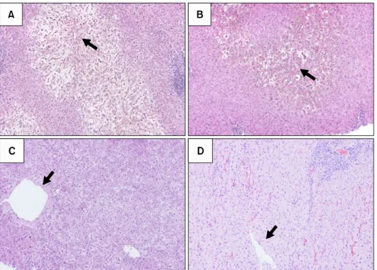
(4) Korean J TransplantㆍDecember 2019ㆍVolume 33ㆍIssue 4. high early graft mortality rate (50%) in patients with. periportal area (Fig. 1D).. functional cholestasis, with total bilirubin >20 mg/dL. In the meantime, the concept of early allograft dys-. after post-LDLT day 7, with very high sensitivity and. function (EAD) characterized by high early amino-. specificity. Huge ascites as a manifestation of graft. transferase levels, persistent cholestasis, and coagulop-. dysfunction did not negatively impact graft survival.. athy in the first week after LT in the use of ex-. Although GV did not affect the occurrence of primary. tended-criteria donors for deceased donor liver trans-. graft dysfunction, model for end-stage liver disease. plantation has been accepted in Western countries. (MELD) score >15, the presence of portosystemic. [36-38]. Olthoff et al. [39] proposed a definition of. shunt, donor age >45 years, portal venous pressure >. EAD in LDLT as the presence of jaundice with bilirubin. 20 mmHg at the end of surgery, and operative blood. >10 mg/dL or coagulopathy with an INR >1.6 on day. loss >10 L were associated with primary graft dys-. 7 without technical complications. The following A2ALL. function in LDLT. The proposed typical pathological. study [40] showed that EAD, with a five-fold risk of. findings of primary graft dysfunction including SFSS. short-term graft loss, was associated with a left lobe. were ballooned hepatocytes with cholestasis in the per-. graft, smaller graft weight, higher preoperative bilir-. icentral venous zone forming a demarcation line with. ubin, higher portal pressure, higher donor age, and. healthy hepatocytes in the periportal zone. Higher se-. higher donor body mass index. The reasons for graft size. rum peak total bilirubin level in primary graft dysfunc-. for EAD in this study might be attributed to nation-wide. tion was also associated with the shift in the ballooned. studies including LDLT centers with small case. hepatocyte borderline with cholestasis from the central. numbers. The Kyoto group [41] evaluated the impact of. venous to portal areas (Fig. 1A and B). Pathological. EAD criteria, reporting that 22 patients (8.5%) satisfied. findings in primary graft dysfunction can be dis-. both criteria. Graft survival was significantly decreased. tinguished from cholestatic lobular hepatitis C by. by the coexistence of both factors (68.2%), compared. pan-lobular hepatocyte ballooning (Fig. 1C) and acute. to only bilirubin >10 mg/dL (24.3%), INR >1.6. rejection with mixed cellular infiltrations around the. (37.5%), and neither (11.9%); however, SFS grafts. Fig. 1. Severe hepatocyte ballooning with cholestasis around the pericentral venous area (A) and the recovering phase (B), representing early allograft dysfunction (EAD) including small-for-size syndrome. The pathological findings with EAD can be distinguished from those of cholestatic lobular hepatitis C with panlobular hepatocyte ballooning (C) and acute rejection with mixed cellular infiltrations around the periportal area (D). Black arrows indicate central veins.. 68.
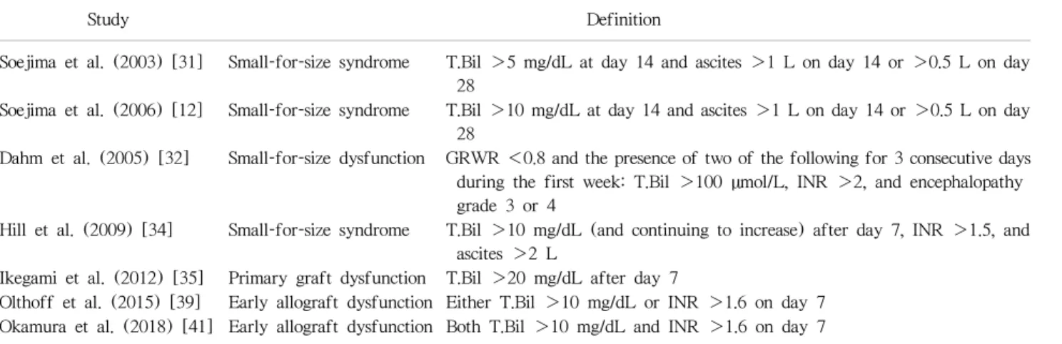
(5) Ikegami T, et al. SFSG, SFSS, and EAD in LDLT. Table 1. Definitions of early allograft dysfunction including small-for-size syndrome Study. Definition. T.Bil >5 mg/dL at day 14 and ascites >1 L on day 14 or >0.5 L on day 28 Soejima et al. (2006) [12] Small-for-size syndrome T.Bil >10 mg/dL at day 14 and ascites >1 L on day 14 or >0.5 L on day 28 Dahm et al. (2005) [32] Small-for-size dysfunction GRWR <0.8 and the presence of two of the following for 3 consecutive days during the first week: T.Bil >100 µmol/L, INR >2, and encephalopathy grade 3 or 4 Hill et al. (2009) [34] Small-for-size syndrome T.Bil >10 mg/dL (and continuing to increase) after day 7, INR >1.5, and ascites >2 L Ikegami et al. (2012) [35] Primary graft dysfunction T.Bil >20 mg/dL after day 7 Olthoff et al. (2015) [39] Early allograft dysfunction Either T.Bil >10 mg/dL or INR >1.6 on day 7 Okamura et al. (2018) [41] Early allograft dysfunction Both T.Bil >10 mg/dL and INR >1.6 on day 7. Soejima et al. (2003) [31]. Small-for-size syndrome. T.Bil, total bilirubin; GRWR, graft recipient weight ratio; INR, international normalized ratio.. with GRWR <0.8 did not negatively impact graft surviv-. and decreased portal inflow via splenic outflow. The. al rates. The Kyoto EAD criteria in LDLT might be rea-. possible challenge in splenic artery ligation is the poten-. sonable for graft mortality in LDLT; therefore, choles-. tial for bleeding and pancreas injury due to a deeply lo-. tasis and coagulopathy should be the primary manifes-. cated splenic artery beneath the pancreas. The Kyushu. tations related to graft mortality in EAD including SFSS.. group has performed simultaneous splenectomy during. The various definitions for EAD including SFSS are listed. LDLT with the use of vessel sealing devices and en-. in Table 1.. do-stapling devices to perform tie-less and blood-less splenectomy, with more potent decompression of portal. HOW TO OVERCOME EAD AFTER LDLT. inflow than that of splenic artery ligation [48-50]. The rationale for splenectomy is based on the fact that spleen. The basic concept of EAD including SFSS is represented. is a major supplier of vasoconstrictive molecules, in-. by the absence of technical complications, and inflow. cluding endothelin, to the liver; thus, splenectomy re-. and outflow issues as previously described [42-44].. sults in hepatic vasodilatation [51,52]. Ikegami et al.. The major donor and recipient factors associated with. [50] usually performed splenic artery ligation before. EAD after DLLT include GRWR <0.8, donor age >45. splenectomy for easier and safer handling of enlarged. years, MELD score >20, recipient with sarcopenia and. spleens in end-stage liver disease. However, splenec-. unable to walk independently, posttransplant portal. tomy is not globally recognized as a standard inflow. pressure >15 mmHg, and post-LDLT complications. modulation procedure in LDLT, except for the Kyushu. including sepsis [17-20,30-35,42-44]. Because man-. and Kyoto groups, due to bleeding complications, portal. agement of recipients with end-stage liver disease is. and splenic venous thrombus, and post-splenectomy. difficult, donor and graft selection with inflow modu-. sepsis. Recently, the Asan group reported innovative. lation to control portal pressure are the only options to. splenic devascularization in LDLT, with sufficient portal. overcome EAD.. decompression and fewer complications compared to. Among inflow modulation methods, splenic artery li-. those for splenectomy [53].. gation is widely recognized as the safest intervention. The creation of portosystemic shunts for portal de-. with optimal outcomes [34,45-47]. The benefit of. compression was first reported in the early to middle. splenic artery ligation is attributed to increased hepatic. 2000s, with optimal outcomes in LDLT using extremely. arterial inflow due to obstruction of the splenic artery. small grafts [54-56]. However, because of unstable. 69.
(6) Korean J TransplantㆍDecember 2019ㆍVolume 33ㆍIssue 4. portal inflow due to liver regeneration in LDLT and por-. function better than SFSS. Graft selection to maintain an. tal steal phenomena leading to graft mortality, no. expected GRWR >0.8, and full venous drainage and in-. Japanese institute has recently performed this procedure. flow modulation using splenic artery ligation may become. [44]. We experienced a case requiring an extra-small. the standard.. graft with expected and actual GV/SLV of 36% and 23%, respectively, from a 20-year-old donor into a pa-. ACKNOWLEDGMENTS. tient with hepatocellular carcinoma, in which an end-to-side portocaval shut after splenectomy was cre-. Conflict of Interest. ated to modulate excessive portal inflow to the small. No potential conflict of interest relevant to this article. graft [57]. The graft showed hepatofugal portal flow 4. was reported.. days after surgery, followed by immediate re-laparotomy for shunt ligation, resulting in a good post-. Author Contributions. operative course. We should have recognized that this. Conceptualization: TI, JMK, MM. Project admin-. young and soft graft had an expected GV/SLV of 36%. istration: DHJ, DSK, YS, JWJ, SGL. Visualization: TI,. to accommodate sufficient portal flow and that the. TY. Writing - original draft: TI. Writing - review & ed-. GV/SLV of 23% was a temporary presentation due to. iting: TI, JMK.. dehydration [26-29]. The generally recommended strategies for graft se-. Additional Contributions. lection and inflow modulation might include the use of. We appreciate great contributions with discussions for. a modified right lobe graft with GRWR >0.8 and full. years regarding this main topic for Kyung-Suk Suh,. venous drainage with splenic artery ligation. However,. Kwang-Woong Lee, and Nam-Joon Yi from Seoul. special techniques in LDLT could be performed in ex-. National University, Gi-won Song and Ki-Hun Kim from. perienced LDLT centers for future development includ-. Asan Medical Center, Soon-Il Kim, Myoung Soo Kim,. ing the use of smaller grafts including the left lobe or. and Dong Jin Joo from Yonsei University Health System,. posterior segment, the use of much larger grafts in-. and Koo-Jeong Kang from Keimyung University Dongsan. cluding dual grafts or extended right lobe graft, and. Medical Center.. technically demanding portal inflow modulations including tie-less splenectomy and creation of portosystemic shunts.. CONCLUSION. REFERENCES 1. Raia S, Nery JR, Mies S. Liver transplantation from live donors. Lancet 1989;2:497. 2. Ozawa K, Uemoto S, Tanaka K, Kumada K, Yamaoka. The historical changes over more than 25 years in LDLT. Y, Kobayashi N, et al. An appraisal of pediatric liver. include those from pediatric to adult patients, from a. transplantation from living relatives: initial clinical expe-. possible chance to generally accepted stable outcomes,. riences in 20 pediatric liver transplantations from living. and right lobe grafts with GRWR >0.8 becoming main-. relatives as donors. Ann Surg 1992;216:547-53.. stream in LDLT, with worldwide expansion. We should. 3. Kawasaki S, Makuuchi M, Matsunami H, Hashikura Y,. recognize a SFS graft expected GRWR cutoff of 0.8. Because volume-related factors may significantly affect graft survival in LDLT due to of institutional guidelines. Ikegami T, Chisuwa H, et al. Preoperative measurement of segmental liver volume of donors for living related liver transplantation. Hepatology 1993;18:1115-20. 4. Hashikura Y, Makuuchi M, Kawasaki S, Matsunami H,. based on a GRWR >0.8, EAD characterized by prolonged. Ikegami T, Nakazawa Y, et al. Successful living-related. cholestasis and coagulopathy on day 7 after LDLT may. partial liver transplantation to an adult patient. Lancet. fit the clinical presentations of post-LDLT graft dys-. 1994;343:1233-4.. 70.
(7) Ikegami T, et al. SFSG, SFSS, and EAD in LDLT. 5. Lo CM, Fan ST, Chan JK, Wei W, Lo RJ, Lai CL.. 17. Goldaracena N, Sapisochin G, Spetzler V, Echeverri J,. Minimum graft volume for successful adult-to-adult living. Kaths M, Cattral MS, et al. Live donor liver trans-. donor liver transplantation for fulminant hepatic failure.. plantation with older (≥50 years) versus younger (<50. Transplantation 1996;62:696-8.. years) donors: does age matter? Ann Surg 2016;263:. 6. Emond JC, Renz JF, Ferrell LD, Rosenthal P, Lim RC,. 979-85.. Roberts JP, et al. Functional analysis of grafts from living. 18. Botha JF, Langnas AN, Campos BD, Grant WJ, Freise. donors: implications for the treatment of older recipients.. CE, Ascher NL, et al. Left lobe adult-to-adult living. Ann Surg 1996;224:544-54.. donor liver transplantation: small grafts and hemi-. 7. Lo CM, Fan ST, Liu CL, Wei WI, Lo RJ, Lai CL, et al. Adult-to-adult living donor liver transplantation using. portocaval shunts in the prevention of small-for-size syndrome. Liver Transpl 2010;16:649-57.. extended right lobe grafts. Ann Surg 1997;226:261-70.. 19. Ogura Y, Hori T, El Moghazy WM, Yoshizawa A, Oike. 8. Kiuchi T, Kasahara M, Uryuhara K, Inomata Y, Uemoto. F, Mori A, et al. Portal pressure <15 mm Hg is a key. S, Asonuma K, et al. Impact of graft size mismatching. for successful adult living donor liver transplantation uti-. on graft prognosis in liver transplantation from living. lizing smaller grafts than before. Liver Transpl 2010;. donors. Transplantation 1999;67:321-7.. 16:718-28.. 9. Inomata Y, Uemoto S, Asonuma K, Egawa H. Right lobe. 20. Ikegami T, Yoshizumi T, Sakata K, Uchiyama H,. graft in living donor liver transplantation. Transplantation. Harimoto N, Harada N, et al. Left lobe living donor liver. 2000;69:258-64.. transplantation in adults: what is the safety limit? Liver. 10. Uemura T, Wada S, Kaido T, Mori A, Ogura Y, Yagi. Transpl 2016;22:1666-75.. S, et al. How far can we lower graft-to-recipient weight. 21. Macshut M, Kaido T, Yao S, Yagi S, Ito T, Kamo N,. ratio for living donor liver transplantation under modulation. et al. Older donor age is a risk factor for negative outcomes. of portal venous pressure? Surgery 2016;159:1623-30. 11. Nishizaki T, Ikegami T, Hiroshige S, Hashimoto K,. after adult living donor liver transplantation using smallfor-size grafts. Liver Transpl 2019;25:1524-32.. Uchiyama H, Yoshizumi T, et al. Small graft for living. 22. Kaido T, Ogawa K, Fujimoto Y, Ogura Y, Hata K, Ito. donor liver transplantation. Ann Surg 2001;233:575-80.. T, et al. Impact of sarcopenia on survival in patients. 12. Soejima Y, Taketomi A, Yoshizumi T, Uchiyama H,. undergoing living donor liver transplantation. Am J. Harada N, Ijichi H, et al. Feasibility of left lobe living. Transplant 2013;13:1549-56.. donor liver transplantation between adults: an 8-year,. 23. Kelly DM, Miller C. Understanding the splenic con-. single-center experience of 107 cases. Am J Transplant. tribution to portal flow: the role of splenic artery ligation. 2006;6(5 Pt 1):1004-11.. as inflow modification in living donor liver transplantation.. 13. Sugawara Y, Makuuchi M, Takayama T, Imamura H,. Liver Transpl 2006;12:1186-8.. Dowaki S, Mizuta K, et al. Small-for-size grafts in liv-. 24. Urata K, Kawasaki S, Matsunami H, Hashikura Y,. ing-related liver transplantation. J Am Coll Surg. Ikegami T, Ishizone S, et al. Calculation of child and. 2001;192:510-3.. adult standard liver volume for liver transplantation.. 14. Hwang S, Lee SG, Lee YJ, Sung KB, Park KM, Kim. Hepatology 1995;21:1317-21.. KH, et al. Lessons learned from 1,000 living donor liver. 25. Marcos A, Orloff M, Mieles L, Olzinski AT, Renz JF,. transplantations in a single center: how to make living. Sitzmann JV. Functional venous anatomy for right-lobe. donations safe. Liver Transpl 2006;12:920-7.. grafting and techniques to optimize outflow. Liver Transpl. 15. Suh KS, Suh SW, Lee JM, Choi Y, Yi NJ, Lee KW.. 2001;7:845-52.. Recent advancements in and views on the donor operation. 26. Hiroshige S, Shimada M, Harada N, Shiotani S, Ninomiya. in living donor liver transplantation: a single-center study. M, Minagawa R, et al. Accurate preoperative estimation. of 886 patients over 13 years. Liver Transpl 2015;. of liver-graft volumetry using three-dimensional com-. 21:329-38.. puted tomography. Transplantation 2003;75:1561-4.. 16. Chan SC, Lo CM, Ng KK, Fan ST. Alleviating the burden. 27. Radtke A, Sotiropoulos GC, Nadalin S, Molmenti EP,. of small-for-size graft in right liver living donor liver. Schroeder T, Lang H, et al. Preoperative volume pre-. transplantation through accumulation of experience. Am. diction in adult living donor liver transplantation: how. J Transplant 2010;10:859-67.. much can we rely on it? Am J Transplant 2007;7:672-9.. 71.
(8) Korean J TransplantㆍDecember 2019ㆍVolume 33ㆍIssue 4. 28. Yoneyama T, Asonuma K, Okajima H, Lee KJ, Yamamoto. 39. Olthoff KM, Emond JC, Shearon TH, Everson G, Baker. H, Takeichi T, et al. Coefficient factor for graft weight. TB, Fisher RA, et al. Liver regeneration after living donor. estimation from preoperative computed tomography volu-. transplantation: adult-to-adult living donor liver trans-. metry in living donor liver transplantation. Liver Transpl 2011;17:369-72.. plantation cohort study. Liver Transpl 2015;21:79-88. 40. Pomposelli JJ, Goodrich NP, Emond JC, Humar A, Baker. 29. Kayashima H, Taketomi A, Yonemura Y, Ijichi H, Harada. TB, Grant DR, et al. Patterns of early allograft dysfunc-. N, Yoshizumi T, et al. Accuracy of an age-adjusted for-. tion in adult live donor liver transplantation: the A2ALL. mula in assessing the graft volume in living donor liver. experience. Transplantation 2016;100:1490-9.. transplantation. Liver Transpl 2008;14:1366-71.. 41. Okamura Y, Yagi S, Sato T, Hata K, Ogawa E, Yoshizawa. 30. Ben-Haim M, Emre S, Fishbein TM, Sheiner PA, Bodian. A, et al. Coexistence of bilirubin ≥10 mg/dL and pro-. CA, Kim-Schluger L, et al. Critical graft size in. thrombin time-international normalized ratio ≥1.6 on day. adult-to-adult living donor liver transplantation: impact. 7: a strong predictor of early graft loss after living donor. of the recipient’s disease. Liver Transpl 2001;7:948-53.. liver transplantation. Transplantation 2018;102:440-7.. 31. Soejima Y, Shimada M, Suehiro T, Hiroshige S, Ninomiya. 42. Kiuchi T, Tanaka K, Ito T, Oike F, Ogura Y, Fujimoto. M, Shiotani S, et al. Outcome analysis in adult-to-adult. Y, et al. Small-for-size graft in living donor liver trans-. living donor liver transplantation using the left lobe. Liver. plantation: how far should we go? Liver Transpl 2003;. Transpl 2003;9:581-6.. 9:S29-35.. 32. Dahm F, Georgiev P, Clavien PA. Small-for-size syn-. 43. Ikegami T, Shimada M, Imura S, Arakawa Y, Nii A,. drome after partial liver transplantation: definition,. Morine Y, et al. Current concept of small-for-size grafts. mechanisms of disease and clinical implications. Am J. in living donor liver transplantation. Surg Today 2008;. Transplant 2005;5:2605-10.. 38:971-82.. 33. Kulik U, Lehner F, Klempnauer J, Borlak J. Primary. 44. Yagi S, Uemoto S. Small-for-size syndrome in living do-. non-function is frequently associated with fatty liver al-. nor liver transplantation. Hepatobiliary Pancreat Dis Int. lografts and high mortality after re-transplantation. Liver. 2012;11:570-6.. Int 2017;37:1219-28.. 45. Lo CM, Liu CL, Fan ST. Portal hyperperfusion injury. 34. Hill MJ, Hughes M, Jie T, Cohen M, Lake J, Payne WD,. as the cause of primary nonfunction in a small-for-size. et al. Graft weight/recipient weight ratio: how well does. liver graft-successful treatment with splenic artery. it predict outcome after partial liver transplants? Liver Transpl 2009;15:1056-62.. ligation. Liver Transpl 2003;9:626-8. 46. Cheng YF, Huang TL, Chen TY, Concejero A, Tsang. 35. Ikegami T, Shirabe K, Yoshizumi T, Aishima S, Taketomi. LL, Wang CC, et al. Liver graft-to-recipient spleen size. YA, Soejima Y, et al. Primary graft dysfunction after. ratio as a novel predictor of portal hyperperfusion syn-. living donor liver transplantation is characterized by de-. drome in living donor liver transplantation. Am J Transplant. layed functional hyperbilirubinemia. Am J Transplant 2012;12:1886-97.. 2006;6:2994-9. 47. Humar A, Beissel J, Crotteau S, Cohen M, Lake J, Payne. 36. Hoyer DP, Paul A, Gallinat A, Molmenti EP, Reinhardt. WD. Delayed splenic artery occlusion for treatment of. R, Minor T, et al. Donor information based prediction. established small-for-size syndrome after partial liver. of early allograft dysfunction and outcome in liver. transplantation. Liver Transpl 2009;15:163-8.. transplantation. Liver Int 2015;35:156-63.. 48. Ikegami T, Shirabe K, Soejima Y, Yoshizumi T, Uchiyama. 37. Lee DD, Singh A, Burns JM, Perry DK, Nguyen JH,. H, Yamashita Y, et al. Strategies for successful left-lobe. Taner CB. Early allograft dysfunction in liver trans-. living donor liver transplantation in 250 consecutive adult. plantation with donation after cardiac death donors results. cases in a single center. J Am Coll Surg 2013;216:353-62.. in inferior survival. Liver Transpl 2014;20:1447-53.. 49. Yoshizumi T, Taketomi A, Soejima Y, Ikegami T,. 38. Wadei HM, Lee DD, Croome KP, Mai ML, Golan E,. Uchiyama H, Kayashima H, et al. The beneficial role. Brotman R, et al. Early allograft dysfunction after liver. of simultaneous splenectomy in living donor liver trans-. transplantation is associated with short- and long-term. plantation in patients with small-for-size graft. Transpl. kidney function impairment. Am J Transplant 2016;16: 850-9.. 72. Int 2008;21:833-42. 50. Ikegami T, Toshima T, Takeishi K, Soejima Y, Kawanaka.
(9) Ikegami T, et al. SFSG, SFSS, and EAD in LDLT. H, Yoshizumi T, et al. Bloodless splenectomy during liver. a new transplant technique. Lancet 2002;359:406-7.. transplantation for terminal liver diseases with portal. 55. Troisi R, Ricciardi S, Smeets P, Petrovic M, Van Maele. hypertension. J Am Coll Surg 2009;208:e1-4.. G, Colle I, et al. Effects of hemi-portocaval shunts for. 51. Uehara H, Akahoshi T, Kawanaka H, Hashimoto N, Nagao. inflow modulation on the outcome of small-for-size grafts. Y, Tomikawa M, et al. Endothelin-1 derived from. in living donor liver transplantation. Am J Transplant. spleen-activated Rho-kinase pathway in rats with secon-. 2005;5:1397-404.. dary biliary cirrhosis. Hepatol Res 2012;42:1039-47.. 56. Yamada T, Tanaka K, Uryuhara K, Ito K, Takada Y,. 52. Kawanaka H, Akahoshi T, Kinjo N, Iguchi T, Ninomiya. Uemoto S. Selective hemi-portocaval shunt based on por-. M, Yamashita YI, et al. Effect of laparoscopic splenectomy. tal vein pressure for small-for-size graft in adult living. on portal haemodynamics in patients with liver cirrhosis. donor liver transplantation. Am J Transplant 2008;8:. and portal hypertension. Br J Surg 2014;101:1585-93.. 847-53.. 53. Moon DB, Lee SG, Hwang S, Ahn CS, Kim KH, Ha. 57. Ikegami T, Soejima Y, Taketomi A, Sanefuji K,. TY, et al. Splenic devascularization can replace splenec-. Kayashima H, Harada N, et al. Living donor liver trans-. tomy during adult living donor liver transplantation: a his-. plantation with extra-small graft: inflow modulation using. torical cohort study. Transpl Int 2019;32:535-45.. splenectomy and temporary portocaval shunt. Hepatogas-. 54. Boillot O, Delafosse B, Méchet I, Boucaud C, Pouyet. troenterology 2008;55:670-2.. M. Small-for-size partial liver graft in an adult recipient:. 73.
(10)
수치
관련 문서
Donor safety in living donor liver transplantation: The Korean organ transplantation registry study.
KOTRY, Korean Organ Transplantation Registry; LDLT, living donor liver transplantation; LG, left lobe group; LT, liver trans- plantation; MHV, middle hepatic vein;
In liver transplantation (LT) for small infant patients, graft size matching to the recipient’s abdomen is critically important because implantation of a large-for-size graft
Purpose: In renal transplantation, the donor age and allograft size are important factors for the outcome of the graft and might be indicators of a functioning renal mass..
Pediatric deceased donor liver transplantation with size reduction for recipient-graft size matching
The type of size reduction in the present case is different from that used in making a reduced or hyper-reduced left lateral section graft because the latter does not reduce
Funneling venoplasty for anomalous graft left hepatic vein in living donor liver transplantation using a split left lateral section graft for an infant patient.. Jung-Man Namgoong
A comparative study of using the right versus left graft in adult-to-adult living donor liver transplantation.. Hye-Sung JO, Young-Dong YU,
When deciding the type of partial liver graft from a living donor as salvage LDLT, graft size-to-recipient matching was the most important factor. Abdominal
MR cholangiography for evaluation of hilar branching anatomy in transplantation of the right hepatic lobe from a living donor.. The efficacy of pre-transplant radiologic evalua-
