• 검색 결과가 없습니다.

Pediatric deceased donor liver transplantation with size reduction for recipient-graft size matching

N/A
N/A
Protected

Academic year: 2021

Share "Pediatric deceased donor liver transplantation with size reduction for recipient-graft size matching"

Copied!
5
0
0

로드 중.... (전체 텍스트 보기)

전체 글

(1)

Pediatric deceased donor liver transplantation with in situ size reduction for recipient-graft size matching

Jung-Man Namgoong

1

, Shin Hwang

1

, Dae-Yeon Kim

1

, Tae-Yong Ha

1

, Gi-Won Song

1

, Dong-Hwan Jung

1

, Kyung Mo Kim

2

, Seak Hee Oh

2

1

Department of Surgery, Asan Medical Center, University of Ulsan College of Medicine, Seoul, Korea,

2

Department of Pediatrics, Asan Medical Center, University of Ulsan College of Medicine, Seoul, Korea

Case Report

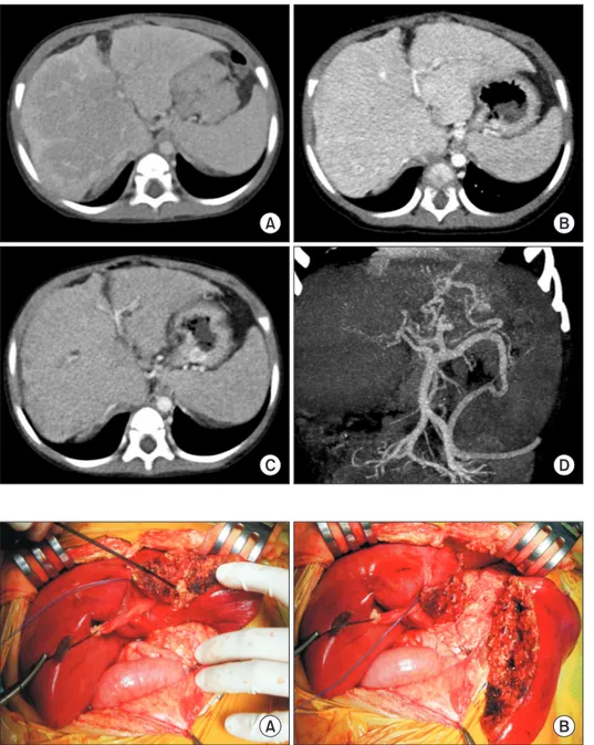
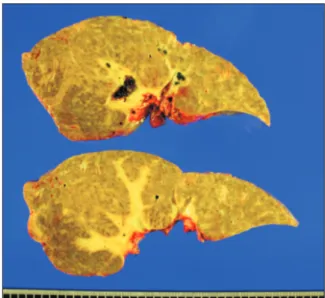
We present a case of pediatric deceased donor liver transplantation using a reduced whole liver graft in a 25-month-old boy weighing 12.7 kg. After he had undergone Kasai portoenterostomy for biliary atresia, his general condition deteriorated progressively. He was enrolled on the waiting list for liver transplantation with Pediatric End-stage Liver Disease score of 15. The donor was a 51-month- old boy with body weight of 20 kg. The donor-to-recipient body weight ratio was 158%. The liver graft appeared to be larger than the recipient’s abdominal cavity. Thus, we planned to do in situ size reduction. Recipient surgery was performed following standard proce- dures. We performed graft outflow vein reconstruction using a modified piggyback technique like the double inferior vena cava meth- od. Since the portal vein was hypoplastic, a side-to-side anastomosis technique was used. We also performed intraoperative portogram to embolize venous collaterals. After completing the graft implantation, we found that the liver graft was too large to be accommodat- ed within the abdomen. After in situ resection of the left lateral section parenchyma, we successfully performed primary closure of the abdominal wound. This patient experienced episodes of acute rejection. He has been doing well for four years after the transplanta- tion.

Key Words: Large-for-size graft; Pediatric transplantation; Graft-recipient weight ratio; Reduced graft; Left lateral section

INTRODUCTION

Liver transplantation (LT) is a life-saving procedure for chil- dren with end-stage liver disease. The success of LT is attribut- ed to the development of effective therapies for immunosup- pression and evolution of surgical techniques that have allowed many more children to receive LT [1-3]. Because most pediatric recipients are under three years of age, there is a significant shortage of size-appropriate whole organs for these patients [3].

Therefore, innovative surgical techniques have been developed to expand the procedure to a wider number of patients, includ- ing split deceased donor liver grafts, partial living donor liver

graft, reduced and hyper-reduced liver grafts, and whole liver grafts.

Living donor liver transplantation (LDLT) has been fre- quently performed because of serious deceased donor shortage in Korea. Types of pediatric LT performed are in the order of LDLT, split LT, whole liver deceased donor LT, and reduced deceased donor LT. In real-world practice in Korea, if a whole liver from an adult deceased donor is too large for an adult patient, split LT for two adult patients is preferred instead of performing reduced LT [4,5]. In contrast, if a whole liver from a pediatric deceased donor is too large for a pediatric patient, there is no practical choice other than to perform reduced LT.

Considering that the incidence of pediatric deceased donors is very low, the actual incidence of reduced LT for pediatric pa- tients is much more lower in Korea. We herein present a case of reduced LT done in a 25-month-old recipient using a liver graft from a 51-month-old deceased donor.

CASE

The recipient was a 25-month-old boy who had undergone Kasai portoenterostomy at the age of 2 months for biliary

Received: September 29, 2020, Revised: October 2, 2020, Accepted: October 3, 2020

Corresponding author: Shin Hwang

Department of Surgery, Asan Medical Center, University of Ulsan College of Medicine, 88 Olympic-ro 43-gil, Songpa-gu, Seoul 05505, Korea

Tel: +82-2-3010-3930, Fax: +82-2-3010-6701, E-mail: shwang@amc.seoul.kr ORCID: https://orcid.org/0000-0002-9045-2531

Copyright Ⓒ The Korean Association of Hepato-Biliary-Pancreatic Surgery

This is an Open Access article distributed under the terms of the Creative Commons Attri- bution Non-Commercial License (http://creativecommons.org/licenses/by-nc/4.0) which permits unrestricted non-commercial use, distribution, and reproduction in any medium, provided the original work is properly cited.

(2)

atresia. His height and body weight were 89 cm and 12.7 kg, respectively. His general condition and liver function deteri- orated progressively. Thus, LT was planned (Fig. 1). He was enrolled on the waiting list of LT of the Korean Network for Organ Sharing with a Pediatric End-stage Liver Disease score of 15 (total bilirubin level of 6.4 mg/dL, albumin level of 2.5 g/

dL, prothrombin international normalized ratio of 1.37, and growth failure).

After some waiting, a 51-month-old boy donor with height of 105 cm and body weight of 20 kg was allocated probably be- cause their body weight ratio was between 50% and 200% (20 kg in donor compared to 12.7 kg in recipient, a ratio of 158%).

The whole liver weighed 915 g on the back table, which had a graft-to-recipient weight ratio (GRWR) of 7.2%. The liver ap-

peared to be larger than the recipient’s abdominal cavity. Thus, we planned to perform in situ size reduction if necessary.

We performed the recipient surgery according to standard procedures of pediatric whole LT. After recipient hepatectomy, we temporarily placed the whole liver graft in the abdomen for size matching. We then began the actual graft implantation.

We performed graft outflow vein reconstruction using a mod- ified piggyback technique by making a large triangular orifice as in a double inferior vena cava method. Since the portal vein was hypoplastic, we used a side-to-side anastomosis technique to prevent anastomotic stenosis. The hepatic artery was recon- structed under surgical microscopy. We performed hepaticoje- junostomy using the preformed Roux-en-Y jejunal limb.

After completing graft implantation procedures, we found

A B

C D

Fig. 1. Pretransplant computed tomography (CT) taken at 1 year (A), 6 months (B), and 1 month (C) before transplantation showing progression of liver cirrhosis. Hypoplastic portal vein with development of collateral veins were visible at the CT scan taken at 1 month before transplantation (D).

Fig. 2. Intraoperative photographs showing in situ size reduction of the whole liver graft.

The left lateral section parenchyma was transected (A) and removed (B) to perform primary closure of the abdominal wound.

A B

(3)

that the liver graft was too large to be accommodated within the abdomen. Since the left liver was large, we decided to per- form in situ resection of the left lateral section parenchyma (Fig.

2). By using a Cavitron ultrasonic aspirator without hepatic in- flow occlusion, we resected the left lateral section meticulously.

The resected parenchyma of the left lateral section weighed 122 g.

Thus, the weight of the implanted right trisection graft was arith- metically 793 g, having a GRWR of 6.2%. The recipient’s native liver weighed 506 g (Fig. 3), which had a GRWR of 4.0%. Thus, we decided to perform primary closure of the abdominal wound.

There were some venous collaterals because of portal hy- pertension. Thus, we performed direct portography through a small branch of the superior mesenteric vein, in which the por- tal blood flow was well preserved. We embolized some collater- al veins from the splenic hilum with coils to prevent the risk of portal steal phenomenon (Fig. 4).

Posttransplant peak values of the liver function test during the first week after transplantation were: serum aspartate transaminase of 330 IU/L, alanine transaminase of 391 IU/L, and total bilirubin of 3.4 mg/dL. Follow-up computed tomog- raphy (CT) scan taken at 4 days posttransplant showed paren- chymal ischemia at the right posterior section (Fig. 5), which might have been associated with extrinsic liver compression by the tight abdominal wall.

Two weeks after the transplantation, a follow-up CT scan

showed resolution of the ischemic portion at the right posteri- or section (Fig. 5). However, ascites developed. Since his liver enzymes were elevated again, we performed a percutaneous liver core biopsy at 18 days posttransplant, in which the pathol-

Fig. 4. Findings of intraoperative direct portography. The reconstructed portal vein showed good blood flow (A). Some collateral veins form the splenic hilum were identified (B, C) and completely embolized with coils (D).

A B

C D

Fig. 3. Gross photograph of the explanted native liver, showing

hepatomegaly combined with advanced liver cirrhosis.

(4)

ogy finding was consistent with acute cellular rejection, with a rejection activity index of 6. The liver function gradually recovered after immunosuppression was augmented with ste- roids. Thereafter, this patient experienced two episodes of bi- opsy-proven mild acute cellular rejection, one at 4 months and one at 16 months posttransplant. The patient has been doing well for four years after the transplantation.

DISCUSSION

Although experience with LT for infant and toddler patients has been accumulating worldwide, large-for-size graft-related problems still remain as big huddles for successful LT. The main problems of large-for-size grafts include the risk of ab- dominal compartment syndrome because of the recipient’s small abdominal cavity, discrepancies in vessel size, insuffi- cient portal circulation, and insufficient tissue oxygenation [6- 10]. Size reduction of the graft is a definite treatment to prevent these critical problems.

In infant patients, the target of graft size reduction is to make the estimated GRWR less than 4% [11]. Considering that a normal adult liver has a GRWR of 2%, this target value may be theoretically slightly lower than 4% for toddler patients. In the present case, the size of the recipient’s native liver had a GRWR of 4.0%, indicating significant hepatomegaly. The size of the whole liver graft had a GRWR of 7.2% before size reduction and

a GRWR of 6.2% after size reduction. Although the reduced liver graft was 55% greater than the recipient’s native liver, the ratio of the graft thickness-to-anteroposterior diameter in the recipient’s abdominal cavity was slightly greater than 1.0. The graft was successfully accommodated within the abdomen with primary wound closure. The type of size reduction in the present case is different from that used in making a reduced or hyper-reduced left lateral section graft because the latter does not reduce graft thickness [6,11].

Implantation of a large-for-size graft can induce various vas- cular complications. The anastomosis site of the graft hepatic vein could be compressed or twisted because of graft compres- sion by the tight abdominal wall, which can lead to hepatic vein outflow obstruction. To prevent such detrimental effects from large-for-size graft implantation, we intentionally made the graft hepatic outflow pathway as large as we could, which was anastomosed through a mixture of the modified piggy- back technique and the double inferior vena cava anastomosis.

If a significant hepatic vein outflow obstruction happens, vena cava stenting can be a rescue therapy. It is expected to have a favorable long-term patency [12].

The patient in the present case had a hypoplastic portal vein with the development of collateral veins because of advanced liver cirrhosis from biliary atresia. If anastomotic stenosis of the portal vein happens, it can lead to life-threatening graft hypoperfusion. To secure portal perfusion, we used two meth-

A B

C D

Fig. 5. Posttransplant computed tomo- graphy (CT) scan follow-up. The CT scan taken 4 days after transplantation showed parenchymal ischemia at the right posterior section (A, B). The CT scan taken 14 days after transplantation showed resolution of the p arenchy mal ischemia (C , D).

However, swelling of the liver graft and

ascites developed because of acute cellular

rejection.

(5)

ods. The first method was using the side-to-side anastomosis technique to prevent anastomotic stenosis of the portal vein.

We made a deep longitudinal incision in the 6 o’clock direction of the graft portal vein and the 12 o’clock direction of the re- cipient portal vein. We then used continuous sutures to unify these two portal veins. This method generated an enlarged portal vein conduit from the superior mesenteric vein-splenic vein confluence to the graft hilar portal vein confluence [13].

The second was complete interruption of the venous collaterals through intraoperative portography and coil embolization [14], which was primarily developed to cope with the small-for-size graft syndrome in adult LDLT. In clinical practice, we have tried to occlude the collateral veins as much as possible during LT operation in both adult and pediatric patients.

In situ size reduction through non-anatomical resection of the left lateral section parenchyma is an easy and intuitive method that can be performed without hepatic inflow occlu- sion. It was applied to the whole liver graft in the present case.

It can also be applied to LDLT or split LT using a left lateral section graft with a long flat fish-like anatomy, as in making a reduced left lateral section graft.

If the liver graft is too large to be accommodated within the abdominal cavity, staged closure methods should be used to prevent abdominal compartment syndrome, as used in infants with gastroschisis [15,16]. If the liver graft bulges out promi- nently, coverage with a silo is necessary. If the liver graft bulges out only slightly, temporary closure with a mesh is reasonable.

In conclusion, in situ size reduction by means of non-ana- tomical hepatic resection is a useful option to prevent large- for-size graft-related problems in pediatric LT.

CONFLICT OF INTEREST

No potential conflict of interest relevant to this article was reported.

ORCID

Jung-Man Namgoong, https://orcid.org/0000-0002-9237-7440 Shin Hwang, https://orcid.org/0000-0002-9045-2531 Dae-Yeon Kim, https://orcid.org/0000-0001-6130-1796 Tae-Yong Ha, https://orcid.org/0000-0001-9932-0212 Gi-Won Song, https://orcid.org/0000-0002-4235-0434 Dong-Hwan Jung, https://orcid.org/0000-0001-5984-023X Kyung Mo Kim, https://orcid.org/0000-0001-7896-6751 Seak Hee Oh, https://orcid.org/0000-0002-9672-8877

AUTHOR CONTRIBUTIONS

Conceptualization: SH. Data curation: JMN, KMK, SHO.

Methodology: DYK, TYH, GWS, DHJ. Visualization: SH.

Writing - original draft: SH, JMN. Writing - review & editing:

SH.

REFERENCES

1. Azouz SM, Diamond IR, Fecteau A. Graft type in pediatric liver transplantation. Curr Opin Organ Transplant 2011;16:494-498.

2. Bartlett A, Rela M. Progress in surgical techniques in pediatric liver transplantation. Pediatr Transplant 2010;14:33-40.

3. Spada M, Riva S, Maggiore G, Cintorino D, Gridelli B. Pediatric liver transplantation. World J Gastroenterol 2009;15:648-674.

4. Hwang S, Lee SG, Park KM, Kim KH, Ahn CS, Moon DB, et al. A case report of split liver transplantation for two adult recipients in Korea. Transplant Proc 2004;36:2736-2740.

5. Jung DH, Hwang S, Song GW, Ahn CS, Moon DB, Kim KH, et al. In situ split liver transplantation for 2 adult recipients: a single-center experience. Ann Transplant 2017;22:230-240.

6. Kanazawa H, Sakamoto S, Fukuda A, Uchida H, Hamano I, Shigeta T, et al. Living-donor liver transplantation with hyperreduced left lateral segment grafts: a single-center experience. Transplantation 2013;95:750-754.

7. Kiuchi T, Kasahara M, Uryuhara K, Inomata Y, Uemoto S, Asonuma K, et al. Impact of graft size mismatching on graft prognosis in liver transplantation from living donors. Transplantation 1999;67:321-327.

8. Kasahara M, Fukuda A, Yokoyama S, Sato S, Tanaka H, Kuroda T, et al. Living donor liver transplantation with hyperreduced left lateral segments. J Pediatr Surg 2008;43:1575-1578.

9. Vanatta JM, Esquivel CO. Status of liver transplantation in infants <

5 kg. Pediatr Transplant 2007;11:5-9.

10. Raices M, Czerwonko ME, Ardiles V, Boldrini G, D’Agostino D, Marcó Del Pont J, et al. Short- and long-term outcomes after live-do- nor transplantation with hyper-reduced liver grafts in low-weight pediatric recipients. J Gastrointest Surg 2019;23:2411-2420.

11. Sakamoto S, Kanazawa H, Shigeta T, Uchida H, Sasaki K, Hamano I, et al. Technical considerations of living donor hepatectomy of seg- ment 2 grafts for infants. Surgery 2014;156:1232-1237.

12. Donaldson J, Obuchowski NA, Le RT, Lomaglio L, Unger RH, Bayona MDP, et al. Stenting for inferior vena cava stenosis after liver trans- plant. AJR Am J Roentgenol 2019;213:1381-1387.

13. Namgoong JM, Hwang S, Ahn CS, Kim KM, Oh SH, Kim DY, et al.

Portal vein reconstruction using side-to-side unification technique for infant-to-infant deceased donor whole liver transplantation. Ann Hepatobiliary Pancreat Surg 2020;24:445-453.

14. Kim JH, Ko GY, Sung KB, Yoon HK, Kim KR, Moon DB, et al.

Transvenous variceal embolization during or after living-donor liver transplantation to improve portal venous flow. J Vasc Interv Radiol 2009;20:1454-1459.

15. Kidd JN Jr, Jackson RJ, Smith SD, Wagner CW. Evolution of staged versus primary closure of gastroschisis. Ann Surg 2003;237:759-764;

discussion 764-765.

16. Namgoong JM, Hwang S, Kim DY, Song GW, Ahn CS, Kim KM,

et al. Pediatric split liver transplantation using a hyperreduced left

lateral segment graft in an infant weighing 4 kg. Korean J Transplant

2020;34:204-209.

수치

Fig. 1. Pretransplant computed tomography  (CT) taken at 1 year (A), 6 months (B), and 1  month (C) before transplantation showing  progression of liver cirrhosis
Fig. 4. Findings of intraoperative direct  portography. The reconstructed portal vein  showed good blood flow (A)
Fig. 5. Posttransplant computed tomo- tomo-graphy (CT) scan follow-up. The CT scan  taken 4 days after transplantation showed  parenchymal ischemia at the right posterior  section (A, B)

참조

관련 문서

Particle size and size distribution of ethylene -modified polystyrene with different EAA concentration at 140% degree of neutralization of EAA. Latex Particle Size

 Communication cost = input file size + 2 × (sum of the sizes of all files passed from Map processes to Reduce processes) + the sum of the output sizes of the Reduce

The lateral size of the defect obtained from the deformation amount and the defect detection test using the shearing interferometer, width and depth of

Fourth, the following items from the preliminary protocol on CPR were added or revised for the final protocol: use of oropharyngeal airway of proper size,

From the results of this study, we concluded that two different sized graft materials have positive effects on new bone formation.. Additionally, smaller

In 4-week group, the group filled with bone graft with decortication revealed larger new bone formation area than shown in the group that had a defect area

: The size of the opening in the choke determines the flow rate, because the pressure upstream is determined primarily by the well FTP, and the pressure downstream

The result of using PLGA nanofiber membrane with bone graft material showed that the PLGA nanofiber membrane in the experimental group of 2 weeks were ten times more new