© 2015 The Korean Ophthalmological Society
This is an Open Access article distributed under the terms of the Creative Commons Attribution Non-Commercial License (http://creativecommons.org/licenses /by-nc/3.0/) which permits unrestricted non-commercial use, distribution, and reproduction in any medium, provided the original work is properly cited.
Original Article
Assessment of Patient Pain Experience during Intravitreal 27-Gauge Bevacizumab and 30-Gauge Ranibizumab Injection
Mete Güler1, Burak Bilgin2, Musa Çapkın1, Ali Şimşek1, Şemsettin Bilak1
1Department of Ophthalmology, Adıyaman University School of Medicine, Adiyaman, Turkey
2Department of Ophthalmology, Adıyaman University Education and Research Hospital, Adiyaman, Turkey
Purpose: To compare pain scores of patients during intravitreal 27-gauge bevacizumab and 30-gauge ranibi- zumab injection procedures.
Methods: Seventy eyes of 70 patients who had not previously undergone intravitreal anti-vascular endothelial growth factor therapy were included in this study. Thirty-five patients received ranibizumab and 35 patients received bevacizumab. The diagnoses of the patients were: 27 age related macular degeneration, 15 diabetic macular edema, 9 diabetic vitreous hemorrhage, 6 central retinal vein occlusion, 11 branch retinal vein occlu- sion and 2 central serous chorioretinopathy. Bevacizumab (1.25 mg/0.05 mL) was injected into the vitreous cavity using a 27-gauge needle, and ranibizumab (0.5 mg/0.05 mL) was injected with 30-gauge needle. Pa- tients were asked just after the injection to rate their perceived pain during the injection using the visual ana- logue scale (VAS) of 0 (no pain) to 10 (unbearable/ worst pain). The average of these scores was used as the primary outcome.
Results: The VAS pain scores in the ranibizumab and bevacizumab groups were 1.06 ± 0.91 (range, 0 to 3) and 1.94 ± 1.55 (range, 0 to 7), respectively, a significant difference (p = 0.005). Patients <65 and ≥65 years of age in both the ranibizumab and bevacizumab groups were then compared. For patients <65, there was a signifi- cant difference in the average VAS pain scores between groups (p = 0.003). However, for patients ≥65 years, there was not a significant difference in the average VAS pain scores between groups (p = 0.238). Female and male patients in both ranibizumab and bevacizumab groups were also compared. For female patients, there was a significant difference in the average VAS pain scores between groups (p = 0.016), although not for male patients (p = 0.078).
Conclusions: Thirty-gauge intravitreal injection is more comfortable than 27-gauge injection. Injection of bevaci- zumab with 30-gauge needle syringes may be more tolerable for patients.
Key Words: Bevacizumab, Intravitreal injections, Pain, Ranibizumab, Visual analogue scale
Recent clinical trials regarding intravitreal injection of anti-vascular endothelial growth factor (VEGF) agents have shown excellent results in the treatment of intraocular neovascular disorders, macular edema, neovascular glau- coma and various corneal neovascular diseases [1-4]. Over the past decade, intravitreal injection of anti-VEGF agents have gained tremendous acceptance among retinal special-
Received: September 25, 2014 Accepted: November 17, 2014
Corresponding Author: Mete Güler, MD. Department of Ophthalmolo- gy, Adiyaman University School of Medicine, Altinsehir Street, 3005th Road, No. 13, 02040 Adiyaman, Turkey. Tel: 90-416-223-3800, Fax: 90- 416-223-1690, E-mail: [email protected]
This study has been presented in 7th Mediterretina International Meet- ing, 17-20 April 2014, Istanbul, Turkey.
ists and become one of the most common intraocular pro- cedures [5]. Minimizing patient pain is critical during in- travitreal injection. Patient discomfort at the time of injection can lead to sudden movements of the eye, which can be associated with intraocular complications. In addi- tion, patients with diseases such as cystoid macular edema and age-related macular degeneration that require multiple intravitreal injections may be less likely to continue intrav- itreal injection after a bad experience [6].
Bevacizumab and ranibizumab are currently the most commonly used anti-VEGF agents. Ranibizumab (Lucen- tis; Genentech, San Francisco, CA, USA), a fragment of a humanized monoclonal antibody against all VEGF iso- forms, is prepared as a single-use glass vial designed to provide 0.05 mL for intravitreal injection with a 30-gauge needle [3]. Bevacizumab is a humanized monoclonal anti- body that binds all isoforms of VEGF and interferes with receptor binding to inhibit its signal [2]. Currently, intrav- itreal injection of bevacizumab is off label and does not have any commercially available form. For clinical use, it has been dispensed into single-use syringes with 26- to 30-gauge needles [7,8].
Despite the common use of intravitreal injections, there have been only a few studies that have investigated injec- tion-related pain [8,9]. Thus, the aim of this study was to compare pain scores of patients during intravitreal 27-gauge bevacizumab and 30-gauge ranibizumab (Lucen- tis) injection procedures.
Materials and Methods
This prospective, interventional, non-randomized, compar- ative study was carried out at Department of Ophthalmology, Adıyaman University. All procedures followed the Declara- tion of Helsinki, and written informed consent was obtained from all patients. They were all informed of the off-label use of the bevacizumab and its potential risks and benefits.
Seventy eyes of 70 patients who had not previously un- dergone intravitreal anti-VEGF therapy were included in this study. Exclusion criteria were history of previous eye surgery other than for cataract, herpetic eye disease, glau- coma, uveitis, active conjunctivitis, keratitis and bullous keratopathy. Patients with poor cooperation in using the visual analogue scale (VAS) and/or using systemic analge- sics or sedative medications were also excluded.
Thirty-five patients received ranibizumab and 35 pa- tients received bevacizumab. Patients were first given topi- cal anesthetic proparacaine 0.5% (Alcaine, Alcon-Cou- vreur, Puurs, Belgium) and povidone-iodine 5% drops.
After 5% povidone-iodine was applied to the operating field, an eyelid speculum was positioned in order to stabi- lize the eyelids. Injections were performed one minute af- ter instillation of the proparacaine drop. Bevacizumab (1.25 mg/0.05 mL) was previously dispensed into single-use 27-gauge needle syringes using an aseptic technique and was injected into the vitreous cavity through the pars pla- na at the inferotemporal quadrant, 3.5 to 4.0 mm posterior to the limbus. Ranibizumab (0.5 mg/0.05 mL) was injected with a 30-gauge needle using the same method. All injec- tions were performed in the operating room.
Patients were asked just after the injection to rate their perceived pain during the injection on a VAS of 0 (no pain) to 10 (unbearable/worst pain). The average of these scores was used as the primary outcome.
Statistical analyses
A Mann-Whitney U-test was used to compare the VAS values, and an independent sample t-test was used to com- pare the ages between the two groups. Sexes were com- pared with a chi-square test. A p-value less than 0.05 was considered statistically significant.
Results
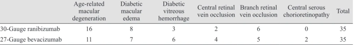
The diagnoses of the patients in the study are summa- rized in Table 1. The mean age was 60.43 ± 12.13 (range, 42 to 83) in the ranibizumab group and 64.86 ± 10.04 (range, 41 to 81) in the bevacizumab group. The mean age of the groups was similar (p = 0.151). In the ranibizumab group, 16 patients were male and 19 were female. In the bevaci- zumab group, 18 patients were male and 17 were female.
The sex distribution between groups was also similar (p = 0.811). The VAS pain scores in the ranibizumab and beva- cizumab groups were 1.06 ± 0.91 (range, 0 to 3) and 1.94 ± 1.55 (range, 0 to 7), respectively (Fig. 1), a significant dif- ference (p = 0.005).
Patients were divided into two groups according to age:
<65 and ≥65 years [9]. In the ranibizumab group, 16 pa- tients were <65 and 19 patients were ≥65. There was not a
significant difference in the average VAS pain scores be- tween groups (p = 0.145). The VAS pain scores in the younger and older groups were 0.81 ± 0.91 and 1.27 ± 0.87, respectively. In the bevacizumab group, 14 patients were
<65 and 21 patients were ≥65. There was not a significant difference in the average VAS pain scores between groups (p = 0.339). VAS pain scores in younger and older groups were 2.29 ± 1.49 and 1.76 ± 1.61, respectively.
Patients <65 years of age in both the ranibizumab (16 patients) and bevacizumab (14 patients) groups were com- pared. There was a significant difference in the average VAS pain scores between groups (p = 0.003). The VAS pain scores in the ranibizumab and bevacizumab groups were 0.81 ± 0.91 and 2.29 ± 1.49, respectively.
Patients ≥65 years of age in both the ranibizumab (19 patients) and bevacizumab (21 patients) group were com- pared. There was not a significant difference in the average VAS pain scores between groups (p = 0.238). The VAS pain scores in the ranibizumab and bevacizumab groups were 1.27 ± 0.87 and 1.76 ± 1.61, respectively.
Patients were divided into two groups according to sex.
In the ranibizumab group, there was not a significant dif-
ference in the average VAS pain scores between sexes (p = 0.481). The VAS pain scores in the male and female groups were 0.93 ± 0.77 and 1.16 ± 1.01, respectively. Nor was there a significant difference between sexes in average VAS pain score in the bevacizumab group (p = 0.163). The VAS pain scores in the male and female groups were 1.61 ± 1.29 and 2.35 ± 1.77, respectively.
Female patients in both the ranibizumab (19 patients) and bevacizumab (17 patients) groups were compared.
There was a significant difference in the average VAS pain scores between groups (p = 0.016). The female VAS pain scores in the ranibizumab and bevacizumab groups were 1.16 ± 1.01 and 2.35 ± 1.77, respectively.
Male patients in both the ranibizumab (16 patients) and bevacizumab (18 patients) groups were compared. There was not a significant difference in the average VAS pain scores between groups (p = 0.078). The male VAS pain scores in the ranibizumab and bevacizumab groups were 0.93 ± 0.77 and 1.61 ± 1.29, respectively.
Discussion
VAS is frequently used as an assessment instrument to evaluate the analgesic effects of various therapies and to detect minute pain changes during analgesic administra- tion. VAS is a simple tool to use on anyone cognitively ca- pable of understanding the parameters and responding to clinician instructions. Indeed, its popularity is frequently attributed to the ease and convenience in a fast-paced clin- ical setting [10]. VAS has been widely used in ophthalmo- logic research [11-14]. We evaluated the pain experiences of the patients with VAS because of its easy and quick use.
Moisseiev et al. [8] evaluated the correlation between pain associated with intravitreal bevacizumab injection and the location of the injection. They did not find any sta- tistically significant difference in terms of pain experience between anatomical quadrants. In order to achieve stan- dardization, we performed all injections into the infero- Table 1. Diagnoses of the patients that underwent intravitreal 30-gauge ranibizumab and 27-gauge bevacizumab injections
Age-related macular degeneration
Diabetic macular edema
Diabetic vitreous hemorrhage
Central retinal
vein occlusion Branch retinal
vein occlusion Central serous chorioretinopathy Total
30-Gauge ranibizumab 16 8 3 2 6 0 35
27-Gauge bevacizumab 11 7 6 4 5 2 35
30-Gauge ranibizumab 27-Gauge bevacizumab 6
4
2
0
Visual analog scale
Fig. 1. Box-plot showing the distribution of mean visual analog scale values in the 30-gauge ranibizumab and 27-gauge bevaci- zumab groups. The black lines in the diagram illustrate the medi- an values of the groups.
temporal quadrant. Knecht et al. [15] compared tunneled scleral intravitreal injection with straight scleral intravitre- al injection in terms of short-term intraocular pressure changes, occurrence and amount of vitreous reflux, and patient discomfor. They did not find a difference in patient discomfort or intraocular pressure increase after the injec- tion between groups. We used a straight injection tech- nique in all patients.
Green-Simms et al. [7] surveyed the intravitreal injec- tion technique practice patterns of retinal specialists in the United States. They found that a majority of the survey participants used a 30-gauge needle for the intravitreal in- jection of ranibizumab (78%) and bevacizumab (60%).
The studies that have evaluated pain scores of patients during intravitreal injection with different needle calibers have reported contrasting results. Rodrigues et al. [16] re- ported that the patients injected with a 26- or 27-gauge needle experienced more pain compared to those injected with 29- or 30-gauge needles. Eaton et al. [17] reported that injections with a 33-gauge device were significantly faster, but there was no significant difference in the levels of pain between a 33-gauge device and a standard 30-gauge nee- dle. Rifkin and Schaal [9] used 27- and 30-gauge needles for injection and determined that the caliber of the needle did not significantly affect the pain score. We found that intravitreal injection with a 30-gauge needle was less pain- ful for the patients. According to our experience, it is easi- er and safer to pierce the sclera with a 30-gauge needle than a 27-gauge needle during intravitreal injection. One patient, a 43-year-old woman diagnosed with diabetic mac- ular edema, experienced a complication during the intrav- itreal 27-gauge bevacizumab injection. The patient report- ed extreme pain during the intravitreal injection as the reason for head movement. The tip of the 27-guage needle touched the posterior capsule of the crystalline lens. Cata- ract development was detected at the control visit. The cat- aract was removed with a phacoemulsification technique, and a foldable IOL was placed into the sulcus.
A 27-gauge needle has a diameter of 413 µm, and a 30-gauge needle has a diameter of 311-µ [9]. Pulido et al.
[18] reported that 27-gauge needles require almost twice the force to penetrate the sclera than 30- or 31-gauge nee- dles. This may explain why patients experienced less pain with the 30-gauge needle in our study.
Rifkin and Schaal [9] demonstrated that the pain score was not significantly related to diagnosis. Moisseiev et al.
[8] also did not find a significant difference in the pain scores between any of the indications for the injection. We did not evaluate the relationship between the diagnosis and pain scores in this study.
Rifkin and Schaal [9] determined that patients aged >65 years reported a lower average pain score than those aged
<65 years. Moisseiev et al. [8] found no correlation be- tween pain score and patient age. To minimize the age fac- tor, we formed two groups with similarly aged patients.
We conducted a subgroup analysis according to two age groups: <65 and ≥65. Patients <65 years of age in the ran- ibizumab group reported a lower average pain score than those in the bevacizumab group. Patients ≥65 years old in the ranibizumab and bevacizumab groups were also com- pared, demonstrating no significant difference in average VAS pain scores.
Rifkin and Schaal [9] showed that female sex was asso- ciated with a lower post-injection pain score. Moisseiev et al. [8] demonstrated no statistically significant difference in pain score according to sex. In our study, there were no pain score differences between the sexes in the ranibizum- ab and bevacizumab groups. We also conducted a sub- group analysis for each sex. Female patients in the ranibi- zumab group reported a lower average pain score than those in the bevacizumab group. There was not a signifi- cant difference in the average pain score in male patients between the ranibizumab and bevacizumab groups.
Our study had some limitations. Forming the groups with same drug might have been more appropriate. Senso- ry innervation of the eye is provided by the peripheral ax- ons of the primary sensory neurons located in the trigemi- nal ganglion [19]. The sensory nerves enter the eyeball mainly through the ciliary nerves and innervate all ocular tissues with the exception of the lens and the retina [19].
Since the retina has no nociceptors, and an equal volume of the drug was injected in both groups, we assumed that the pain sensation during intravitreal injection was mainly due to the caliber of the needle and so separated the pa- tients accordingly. Another limitation of the study was the subjective character of the VAS. However, since there is not a quantitative technique to evaluate the amount of pain, the VAS was our best option.
In conclusion a 30-gauge intravitreal injection is more comfortable than a 27-gauge injection. Injection of bevaci- zumab with a 30-gauge needle syringe may be more toler- able for patients.
Conflict of Interest
No potential conflict of interest relevant to this article was reported.
References
1. Rosenfeld PJ, Brown DM, Heier JS, et al. Ranibizumab for neovascular age-related macular degeneration. N Engl J Med 2006;355:1419-31.
2. Avery RL, Pearlman J, Pieramici DJ, et al. Intravitreal bev- acizumab (Avastin) in the treatment of proliferative diabet- ic retinopathy. Ophthalmology 2006;113:1695.
3. Pieramici DJ, Rabena M, Castellarin AA, et al. Ranibi- zumab for the treatment of macular edema associated with perfused central retinal vein occlusions. Ophthalmology 2008;115:e47-54.
4. Park SC, Su D, Tello C. Anti-VEGF therapy for the treat- ment of glaucoma: a focus on ranibizumab and bevacizum- ab. Expert Opin Biol Ther 2012;12:1641-7.
5. Peyman GA, Lad EM, Moshfeghi DM. Intravitreal injec- tion of therapeutic agents. Retina 2009;29:875-912.
6. Tewari A, Shah GK, Dhalla MS, Blinder KJ. Surface anes- thesia for office-based retinal procedures. Retina 2007;27:
804-5.
7. Green-Simms AE, Ekdawi NS, Bakri SJ. Survey of intrav- itreal injection techniques among retinal specialists in the United States. Am J Ophthalmol 2011;151:329-32.
8. Moisseiev E, Regenbogen M, Bartfeld Y, Barak A. Evalua- tion of pain in intravitreal bevacizumab injections. Curr Eye Res 2012;37:813-7.
9. Rifkin L, Schaal S. Factors affecting patients’ pain intensi- ty during in office intravitreal injection procedure. Retina 2012;32:696-700.
10. Reed MD, Van Nostran W. Assessing pain intensity with the visual analog scale: a plea for uniformity. J Clin Phar-
macol 2014;54:241-4.
11. Aslankurt M, Aslan L, Baskan AM, et al. Pain and cooper- ation in patients having dominant-side or nondominant-side phacoemulsification. J Cataract Refract Surg 2014;40:199- 202.
12. Mirshahi A, Lashay A, Roozbahani M, et al. Pain score of patients undergoing single spot, short pulse laser versus conventional laser for diabetic retinopathy. Graefes Arch Clin Exp Ophthalmol 2013;251:1103-7.
13. Chen D, Lian Y, Li J, et al. Monitor corneal epithelial heal- ing under bandage contact lens using ultrahigh-resolution optical coherence tomography after pterygium surgery.
Eye Contact Lens 2014;40:175-80.
14. Narvaez J, Wessels I, Bacon G, et al. Prospective random- ized evaluation of short-term complications when using buffered or unbuffered lidocaine 1% with epinephrine for blepharoplasty surgery. Ophthal Plast Reconstr Surg 2010;
26:33-5.
15. Knecht PB, Michels S, Sturm V, et al. Tunnelled versus straight intravitreal injection: intraocular pressure changes, vitreous reflux, and patient discomfort. Retina 2009;29:
1175-81.
16. Rodrigues EB, Grumann A Jr, Penha FM, et al. Effect of needle type and injection technique on pain level and vitre- al reflux in intravitreal injection. J Ocul Pharmacol Ther 2011;27:197-203.
17. Eaton AM, Gordon GM, Wafapoor H, et al. Assessment of novel guarded needle to increase patient comfort and de- crease injection time during intravitreal injection. Ophthal- mic Surg Lasers Imaging Retina 2013;44:561-8.
18. Pulido JS, Zobitz ME, An KN. Scleral penetration force re- quirements for commonly used intravitreal needles. Eye 2007;21:1210-1.
19. Levin LA, Nilsson SF, Hoeve JV, Wu S. Sensory innerva- tion of the eye. In: Levin LA, Nilsson SF, Hoeve JV, Wu S.
Adler’s physiology of the eye. 11th ed. London: Elsevier;
2011. p. 363-84.