다초점맥락막염과 연관된 맥락막신생혈관에서 유리체내 베바시주맙 주사
Intravitreal Bevacizumab Injection for Choroidal Neovascularization Associated with Multifocal Choroiditis
조인환1,2, 박운철1,2, 유형곤1,2
In Hwan Cho
1,2, Un Chul Park
1,2, Hyeong Gon Yu
1,21서울대학교 의과대학 안과학교실, 2서울대학교병원 의생명연구원 망막변성연구소
1
Department of Ophthalmology, Seoul National University College of Medicine, Seoul, Korea
2
Retinal Degeneration Research Lab, Biomedical Research Institute, Seoul National University Hospital, Seoul, Korea
Purpose: To evaluate the long-term efficacy and safety of intravitreal bevacizumab injections for choroidal neovascularization (CNV)
associated with multifocal choroiditis (MFC).Methods: This is a retrospective case series study. Patients who were treated with intravitreal bevacizumab injections for CNV associated
with MFC from January 2004 to August 2016 were included. The characteristics of patients, change in best corrected visual acuity, and central subfield thickness (CST) were reviewed.Results: Among 34 MFC eyes (22 patients), 6 eyes (6 patients, 17.6%) had CNV associated with MFC. The mean age of patients was 41
(26-64) years and four of six patients (66%) were female. The mean duration of follow-up was 101.5 (36-178) months and the mean num- ber (range) of intravitreal bevacizumab injections was 2.3 (1-6). Three of six (50%) eyes showed evident inflammation and were treated with immunosuppression agents. Visual acuity improved in five of six (83%) eyes and CST decreased in all eyes at final visit. One eye with initial poor visual acuity (2/100) continued to have poor final visual acuity (2/100) with scar changes. No adverse events were reported.Conclusions: Intravitreal bevacizumab injections were safe and effective at improving visual acuity and decreasing CST over long-term
follow-up in a small series of patients with CNV secondary to MFC.Keywords: Bevacizumab; Choroidal neovascularization; Multifocal choroiditis
Address reprint requests to Hyeong Gon Yu, MD, PhD
Department of Ophthalmology, Seoul National University Hospital, #101 Daehak-ro, Jongno-gu, Seoul 03080, Korea
Tel: 82-2-2072-2437, Fax: 82-2-741-3187 E-mail: [email protected]
Received: 2016. 9. 22 Revised: 2016. 10. 6 Accepted: 2016. 10. 13
Introduction
Multifocal choroiditis (MFC) is chronic ocular inflammation commonly characterized by the presence of vitritis and mul-
tiple chorioretinal lesions in the posterior pole and/or periph-
ery [1-5]. Although it has been reported that visual prognosis
is relatively good in most patients with MFC, several compli-
cations, including macular edema, choroidal neovasculariza-
tion (CNV), and corticosteroid- related cataract or glaucoma, can occur [6]. Among these complications, CNV has been reported as the most common cause of visual loss, develop- ing in 27% to 32% of MFC patients [1-11]. Many treatment options for CNV associated with MFC have been suggested such as local corticosteroids [6], rapamycin [10], photody- namic therapy (PDT) [12], transpupillary thermotherapy [13], submacular surgery [14], and macular translocation [15]. In general, these treatment options do not achieve significant visual acuity improvement on follow-up. A new treatment option for CNV associated with MFC is needed.
Anti-vascular endothelial growth factors such as bevaci- zumab have demonstrated improvement of visual acuity in treating CNV for neovascular age-related macular degener- ation [16], pathologic myopia [17], and retinal vein occlusion [18]. The development of CNV in MFC may share a final common pathway with these diseases [19]. Therefore, an- ti-vascular endothelial growth factor (anti-VEGF) therapies seem to be a rational treatment approach to CNV associated with MFC. Several studies have reported clinical outcomes after anti-VEGF treatment and demonstrated favorable results after short-term follow-up [20,21]. However, few re- ports have examined long-term results after treatment. The aim of this study was to evaluate the long-term efficacy and safety of intravitreal bevacizumab injections for naïve CNV associated with MFC after more than 3 years of follow-up.
Materials and Methods
Retrospective chart review was conducted for patients with a diagnosis of CNV associated with MFC who received in- travitreal bevacizumab injection from January 2004 through August 2016. The diagnostic criteria for MFC were based on the original description [22]. CNV diagnosis was made by identification of the early hyperfluorescence, with late dye leakage on fluorescein angiography (FA). Patients were treated until the absence of intra- and subretinal fluid was observed in optical coherence tomography (OCT) and the FA showed an absence of leakage. This research followed the te- nets of the Declaration of Helsinki and was approved by the institutional review boards.
Data was collected from medical records and included age at presentation, gender, best-corrected visual acuity (BCVA) on Snellen, evidence of active inflammation, location of
CNV, and central subfield thickness (CST). According to Macular Photocoagulation Study criteria, the location of CNV was defined as extrafoveal (200 µm outside the center of the foveal avascular zone), juxtafoveal (within 1-199 µm of the center of the foveal avascular zone), or subfoveal (under the center of the foveal avascular zone). CST was measured by a macular cube scan of OCT. Duration since CNV inacti- vation from treatment to the last follow-up was recorded. Use of any form of immunosuppression was recorded, as were ocular and systemic adverse events such as vitreous hemor- rhage, retinal detachment, endophthalmitis, and thrombotic event.
Results
A total of 34 eyes (22 patients) diagnosed with MFC were included in this study. Among 34 eyes, 6 eyes (6 patients, 17.6%) had treatment naïve CNV associated with MFC. The mean age of the patients was 41 (26-64) years and four of six patients (66%) were female. The mean follow-up duration was 101.5 (36-178) months and the mean number of intra- vitreal bevacizumab injections was 2.3 (1-6). Among the 6 eyes, 3 (50%) eyes showed evident inflammation and were treated with immunosuppression agents. Among 6 CNV eyes, 3 (50%) eyes had subfoveal CNV. After treatment, 5 of 6 (83%) eyes improved to 20/40 or better at final visit. How- ever, one eye with initial poor visual acuity (2/100) contin- ued to have poor visual acuity (2/100) at final visit with scar changes. CST decreased in all eyes after intravitreal beva- cizumab injections. Durations from CNV regression after treatment to last follow-up was 41.8 (12-84) months. After regression of CNV lesion, it did not recur and was main- tained during long-term follow-up. No ocular or systemic adverse events associated with intravitreal bevacizumab in- jection were reported (Table 1).
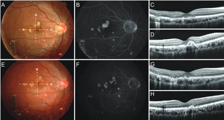
A representative case is included in Fig. 1. A 31-year-old female visited our clinic with the complaint of decreased vi- sual acuity in her right eye. On ophthalmic examination, best corrected visual acuity was 20/40 in the right eye and 20/25 in the left eye with a -1.50 spherical equivalent bilaterally.
Anterior examination revealed no evidence of inflammation.
However, dilated fundus examination revealed vitreous cells
with multifocal chorioretinal lesion in the posterior pole of
the right eye. There was juxtafoveal yellow gray membrane
superior to fovea. Following FA and OCT examination, we confirmed the diagnosis of CNV associated with MFC (Fig.
1A-D).
The patient was treated with prednisolone and cyclospo- rine for control of evident inflammation and received two courses of intravitreal bevacizumab injections for the CNV lesion. BCVA of the right eye improved to 20/25 and was maintained during 30 months of follow-up. FA and OCT examination revealed decrease CNV activity (Fig. 1E-H).
There was no adverse event associated with intravitreal bev- acizumab injection.
Discussion
In this case series, choroidal neovascularization (CNV) as- sociated with multifocal choroiditis (MFC) was successfully treated with intravitreal bevacizumab injections. BCVA improved to 20/40 or better in 5 of 6 eyes (83%) without adverse events. In addition, the decrease of central subfield thickness (CST) was observed in all eyes. After regression, the CNV lesion did not recur and was maintained through long-term follow-up.
Because CNV associated with MFC has been the most common cause of severe visual loss [1-11], several treatment approaches have been tried. Conventional treatment such as corticosteroid and immunosuppressant agents might be beneficial in controlling inflammation, but their safety and effectiveness in CNV continue to be debated [8-10]. Surgery for CNV may provide positive results in some selected cases but requires considerable surgical skill and has high rates of recurrence and complications [11]. Photodynamic therapy has had several positive reports, but this intervention merely stabilizes the visual function and is unable to guarantee sig- nificant improvement of visual acuity [12].
Anti-VEGF agents are a newly emerged treatment option for CNV associated with MFC. This treatment is based on the concept that vascular endothelial growth factors are im- portant in the development of CNV. Shimada et al. [23] re- ported that there was overexpression of vascular endothelial growth factor in samples of active CNV from patients with MFC. Fine et al. [21] reported a case series of 6 eyes and concluded that bevacizumab and ranibizumab were effective at improving visual acuity over 6 months in patients with CNV associated with MFC. Parodi et al. [20] reported intra- Ta bl e 1 . P at ie nt ch ar ac te ris tic s a nd cl in ic al o ut com es o f m ul tif oc al chor oid iti s w ith chor oid al ne ova sc ul ar iz at ion Pa tie nt Se x Ag e La te ral ity Sp he ric al equi va le nt (d io pt er s)
Dur at io n of fo llo w u p (m on th s) Ev ide nt infl amm ati on Im m uno su pp re ssion ag ent Lo ca tio n o f CN V Nu m be r o f in je cti on s BC VA CST Ad ve rs e eve nt
Du ra tio ns si nc e CN V in ac tiv ati on to fi na l v isi t (m on th s) In iti al vis it Fi nal vis it In iti al vis it Fi nal vis it 1 F 26 L - 3. 25 12 0 None None Su bf ov ea l 1 20/ 30 20 /20 270 22 9 None 12 2 F 64 R - 4. 00 92 Ye s Pr ed ni so lone Ju xt afo ve al 1 20 /5 0 20 /4 0 236 221 None 84 3 M 42 L - 0. 50 14 4 None None Su bf ov ea l 2 2/ 10 0 2/ 10 0 255 231 None 67 4 F 39 L - 0. 75 36 Ye s Pr ed ni so lone , az at hi op rin e Su bf ov ea l 2 20 /5 0 20/ 25 503 259 None 24 5 F 31 R -1 .5 0 39 Ye s Pr ed ni so lone , c yc lo sp or ine Ju xt afo ve al 2 20 /4 0 20/ 25 291 236 None 30 6 M 44 R -2 .5 0 17 8 No None Ju xt afo ve al 6 20/ 25 20/ 25 267 24 2 None 34 CN V = ch or oi dal n eo vas cul ar iza tio n; B CV A = b es t c or re ct ed v isu al a cu ity ; C ST = c en tral s ub fo ve al t hi ck ne ss .
vitreal bevacizumab injection was beneficial for juxtafoveal CNV associated with MFC. In our case series, visual acuity improved to 20/40 or better at final visit and CST decreased after intravitreal bevacizumab injections, which is consistent with previous reports. One eye with initial poor visual acuity continued to show poor final visual acuity with scar changes.
This patient already had atrophic changes at initial presenta- tion and these changes might have restricted improvement of visual acuity.
On long-term follow-up (178 months), the regressed CNV lesion did not recur in our case series. The duration from CNV regression to last follow-up (41.8 months) was more than 3 years. These results were consistent with previous studies that reported the recurrence of CNV associated with MFC was rare [6]. Parodi et al [20]. also reported that juxtafoveal CNV associated with MFC stabilized and did not recur after intrav- itreal bevacizumab injection within the first 6 months.
An inflammatory mechanism may play a role in CNV for-
mation. Patients with MFC are at risk for the development of CNV, possibly because of chronic peri-vascular, predominant- ly B-cell lymphocytic infiltration of the choroid, and/or dis- ruption of Bruch’s membrane [24]. In our case series, among 6 eyes with CNV, half had evidence of active inflammation and were successfully treated with anti-inflammatory agents.
In conclusion, intravitreal bevacizumab injection was ef- fective and safe for improving visual acuity and decreasing CST after long-term follow-up in a small series of patients with CNV associated with MFC. Further studies with a larg- er samples size and controls are required to evaluate the clin- ical efficacy of bevacizumab for the management of MFC.
Conflicts of interest There are no conflicts of interest.
A
E
B
F
C
G D
H
Figure 1. Representative case (Patient 5) of choroidal neovascularization (CNV) associated with multifocal choroiditis (MFC). Before intravitre-
al bevacizumab injection (A-D). (A) Color photograph of patient 5 showed multiple chorioretinal lesion in the posterior pole with juxtafoveal yellow gray membrane superior to fovea which is consistent with CNV secondary to MFC . (B) The late phase of fluorescein angiography (FA) of the same patient showed leakage superior to fovea consistent with CNV lesion. (C) The horizontal scan of optical coherence tomography (OCT) showed a photoreceptor layer defect corresponding to the chorioretinal lesions. (D) The vertical scan of OCT showed a hyperreflective area lo- cated above the retinal pigment epithelium (RPE) corresponding to the CNV. After intravitreal bevacizumab injection (E-H). (E) Color photograph showed decrease of juxtafoveal yellow gray membrane superior to fovea. (F) The late phase of FA showed decrease of leakage at the CNV lesion. (G) The horizontal scan of OCT showed progression of photoreceptor layer defect. (H) The vertical scan of OCT showed a decrease of hyperreflec- tive area located above the RPE after intravitreal bevacizumab injection.
References
1. Nozik RA, Dorsch W. A new chorioretinopathy associated with anterior uveitis. Am J Ophthalmol 1973;76:758-62.
2. Dreyer RF, Gass DJ. Multifocal choroiditis and panuveitis. A syn- drome that mimics ocular histoplasmosis. Arch Ophthalmol 1984;102:1776-84.
3. Deutsch TA, Tessler HH. Inflammatory pseudohistoplasmosis.
Ann Ophthalmol 1985;17:461-5.
4. Cantrill HL, Folk JC. Multifocal choroiditis associated with pro- gressive subretinal fibrosis. Am J Ophthalmol 1986;101:170-80.
5. Morgan CM, Schatz H. Recurrent multifocal choroiditis. Ophthal- mology 1986;93:1138-47.
6. Thorne JE, Wittenberg S, Jabs DA, et al. Multifocal choroiditis with panuveitis incidence of ocular complications and of loss of visual acuity. Ophthalmology 2006;113:2310-6.
7. Brown J Jr, Folk JC, Reddy CV, Kimura AE. Visual prognosis of multifocal choroiditis, punctate inner choroidopathy, and the diffuse subretinal fibrosis syndrome. Ophthalmology 1996;103:1100-5.
8. Michel SS, Ekong A, Baltatzis S, Foster CS. Multifocal choroiditis and panuveitis: immunomodulatory therapy. Ophthalmology 2002;109:378-83.
9. Flaxel CJ, Owens SL, Mulholland B, et al. The use of corticoste- roids for choroidal neovascularisation in young patients. Eye 1998;12:266-72.
10. Nussenblatt RB, Coleman H, Jirawuthiworavong G, et al. The treatment of multifocal choroiditis associated choroidal neovas- cularization with sirolimus (rapamycin). Acta Ophthalmol Scand 2007;85:230-1.
11. Olsen TW, Capone A Jr, Sternberg P Jr, et al. Subfoveal choroidal neovascularization in punctate inner choroidopathy. Surgi- cal management and pathologic findings. Ophthalmology 1996;103:2061-9.
12. Spaide RF, Freund KB, Slakter J, et al. Treatment of subfoveal cho- roidal neovascularization associated with multifocal choroiditis and panuveitis with photodynamic therapy. Retina 2002;22:545-9.
13. Pathengay A, Malhotra S, Das T. Pneumatic displacement of sub- retinal haemorrhage followed by transpupillary thermotherapy of choroidal neovascular membrane secondary to multifocal choroiditis. Eye (Lond) 2005;19:929-31.
14. Brindeau C, Glacet-Bernard A, Coscas F, et al. Surgical removal of subfoveal choroidal neovascularization: visual outcome and prognostic value of fluorescein angiography and optical coher- ence tomography. Eur J Ophthalmol 2001;11:287-95.
15. Fujii GY, Humayun MS, Pieramici DJ, et al. Initial experience of inferior limited macular translocation for subfoveal choroidal neovascularization resulting from causes other than age related macular degeneration. Am J Ophthalmol 2001;131:90-100.
16. Spaide RF, Laud K, Fine HF, et al. Intravitreal bevacizumab treat- ment of choroidal neovascularization secondary to age related macular degeneration. Retina 2006;26:383-90.
17. Sarao V, Veritti D, Macor S, Lanzetta P. Intravitreal bevacizumab for choroidal neovascularization due to pathologic myopia: long-term outcomes. Graefes Arch Clin Exp Ophthalmol 2016;254:445-54.
18. Ho M, Liu DT, Lam DS, Jonas JB. Retinal vein occlusions, from basics to the latest treatment. Retina 2016;36:432-48.
19. Dhingra N, Kelly S, Majid MA, et al. Inflammatory choroidal neovascular membrane in posterior uveitis-pathogenesis and treatment. Indian J Ophthalmol 2010;58:3-10.
20. Parodi MB, Iacono P, Mansour A, et al. Intravitreal bevacizumab for juxtafoveal choroidal neovascularization secondary to multi- focal choroiditis. Retina 2013;33:953-6.
21. Fine HF, Zhitomirsky I, Freund KB, et al. Bevacizumab (avastin) and ranibizumab (lucentis) for choroidal neovascularization in multifocal choroiditis. Retina 2009;29:8-12.
22. Dreyer RF, Gass DJ. Multifocal choroiditis and panuveitis. A syn- drome that mimics ocular histoplasmosis. Arch Ophthalmol 1984;102:1776-84.
23. Shimada H, Yuzawa M, Hirose T, et al. Pathological findings of multifocal choroiditis with panuveitis and punctuate inner cho- roidopathy. Jpn J Ophthalmol 2008;52:282-8.
24. Dunlop AA, Cree IA, Hague S, et al. Multifocal choroiditis: clinico- pathologic correlation. Arch Ophthalmol 1998;116:801-3.