Serum Vascular Endothelial Growth Factor and Angiopoietin-2 Are Associated with the Severity of Systemic Inflammation
Rather than the Presence of Hemoptysis in Patients with Inflammatory Lung Disease
Hye Yun Park, Cho Rom Hahm, Kyeongman Jeon, Won-Jung Koh, Gee Young Suh, Man Pyo Chung, Hojoong Kim, O Jung Kwon, and Sang-Won Um
Division of Pulmonary and Critical Care Medicine, Department of Medicine, Samsung Medical Center, Sungkyunkwan University School of Medicine, Seoul, Korea.
Received: April 25, 2011 Revised: June 3, 2011 Accepted: June 9, 2011
Corresponding author: Dr. Sang-Won Um, Division of Pulmonary and Critical Care Medicine, Department of Medicine, Samsung Medical Center, Sungkyunkwan University School of Medicine,
50 Irwon-Dong, Gangnam-gu, Seoul 135-710, Korea.
Tel: 82-2-3410-3429, Fax: 82-2-3410-6956 E-mail: [email protected]
∙ The authors have no financial conflicts of interest.
© Copyright:
Yonsei University College of Medicine 2012 This is an Open Access article distributed under the terms of the Creative Commons Attribution Non- Commercial License (http://creativecommons.org/
licenses/by-nc/3.0) which permits unrestricted non- commercial use, distribution, and reproduction in any medium, provided the original work is properly cited.
Purpose: Vascular endothelial growth factor (VEGF) and angiopoietin-2 (Ang-2) are major mediators of angiogenesis and are induced by tissue inflammation and hypoxia. The purpose of this study was to investigate whether serum VEGF and Ang-2 are associated with the presence of hemoptysis and the extent of systemic inflammation in patients with inflammatory lung diseases. Materials and Meth- ods: We prospectively enrolled 52 patients with inflammatory lung disease be- tween June 2008 and October 2009. Results: The median values of VEGF and Ang-2 were 436 pg/mL and 2383 pg/mL, respectively. There was a significant positive correlation between serum Ang-2 and VEGF levels. VEGF levels were not significantly different according to the presence of hemoptysis. C-reactive pro- tein (CRP) and Ang-2 level were significantly higher in patients without hemopty- sis (n=26) than in those with hemoptysis (n=26; p<0.001 and p<0.001, respective- ly). CRP and arterial oxygen tension (PaO2) were significantly correlated with both serum VEGF (p=0.032 and p=0.016, respectively) and Ang-2 levels (p<0.001 and p=0.041, respectively), after adjusting for other factors. Age and the absence of he- moptysis were factors correlated with serum Ang-2 levels. Conclusion: Our study suggests that serum VEGF and Ang-2 levels are associated with PaO2 and the se- verity of inflammation rather than the presence of hemoptysis in patients with in- flammatory lung diseases. Thus, hemoptysis may not be mediated by increased se- rum levels of VEGF and Ang-2 in patients with inflammatory lung diseases, and further studies are required to determine the mechanisms of hemoptysis.
Key Words: Angiogenesis, angiopoietin-2, hemoptysis, inflammation, vascular endothelial growth factor
INTRODUCTION
Angiogenesis is characterized by the formation of new microvessels from preex-
Patients with hemoptysis (hemoptysis group) were enrolled if they had hemoptysis within 2 weeks before study enroll- ment, and the patients without hemoptysis (no hemoptysis group) were enrolled if they did not have any hemoptysis more than 2 years before study enrollment. All study pa- tients except one were admitted to general ward or emer- gency department for the management of symptomatic be- nign lung diseases. We excluded patients with neoplastic conditions, including lung cancer, vasculitis, and lung dis- ease associated with collagen vascular disease. Serum sam- ples for VEGF and Ang-2, clinical data, such as age, gen- der, smoking status, clinical symptoms, and laboratory data, including peripheral white blood cell (WBC) count, hemo- globin (Hb), prothrombin time (PT), C-reactive protein (CRP), and arterial oxygen tension (PaO2), were obtained at study enrollment.
Determination of sample size
The required sample size was calculated based on VEGF levels according to the presence of hemoptysis in patients with asperilloma.6 Assuming 437±164 pg/mL of VEGF in the hemoptysis group and 162±102 pg/mL in the no he- moptysis group, a sample size of 42 patients (21 per group) was required to detect a 120 pg/mL difference between the two groups with a power of 90% and α error of 0.05 using a two-tailed test.17 Anticipating a potential 20% drop-out rate, we planned to include 52 subjects (26 per group).
Measurement of VEGF and Ang-2
Serum samples from each individual were obtained at the time of study enrollment. Sera were stored at -80°C. Serum VEGF and Ang-2 concentrations were measured in tripli- cate for each sample using a commercial enzyme-linked immunosorbent assay (ELISA) kit (R&D Systems, Minne- apolis, MN, USA). The detection ranges of ELISA kits for VEGF and Ang-2 were 31.2-2000 pg/mL and 46.9-3000 pg/mL, respectively. Therefore, serum was diluted to mea- sure serum Ang-2 levels.
Statistical analysis
Data presented are expressed as either the median and in- terquartile range (IQR, 25th and 75th percentiles) or the number (percentage) of patients. For univariate analysis or relation with hemoptysis, categorical variables were ana- lyzed using a Pearson χ2 test or Fisher’s exact test; continu- ous variables were analyzed using a Mann-Whitney U test.
VEGF and Ang-2 levels did not fit normality assumptions;
isting vasculature and is associated with numerous inflam- matory conditions, such as atherosclerosis, arthritis, reti- nopathy, and tumor growth.1 Previous studies provided evidence that inflammation exists in a mutually dependent association with angiogenesis.2,3
Among the numerous angiogenic factors, vascular endo- thelial growth factor (VEGF) is the most extensively stud- ied and is significantly related to the severity of inflamma- tory lung diseases, such as active tuberculosis, chronic bronchitis, pulmonary aspergilloma, and pulmonary disease caused by cystic fibrosis.4-7 Overexpression of VEGF is also significantly correlated with lung cancer progression and metastasis. Antiangiogenic agents have received wide- spread attention as targets for lung cancer therapy.8,9 Recent studies reported that VEGF elevation correlates significant- ly with the presence of hemoptysis in patients with pulmo- nary aspergilloma.6 Another major factor involved in angio- genesis, angiopoietin-2 (Ang-2), is also induced by hypoxemia10 and plays an important role in initiating vessel sprouting in concert with VEGF.11,12 In contrast with angio- poietin-1, which stabilizes blood vessels, Ang-2 destabiliz- es blood vessels, initiating angiogenic changes instead of regression, and promotes the neovascularization of tumor cells.13-16
The purpose of this study was to investigate whether se- rum VEGF and Ang-2 were associated with the presence of hemoptysis and the level of systemic inflammation in pa- tients with inflammatory lung diseases.
MATERIALS AND METHODS
Study population
The study was approved by the Institutional Review Board of Samsung Medical Center (a 2600-bed University affiliat- ed hospital in Seoul, Korea) and was registered with www.
clinicaltrials.gov (NCT01171768). Informed written con- sent was obtained from all participants.
Patients presented to the Samsung Medical Center for the treatment of benign inflammatory lung diseases, including bronchiectasis, aspergilloma, pneumonia, and post-tubercu- losis destroyed lung, between June 2008 and December 2009. Diagnosis of post-tuberculosis destroyed lung (inactive tuberculosis) was considered when chest computed tomogra- phy (CT) scan showed the findings compatible with fibrotic sequelae of old tuberculosis and all mycobacterial examina- tions of sputum or bronchial washing fluid were negative.
Correlation between serum VEGF or angiopoietin-2 and other parameters
The median VEGF levels were 375 pg/mL in bronchiecta- sis, 472 pg/mL in aspergilloma, 554 pg/mL in post-tubercu- losis destroyed lung, and 451 pg/mL in pneumonia. The median Ang-2 levels were 2444 pg/mL in bronchiectasis, 1689 pg/mL in aspergilloma, 3021 pg/mL in post-tubercu- losis destroyed lung, and 4344 pg/mL in pneumonia.
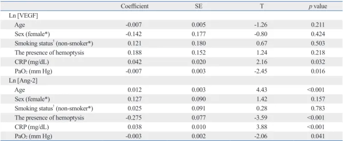
Serum Ang-2 levels were significantly correlated with se- rum VEGF levels (p=0.028) (Fig. 1). Serum VEGF levels demonstrated a significant positive correlation with WBC and a negative correlation with PaO2 levels (Table 3). Age, gender, smoking status, presence of hemoptysis, and CRP levels showed no significant correlation with VEGF levels thus, log-transformed VEGF and Ang-2 (ln VEGF and ln
Ang-2) values were used in analyses. As the VEGF and Ang-2 levels were measured in triplicate from single pa- tient samples, a mixed model was used to analyze associa- tions between parameters. Additionally, to take random ef- fects between subjects into account, we used the ‘random statement’ function in PROC MIXED. All p values are two- sided with p<0.05 considered to indicate statistical signifi- cance. Statistical analyses were performed using the SAS software (ver. 9.1; SAS Institute, Cary, NC, USA).
RESULTS
Baseline clinical and laboratory features
Characteristics of the enrolled patients are listed in Table 1.
In this study, 52 study patients underwent extensive evalua- tions for underlying disease including chest CT scan (n=
46), bronchoscopy (n=21), and sputum bacterial and myco- bacterial examinations (n=44). There were 25 men and 27 women, with a median age of 58 years (range, 47-66). Of the 52 patients, 14 patients (27%) had a history of smoking, and 22 (42%) had a history of tuberculosis treatment. The most common disease in enrolled patients was bronchiecta- sis (62%); 14% had an aspergilloma, and 14% had post-tu- berculosis destroyed lung. Median VEGF and Ang-2 levels were 436 pg/mL (257-724) and 2383 pg/mL (1807-3209), respectively. In total, 5 patients received oxygen therapy (1-2 L/min) on arterial blood gas analysis.
Comparison of clinical laboratory features depending on the presence of hemoptysis
In patients with hemoptysis (n=26), bronchiectasis (54%), aspergilloma (27%), and post-tuberculosis destroyed lung (19%) were observed. In those without hemoptysis (n=26), bronchiectasis (69%), post-tuberculosis destroyed lung (8%), pneumonia (19%), and pulmonary tuberculosis (4%) were observed. Patients with hemoptysis had more signifi- cant history of tuberculosis treatment compared with those without hemoptysis. However, the median CRP and Ang-2 levels were significantly higher in patients without hemop- tysis than in those with hemoptysis (CRP; 0.34 vs. 3.29 mg/dL; p<0.001, and Ang-2; 2017 vs. 2946 pg/mL;
p<0.001). There was no significant difference in age, gen- der, smoking history, presenting symptoms, or laboratory findings (including WBC, Hb, PT, PaO2, serum VEGF lev- els) (Table 2).
Table 1. Baseline Characteristics of Enrolled Patients (n=52) Median (IQR) or No. (%)
Age 58 (47-66)
Sex
Female 25 (48%)
Male 27 (52%)
Smoking status
Current smoker 2 (4%)
Ex-smoker 12 (23%)
Non-smoker 38 (73%)
Comorbidities
Hypertension 8 (15%)
Diabetes mellitus 2 (4%) Prior therapy for tuberculosis 22 (42%) Lung Diseases
Bronchiectasis 32 (62%)
Aspergilloma 7 (14%)
Post-tuberculosis destroyed lung 7 (14%) Pneumonia without bronchiectasis 5 (10%) Pulmonary tuberculosis 1 (2%) Presenting symptoms
Cough 36 (69%)
Dyspnea 15 (29%)
Fever 7 (13%)
Laboratory findings
WBC (#/μL) 8035 (6257-10533)
Hemoglobin (g/dL) 12.5 (11.3-14.0)
PT (INR) 1.05 (0.99-1.13)
CRP (mg/dL) 1.13 (0.24-4.13)
PaO2 (mm Hg) 81.7 (66.1-90.5)
VEGF (pg/mL) 436 (257-724)
Ang-2 (pg/mL) 2383 (1807-3209)
WBC, white blood cell; PT, prothrombin time; CRP, C-reactive protein;
VEGF, vascular endothelial growth factor; Ang-2, angiopoietin-2; IQR, in- terquartile range; INR, international normalized ratio; PaO2, arterial oxygen tension.
of hemoptysis were significantly correlated with serum Ang-2 levels (Table 4).
DISCUSSION
In the present study, serum VEGF levels did not differ ac- cording to the presence of hemoptysis. However, we dem- onstrated that serum VEGF and Ang-2 levels in patients with inflammatory lung diseases correlated significantly with elevations in serum CRP and reduction of PaO2. (Table 3). Ang-2 levels showed significantly positive corre-
lations with age, WBC, and CRP levels, while demonstrat- ing a negative correlation with PaO2 levels (Table 3).
Multivariate analysis using PROC MIXED for repeated measures data was performed to identify factors significant- ly correlated with serum VEGF or Ang-2 levels. CRP lev- els and PaO2 were found to be significant correlated with both serum VEGF (p=0.032 and p=0.016, respectively) and Ang-2 levels (p<0.001 and p=0.041, respectively) after ad- justing for other factors (age, gender, smoking history, and the presence of hemoptysis) (Table 4). Age and the absence
Table 2. Comparison of Clinical Laboratory Features according to the Presence of Hemoptysis
With hemoptysis (n=26) Without hemoptysis (n=26) p value
Age 56 (47-65) 60 (48-66) 0.826
Sex 0.405
Female 11 (42%) 14 (54%)
Male 15 (58%) 12 (46%)
Smoking status 0.252
Current smoker 2 (8%) 0
Ex-smoker 7 (27%) 5 (19%)
Non-smoker 17 (65%) 21 (81%)
Comorbidities
Hypertension 6 (23%) 2 (8%) 0.248
Diabetes mellitus 1 (4%) 1 (4%) 1.000
Prior therapy for tuberculosis 15 (58%) 7 (27%) 0.025
Lung diseases 0.005
Bronchiectasis 14 (54%) 18 (69%)
Aspergilloma 7 (27%) 0
Post-tuberculosis destroyed lung 5 (19%) 2 (8%)
Pneumonia 0 5 (19%)
Pulmonary tuberculosis 0 1 (4%)
Severity of diseases
No. of involved lobes 2 (1-3) 3 (2-4) 0.390
Presenting Symptoms
Cough 16 (62%) 20 (77%) 0.229
Dyspnea 6 (23%) 9 (35%) 0.358
Fever 2 (8%) 5 (19%) 0.419
Laboratory findings
WBC (#/μL) 7645 (6353-9695) 8285 (5973-10638) 0.742
Hemoglobin (g/dL) 13 (11-14) 12.4 (11.8-13.9) 0.615
Platelet (×103/μL) 216 (186-260) 250 (195 - 317) 0.359
PT (INR) 1.06 (1.01-1.14) 1.04 (0.98-1.12) 0.359
aPTT (sec) 36 (31-38) 38 (34-41) 0.039
CRP (mg/dL) 0.34 (0.13-2.09) 3.29 (0.60-7.19) 0.001
PaO2 (mm Hg) 83.5 (75.1-94.2) 74.7 (64.7-87.6) 0.142
VEGF* 389 (270-684) 449 (224-776) 0.934
Ang-2* 2017 (1633-2597) 2946 (2101-4698) <0.001
WBC, white blood cell; PT, prothrombin time; aPTT, activated partial thromboplastin time; CRP, C-reactive protein; VEGF, vascular endothelial growth factor;
Ang-2, angiopoietin-2; INR, international normalized ratio; PaO2, arterial oxygen tension.
*The analysis used a mixed model, accounting for the random effect between subjects (logarithmic transformed VEGF and Ang-2 values were used).
sults also demonstrated that VEGF levels were increased in inflammatory lung diseases, such as bronchiectasis, asper- gilloma, post-tuberculosis destroyed lung, and pneumonia, when compared with published normal values.20,21 Addi- tionally, elevated VEGF levels correlated with severity of inflammation. Significant change in the serum VEGF levels Angiogenesis, the growth of new capillary blood vessels
from pre-existing vasculature, is an important contributor to tissue inflammation and abnormal remodeling in inflamma- tory diseases. Consistent with previous data that levels of VEGF are significantly elevated in inflammatory disorders, including lung inflammatory disease,6,7,18,19 our present re-
0 500 1000 1500 2000
VEGF (pg/mL)
0 2000 4000 6000 8000 10000 0
500 1000 1500 2000
0 2000 4000 6000 8000 10000 0
500 1000 1500 2000
0 2000 4000 6000 8000 10000
Fig. 1. Correlation between serum VEGF and Ang-2 levels. VEGF, vascular endothelial growth factor; Ang-2, angiopoietin-2. p=0.028.
Table 3. Univariate Analysis of Associations between Serum VEGF or Angiopoietin-2 levels and Other Measured Parameters
Variables Ln [VEGF] Ln [Ang-2]
Coefficient SE p value Coefficient SE p value
Age -0.003 0.005 0.524 0.013 0.003 <0.001
Sex (female*) 0.114 0.128 0.375 0.144 0.081 0.077
Smoking status† (non-smoker*) 0.116 0.144 0.423 0.092 0.092 0.317
Comorbidities
Hypertension (No*) -0.159 0.177 0.370 -0.020 0.113 0.862
Diabetes mellitus (No*) -0.050 0.333 0.880 0.260 0.211 0.220
Prior therapy for tuberculosis (No*) 0.089 0.130 0.493 -0.154 0.082 0.061
Presenting symptoms
Cough (No*) 0.247 0.137 0.074 0.196 0.087 0.026
Dyspnea (No*) 0.355 0.139 0.011 0.286 0.087 0.001
Fever (No*) -0.199 0.187 0.288 0.030 0.120 0.801
Hemoptysis (No*) 0.011 0.128 0.934 -0.420 0.074 <0.001
Amount of hemoptysis (cc) -0.0001 0.0003 0.672 0.0003 0.0002 0.121
Laboratory findings
WBC (#/μL) 0.00006 0.00002 0.005 0.000064 0.00001 <0.001
Hemoglobin (g/dL) 0.016 0.017 0.358 -0.004 0.011 0.745
PT (INR) 1.085 0.733 0.141 1.0961 0.462 0.019
CRP (mg/dL) 0.022 0.02 0.160 0.059 0.009 <0.001
PaO2 (mm Hg) -0.006 0.003 0.055 -0.004 0.002 0.027
Ln, natural logarithm; WBC, white blood cell; PT, prothrombin time; CRP, C-reactive protein; VEGF, Vascular endothelial growth factor; Ang-2, angiopoietin-2;
SE, standard error; PaO2, arterial oxygen tension.
Analysis using a mixed model to account for the random effect between subjects (logarithmic transformed VEGF and Ang-2 values were used).
*Reference category.
†Smoking status: smokers (current smoker or ex-smoker) and non-smoker.
Ang-2 (pg/mL) Ang-2 (pg/mL) Ang-2 (pg/mL)
VEGF (pg/mL) VEGF (pg/mL)
ting of various cancers.15,28 To our best knowledge, this is the first reported study to evaluate Ang-2 levels in inflammato- ry lung diseases, including bronchiectasis, post-tuberculosis destroyed lung, and aspergilloma.
During development of inflammatory tissue, hypoxic conditions can promote angiogenesis, leading to induced VEGF and Ang-2 production. Expression of Ang-2 was el- evated in cultured endothelial cells exposed to hypoxia,29-31 while VEGF mRNA expression was increased.32,33 In the present study, PaO2 levels were correlated inversely with VEGF and Ang-2 levels; suggesting that angiogenesis me- diators may be released in response to low PaO2 in patients with inflammatory lung diseases.
Significant associations between VEGF and Ang-2 levels have been noted in earlier studies in patients with various tu- mors,28 diabetes mellitus,34 and asthma.35 The present study demonstrated that serum VEGF and Ang-2 levels had a sig- nificant positive correlation in inflammatory lung diseases.
However, in contrast to a previous study,6 our study did not find a significant association between the presence of hemoptysis and VEGF levels. This lack of an association may be explained by differences in the distribution of dis- ease between the hemoptysis and the no hemoptysis groups.
As shown in Table 2, the prevalence of bronchiectasis (54%), aspergilloma (27%), and post-tuberculosis destroyed lung (19%) in the hemoptysis group differed from the no hemop- tysis group where bronchiectasis (69%), pneumonia (19%), and post-tuberculosis destroyed lung (8%) were prevalent.
with the treatment of inflammatory lung diseases have been reported.4,6 To our best knowledge, however, our present study is the first to demonstrate that serum VEGF levels are positively correlated with serum CRP levels and WBCs. In a previous study on patients undergoing major surgery, there was no significant correlation between increases in se- rum VEGF levels and serum IL-6 or CRP levels.22
Recently, several clinical studies have shown that circu- lating levels of Ang-2 are increased in septic shock patients, as well as non-septic patients with, or at risk of, acute lung injury/acute respiratory distress syndrome; this is related to vascular permeability and pulmonary dysfunction.23-26 In these studies, serum Ang-2 levels were positively correlated with serum CRP levels. In the present study, Ang-2 levels were positively correlated with CRP levels and WBCs, both representing markers of inflammation. Compared with the levels of Ang-2 in previous studies,23,25,27 the median level of Ang-2 in septic shock patients was greater than 4000 pg/
mL; this is significantly higher than the levels seen in the current study (median Ang-2 level of 2383 pg/mL). How- ever, the levels of Ang-2 in the current study are similar to data measured in cancer patients, including lung, breast, and prostate,15,28 and were greater than the levels of Ang-2 in the healthy volunteers without lung disease (1000-1500 pg/mL).28 Our findings suggest that high Ang-2 levels are associated with the severity of inflammatory lung disease in patients without sepsis, and that the Ang-2 levels in inflam- matory diseases are similar to the levels reported in the set-
Table 4. Multivariate Correlations between Serum VEGF or Angiopoietin-2 Levels and Other Measured Parameters
Coefficient SE T p value
Ln [VEGF]
Age -0.007 0.005 -1.26 0.211
Sex (female*) -0.142 0.177 -0.80 0.424
Smoking status† (non-smoker*) 0.121 0.180 0.67 0.503
The presence of hemoptysis 0.188 0.152 1.24 0.218
CRP (mg/dL) 0.042 0.020 2.16 0.032
PaO2 (mm Hg) -0.007 0.003 -2.45 0.016
Ln [Ang-2]
Age 0.012 0.003 4.43 <0.001
Sex (female*) 0.127 0.090 1.42 0.157
Smoking status† (non-smoker*) 0.025 0.091 0.28 0.783
The presence of hemoptysis -0.275 0.077 -3.59 <0.001
CRP (mg/dL) 0.038 0.010 3.88 <0.001
PaO2 (mm Hg) -0.003 0.002 -2.06 0.041
Ln, natural logarithm; T, t statistic value; VEGF, vascular endothelial growth factor; Ang-2, angiopoietin-2; CRP, C-reactive protein; SE, standard error; PaO2, arterial oxygen tension.
Analysis using a mixed model to account for the random effect between subjects (logarithmic transformed VEGF and Ang-2 values were used).
*Reference category.
†Smoking status: smokers (current smoker or ex-smoker) and non-smoker.
This study was supported by the Samsung Medical Cen- ter Clinical Research Development Program grant, #CRS- 109-10-1.
REFERENCES
1. Folkman J. Angiogenesis in cancer, vascular, rheumatoid and oth- er disease. Nat Med 1995;1:27-31.
2. Jackson JR, Seed MP, Kircher CH, Willoughby DA, Winkler JD.
The codependence of angiogenesis and chronic inflammation.
FASEB J 1997;11:457-65.
3. Dvorak HF, Brown LF, Detmar M, Dvorak AM. Vascular permea- bility factor/vascular endothelial growth factor, microvascular hy- perpermeability, and angiogenesis. Am J Pathol 1995;146:1029- 4. Matsuyama W, Hashiguchi T, Matsumuro K, Iwami F, Hirotsu Y, 39.
Kawabata M, et al. Increased serum level of vascular endothelial growth factor in pulmonary tuberculosis. Am J Respir Crit Care Med 2000;162:1120-2.
5. Kanazawa H. Role of vascular endothelial growth factor in the pathogenesis of chronic obstructive pulmonary disease. Med Sci Monit 2007;13:RA189-95.
6. Inoue K, Matsuyama W, Hashiguchi T, Wakimoto J, Hirotsu Y, Kawabata M, et al. Expression of vascular endothelial growth fac- tor in pulmonary aspergilloma. Intern Med 2001;40:1195-9.
7. McColley SA, Stellmach V, Boas SR, Jain M, Crawford SE. Se- rum vascular endothelial growth factor is elevated in cystic fibro- sis and decreases with treatment of acute pulmonary exacerbation.
Am J Respir Crit Care Med 2000;161:1877-80.
8. Donovan EA, Kummar S. Targeting VEGF in cancer therapy.
Curr Probl Cancer 2006;30:7-32.
9. Mattern J, Koomägi R, Volm M. Association of vascular endothe- lial growth factor expression with intratumoral microvessel densi- ty and tumour cell proliferation in human epidermoid lung carci- noma. Br J Cancer 1996;73:931-4.
10. Nilsson I, Shibuya M, Wennström S. Differential activation of vascular genes by hypoxia in primary endothelial cells. Exp Cell Res 2004;299:476-85.
11. Tsigkos S, Koutsilieris M, Papapetropoulos A. Angiopoietins in angiogenesis and beyond. Expert Opin Investig Drugs 2003;12:
933-41.
12. Hashizume H, Falcón BL, Kuroda T, Baluk P, Coxon A, Yu D, et al. Complementary actions of inhibitors of angiopoietin-2 and VEGF on tumor angiogenesis and growth. Cancer Res 2010;
70:2213-23.
13. Pouysségur J, Dayan F, Mazure NM. Hypoxia signalling in cancer and approaches to enforce tumour regression. Nature 2006;441:
437-43.
14. Boussat S, Eddahibi S, Coste A, Fataccioli V, Gouge M, Housset B, et al. Expression and regulation of vascular endothelial growth factor in human pulmonary epithelial cells. Am J Physiol Lung Cell Mol Physiol 2000;279:L371-8.
15. Park JH, Park KJ, Kim YS, Sheen SS, Lee KS, Lee HN, et al. Se- rum angiopoietin-2 as a clinical marker for lung cancer. Chest 2007;132:200-6.
16. Jain RK. Molecular regulation of vessel maturation. Nat Med 2003;9:685-93.
When the VEGF levels were compared between patients with aspergilloma, which were present only in the hemop- tysis group, and those with pneumonia, which were present only in the non-hemoptysis group, no significant difference in VEGF levels was observed. Additionally, when compari- son was restricted to patients with bronchiectasis from both groups, VEGF levels were not significantly correlated with the presence of hemoptysis. The lack of a significant differ- ence in VEGF levels between the hemoptysis and no he- moptysis groups can be partially explained by the different disease distribution in the two groups. In previous reports of associations between VEGF levels and hemoptysis in 21 pulmonary aspergilloma patients,6 the mean serum VEGF levels in 6 patients with hemoptysis and 15 patients without hemoptysis were 437 pg/mL and 162 pg/mL, respectively.
The reported serum VEGF levels were similar to the asper- gilloma patients with hemoptysis in the current study.
The present study has several other limitations. The mea- surements of VEGF and Ang-2 levels were made with se- rum, not lung tissue; it is unclear whether serum VEGF or Ang-2 levels reflect the levels in lung tissue. Also, because the present study was conducted with multiple inflammato- ry lung diseases, the sample size might have influenced the statistical power; this limits the interpretation of the study despite the fact that a formal sample size calculation was performed. Finally, the timing of measurement of serum angiogenesis markers in this study was not uniform in rela- tion to stages of lung injury. Since the level of serum angio- genesis markers could vary in different stages of lung inju- ry, our data should be interpreted conservatively. A larger- scale study in patients with bronchiectasis, aspergilloma, and pneumonia is needed to further clarify the association between hemoptysis and VEGF levels.
In conclusion, serum VEGF and Ang-2 levels were asso- ciated with the PaO2 and the severity of inflammation, rather than the presence of hemoptysis in patients with inflamma- tory lung diseases. Hemoptysis in patients with inflammato- ry lung diseases may not be mediated by increased serum levels of VEGF and Ang-2. Further studies are required to determine the mechanism(s) of hemoptysis in patients with inflammatory lung disease.
ACKNOWLEDGEMENTS
This research was supported by the IN-SUNG Foundation for Medical Research (CA98761).
informative prognostic biomarkers of morbidity and mortality in severe sepsis. Crit Care Med 2011;39:702-10.
27. Orfanos SE, Kotanidou A, Glynos C, Athanasiou C, Tsigkos S, Dimopoulou I, et al. Angiopoietin-2 is increased in severe sepsis:
correlation with inflammatory mediators. Crit Care Med 2007;35:
199-206.
28. Caine GJ, Blann AD, Stonelake PS, Ryan P, Lip GY. Plasma an- giopoietin-1, angiopoietin-2 and Tie-2 in breast and prostate can- cer: a comparison with VEGF and Flt-1. Eur J Clin Invest 2003;
33:883-90.
29. Pugh CW, Ratcliffe PJ. Regulation of angiogenesis by hypoxia:
role of the HIF system. Nat Med 2003;9:677-84.
30. Mandriota SJ, Pyke C, Di Sanza C, Quinodoz P, Pittet B, Pepper MS. Hypoxia-inducible angiopoietin-2 expression is mimicked by iodonium compounds and occurs in the rat brain and skin in re- sponse to systemic hypoxia and tissue ischemia. Am J Pathol 2000;156:2077-89.
31. Pichiule P, Chavez JC, LaManna JC. Hypoxic regulation of angio- poietin-2 expression in endothelial cells. J Biol Chem 2004;279:
12171-80.
32. Bruick RK, McKnight SL. Building better vasculature. Genes Dev 2001;15:2497-502.
33. Semenza GL. Regulation of hypoxia-induced angiogenesis: a chaperone escorts VEGF to the dance. J Clin Invest 2001;108:39- 34. Lim HS, Lip GY, Blann AD. Angiopoietin-1 and angiopoietin-2 in 40.
diabetes mellitus: relationship to VEGF, glycaemic control, endo- thelial damage/dysfunction and atherosclerosis. Atherosclerosis 2005;180:113-8.
35. Kanazawa H, Nomura S, Asai K. Roles of angiopoietin-1 and an- giopoietin-2 on airway microvascular permeability in asthmatic patients. Chest 2007;131:1035-41.
17. Hayes RJ, Bennett S. Simple sample size calculation for cluster- randomized trials. Int J Epidemiol 1999;28:319-26.
18. Kraft A, Weindel K, Ochs A, Marth C, Zmija J, Schumacher P, et al. Vascular endothelial growth factor in the sera and effusions of patients with malignant and nonmalignant disease. Cancer 1999;85:178-87.
19. Alatas F, Alatas O, Metintas M, Ozarslan A, Erginel S, Yildirim H.
Vascular endothelial growth factor levels in active pulmonary tu- berculosis. Chest 2004;125:2156-9.
20. Hyodo I, Doi T, Endo H, Hosokawa Y, Nishikawa Y, Tanimizu M, et al. Clinical significance of plasma vascular endothelial growth factor in gastrointestinal cancer. Eur J Cancer 1998;34:2041-5.
21. Kikuchi K, Kubo M, Kadono T, Yazawa N, IHN H, Tamaki K.
Serum concentrations of vascular endothelial growth factor in col- lagen diseases. Br J Dermatol 1998;139:1049-51.
22. Futami R, Miyashita M, Nomura T, Makino H, Matsutani T, Sasa- jima K, et al. Increased serum vascular endothelial growth factor following major surgical injury. J Nihon Med Sch 2007;74:223-9.
23. Siner JM, Bhandari V, Engle KM, Elias JA, Siegel MD. Elevated serum angiopoietin 2 levels are associated with increased mortali- ty in sepsis. Shock 2009;31:348-53.
24. van der Heijden M, Pickkers P, van Nieuw Amerongen GP, van Hinsbergh VW, Bouw MP, van der Hoeven JG, et al. Circulating angiopoietin-2 levels in the course of septic shock: relation with fluid balance, pulmonary dysfunction and mortality. Intensive Care Med 2009;35:1567-74.
25. van der Heijden M, van Nieuw Amerongen GP, Koolwijk P, van Hinsbergh VW, Groeneveld AB. Angiopoietin-2, permeability oe- dema, occurrence and severity of ALI/ARDS in septic and non- septic critically ill patients. Thorax 2008;63:903-9.
26. Ricciuto DR, dos Santos CC, Hawkes M, Toltl LJ, Conroy AL, Rajwans N, et al. Angiopoietin-1 and angiopoietin-2 as clinically