INTRODUCTION
Community-acquired pneumonia (CAP) is one of the most common respiratory diseases in children and it represents a frequent cause of hospital admission. Streptococcus pneumoniae is the leading cause of CAP in children aged 3 weeks to 5 yr, and it is the major pathogen leading to complicated pneumo- nia such as parapneumonic effusion and empyema (1). Pleu- ral effusion occasionally develops in children with commu- nity-acquired bacterial pneumonia and this can be diagnosed in about 40-50% of patients with bacterial pneumonia (2).
S. pneumoniae is the leading etiologic agent associated with parapneumonic effusions in the children for whom an etio- logic agent was recovered. Despite of its prevalence, there is only limited consensus about its pathogenesis due to the lack of available evidence-based data. Increased vascular perme- ability and leakage may play an important role in the devel- opment of exudative pleural effusions.
Vascular endothelial growth factor (VEGF) is a dimeric 46-kDa protein, and it is an endothelial, cell-specific, multi- functional cytokine that plays an important role in angiogen- esis and vascular permeability (3-5). VEGF has been postu-
lated to be an important mediator in the formation of malig- nant pleural and peritoneal fluid (6). VEGF levels are also known to be elevated in chronic pulmonary diseases such as asthma and cystic fibrosis (7-9). It has recently been report- ed that serum VEGF levels are significantly increased in the patients with active pulmonary tuberculosis and these levels are decreased after successful treatment (10). However in regard to pneumonia, only a few cases have been reported on so far (11, 12), and there has been no data about CAP as classified on the basis of the radiologic type and etiology. Therefore, we investigated the serum levels of VEGF in pediatric patients with CAP according to its radiologic type and etiology to see if the serum VEGF levels are related to the pathogenesis of severe, complicated pneumonia.
MATERIALS AND METHODS Study groups
From 1 May 2003 to 30 June 2004, 29 children with CAP (11 boys and 18 girls aged from 4 to 168 months; mean age:
Seong Hwan Choi, Eun Young Park, Hye Lim Jung, Jae Won Shim, Deok Soo Kim, Moon Soo Park, Jung Yeon Shim
Department of Pediatrics, Kangbuk Samsung Hospital, Sungkyunkwan University School of Medicine, Seoul, Korea
Address for correspondence Jung Yeon Shim, M.D.
Division of Pediatric Allergy & Pulmonology, Department of Pediatrics, Kangbuk Samsung Hospital, Sungkyunkwan University School of Medicine, 108 Peong-dong, Jongro-gu, Seoul 110-746, Korea Tel : +82.2-2001-2207, Fax : +82.2-2001-2199 E-mail : [email protected]
608
Serum Vascular Endothelial Growth Factor in Pediatric Patients with Community-Acquired Pneumonia and Pleural Effusion
This study investigated the serum vascular endothelial growth factor (VEGF) levels in children with community-acquired pneumonia. Serum VEGF levels were mea- sured in patients with pneumonia (n=29) and in control subjects (n=27) by a sand- wich enzyme-linked immunosorbent assay. The pneumonia group was classified into bronchopneumonia with pleural effusion (n=1), bronchopneumonia without pleural effusion (n=15), lobar pneumonia with pleural effusion (n=4), and lobar pneumonia without pleural effusion (n=9) groups based on the findings of chest radiographs. We also measured serum IL-6 levels and the other acute inflamma- tory parameters. Serum levels of VEGF in children with pneumonia were signifi- cantly higher than those in control subjects (p<0.01). Children with lobar pneumo- nia with or without effusion showed significantly higher levels of serum VEGF than children with bronchopneumonia. For lobar pneumonia, children with pleural effu- sion showed higher levels of VEGF than those without pleural effusion. Children with a positive urinary S. pneumonia antigen test also showed higher levels of VEGF than those with a negative result. Serum IL-6 levels did not show significant differ- ences between children with pneumonia and control subjects. Serum levels of VEGF showed a positive correlation with the erythrocyte sedimentation rate in the children with pneumonia. In conclusion, VEGF may be one of the key mediators that lead to lobar pneumonia and parapneumonic effusion.
Key Words : Vascular Endothelial Growth Factor A; Interleukin-6; Pleural Effusion; Community-Acquired Infec- tions; Pneumonia
Received : 6 April 2005 Accepted : 16 December 2005
52 months) and 27 afebrile healthy children were prospecti- vely recruited for this study. The children with CAP had acute respiratory symptoms with fever (temperature ≥38.0℃) and new infiltrates on their chest radiographs. Patients were ex- cluded from the study if any of the following criteria were found: the presence of malignancy, immunodeficiency or con- gestive heart disease; the presence of an alternative diagnosis during the follow-up; the children had been hospitalized in the preceding 72 hr.
Parapneumonic effusion was evaluated with chest radio- graphs and the patient was excluded from the study if the cause of the patient’s illness was identified as other than pneu- monia, or if the pleural fluid was transudate. The patients were classified into bronchopneumonia with pleural effusion (n=1), bronchopneumonia without pleural effusion (n=15), lobar pneumonia (focal consolidation was considered as lobar pneumonia) with pleural effusion (n=4), and lobar pneumo- nia without pleural effusion (n=9). Twenty seven healthy children who visited hospital for routine checks and had no respiratory symptoms were enrolled as the control group (13 boys and 14 girls; age range: 80-132 months; mean age: 119 months). The study design was approved by the ethics com- mittee of the hospital and an informed consent was obtained from all the parents.
Microbiological investigations
To identify the causative organisms, we performed blood and/or pleural fluid culture, rapid urinary Streptococcal pneu- moniae antigen test and detection of antibody to Mycoplasma pneumoniae. Pleural fluid was obtained by thoracentesis from the patients with parapneumonic effusion. Pleural fluid was investigated for biochemistry, the leukocyte count, Gram’s stain and culture for aerobic and anaerobic bacteria. Exudate was defined by the ratio of the protein in pleural fluid/serum
>0.5 and the ratio of LDH in the pleural fluid/serum >0.6.
We did not perform culture, polymerase chain reaction or serologic investigations for virus.
The urine samples were analysed for the presence of S. pneu- moniae cell-wall antigen with using the rapid urine S. pneumo- niae antigen assay kit (Binax NOW�, Portland, ME, U.S.A.).
This test device contains an immunochromatographic mem- brane that is used to detect soluble pneumococcal antigen in human urine. Blood samples were drawn on days 1 and 14 of treatment for the detection of antibodies to Mycoplas- ma pneumoniae, and the infection was defined as mycoplasma pneumonia when a fourfold increase of the antibody titer was detected during the convalescent period.
Measurement of VEGF and IL-6
On the first day of admission, venous blood was drawn and centrifuged at 300×g for 10 min at 4℃. The samples were stored at -70℃until the measurements of VEGF and IL-6
were performed with using enzyme linked immunosorbent assay kits (R&D Systems, Minneapolis, MN, U.S.A.).
In brief, 200 L of the cell supernatant was incubated with 50 L of assay diluent for 2 hr at room temperature in a 96- well plate coated with a monoclonal antibody against VEGF or IL-6. After three washes, a conjugate that consisted of a polyclonal VEGF or IL-6 antibody and horseradish peroxidase was then added, and this was allowed to incubate for 2 hr at room temperature. After addition of a color reagent, the absor- bance was measured at a wavelength of 450 nm in a Ther- mo-Max microplate reader. For standardization, serial dilu- tions of recombinant human VEGF or IL-6 were assayed at the same time. The detection limit was 3.12 pg/mL for IL-6 and 31.2 pg/mL for VEGF.
Tests for the complete blood cell count, the erythrocyte sed- imentation rate (ESR), and the C-reactive protein (CRP) lev- els were performed at the same time.
Statistical analysis
The results were expressed as means±standard error of the mean. All the statistics were performed by using the SPSS version 11.0 software program. Comparison of the VEGF and IL-6 concentrations between the study groups were analyzed by the Mann-Whitney rank sum test and p values <0.05 were considered as significant. The correlations were determined by Spearman rank correlation test.
RESULTS Patient characteristics
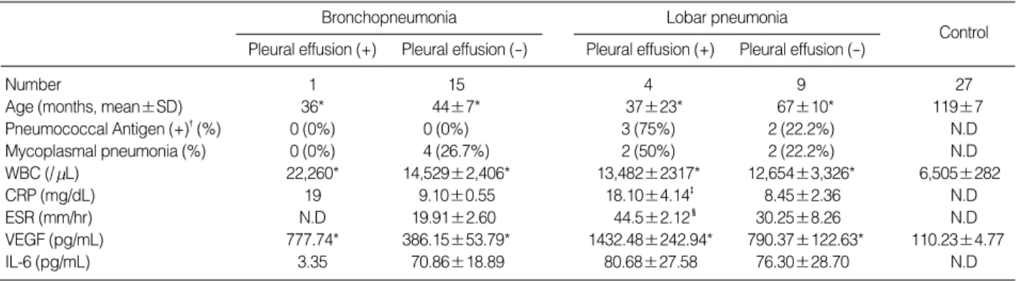
The characteristics of the enrolled children are presented in Table 1. Out of the 29 children with pneumonia, there were 16 children with bronchopneumonia (1 with parapneu- monic effusion) and 13 children with lobar pneumonia (4 with parapneumonic effusion). Thoracentesis was performed in 4 patients with parapneumonic effusion except for one patient with bronchopneumonia who had only a scanty amount of fluid. All the pleural fluids were confirmed as exudates.
The mean age of the control group was higher compared to the pneumonia groups. However, age did not influence the serum VEGF concentrations. Neither our previous study (13) nor this study showed any correlations between serum VEGF levels and the subjects’ age in control groups. The mean white blood cell counts in the patients with pneumo- nia were higher than those in the control subjects, but showed no differences among the pneumonia groups. The mean level of C-reactive protein was significantly increased in the patients having lobar pneumonia with effusion compared to the pati- ents having lobar pneumonia without effusion and the patients having bronchopneumonia with effusion. The mean erythro- cyte sedimentation rate was significantly increased in the
patients having lobar pneumonia with effusion compared to the other groups, and the ESR was also significantly increased in the patients having lobar pneumonia without effusion com- pared to the patients having bronchopneumonia with effusion.
The urinary Streptococcus pneumoniae antigen test was positive in 5 of 13 patients with lobar pneumonia, and this test was negative in all the patients with bronchopneumonia. Among those five patients with positive tests, three patients had para- pneumonic effusions. There were no culture-confirmed cases.
There were 8 patients with mycoplasma pneumonia. Four of them had bronchopneumonia and two patients had lobar pneumonia without effusion and two patients had lobar pne- umonia with effusion. The serum levels of IL-6 showed no significant differences among the pneumonia groups.
Serum VEGF and IL-6 levels according to the radiologic types of pneumonia and the etiologies
The mean concentration of serum VEGF was 669.42±
85.28 pg/mL in the patients with pneumonia, and this was significantly increased compared to the control subjects (110.23
±4.77 pg/mL, p<0.01). The mean level of serum VEGF in the patients with lobar pneumonia was 987.95±133.10 pg/
mL, and this was significantly higher than that in the patients with bronchopneumonia (410.62±55.96 pg/mL, p<0.01) (Fig. 1).
In the patients with lobar pneumonia, the serum VEGF levels of those with pleural effusion (1432.48±242.94 pg/
mL) were also significantly higher than in those patients without pleural effusion (790.37±122.63 pg/mL, p=0.021).
Likewise, in the patients with lobar pneumonia, the mean level of serum VEGF in those patients with a positive urine S. pneumoniae antigen test (1153.73±95.35 pg/mL) was significantly increased compared to those patients with neg- ative results (702.06±172.15 pg/mL, p=0.047) (Fig. 2). For the patients with mycoplasma pneumonia, they did not show any significant differences in the serum VEGF and IL-6 levels compared to those patients without mycoplasma pneumonia
Bronchopneumonia Pleural effusion (+) Pleural effusion (-)
Control Lobar pneumonia
Pleural effusion (+) Pleural effusion (-)
Number 1 15 4 9 27
Age (months, mean±SD) 36* 44±7* 37±23* 67±10* 119±7
Pneumococcal Antigen (+)�(%) 0 (0%) 0 (0%) 3 (75%) 2 (22.2%) N.D
Mycoplasmal pneumonia (%) 0 (0%) 4 (26.7%) 2 (50%) 2 (22.2%) N.D
WBC (/ L) 22,260* 14,529±2,406* 13,482±2317* 12,654±3,326* 6,505±282
CRP (mg/dL) 19 9.10±0.55 18.10±4.14� 8.45±2.36 N.D
ESR (mm/hr) N.D 19.91±2.60 44.5±2.12� 30.25±8.26 N.D
VEGF (pg/mL) 777.74* 386.15±53.79* 1432.48±242.94* 790.37±122.63* 110.23±4.77
IL-6 (pg/mL) 3.35 70.86±18.89 80.68±27.58 76.30±28.70 N.D
Table 1.Patient profiles and laboratory findings in each group
Data are presented as mean±SEM. N.D, Not done.
*p<0.05 compared with control group; �Urine streptococcal pneumonia antigen test positive; �p<0.05 compared with lobar pneumonia, and broncho- pneumonia without pleural effusion group; �p<0.05 compared with bronchopneumonia without pleural effusion group.
VEGF concentration (pg/mL)
2,000
1,500
1,000
500
0
Pneumonia Control
p<0.05
Fig. 1.(A) Comparison of the serum VEGF concentrations between the pneumonia and control groups. Serum concentrations of VEGF in the children with pneumonia are much higher than those VEGF levels in the control subjects (p<0.01). (B) Comparison of the serum VEGF concentrations according to the types of pneumonia on the basis of the chest radiographs. The serum levels of VEGF in children with lobar pneumonia are significantly higher than those VEGF levels in the children with bronchopneumonia (p<0.01).
A
VEGF concentration (pg/mL)
2,000
1,500
1,000
500
0
Bronchopneumonia Lobar pneumonia p<0.05
B
(data not shown).
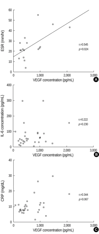
For the serum IL-6 levels, there were no significant differ- ences between the patients with bronchopneumonia and lobar pneumonia, and between the patients with effusion and without effusion, and nor were there significant differ- ences between the subjects who were positive or negative on the urinary S. pneumoniae antigen test (data not shown). The serum levels of VEGF in the patients with pneumonia showed positive correlation with their erythrocyte sedimentation rates (p=0.024), but there was no correlation with the serum IL-6 and CRP levels (Fig. 3). The white blood cell counts and the duration of fever showed no correlations with the serum VEGF levels (data not shown).
DISCUSSION
In this study, we have demonstrated that the serum VEGF concentrations were elevated in the children with pneumonia, and especially in those children who had lobar pneumonia
VEGF concentration (pg/mL)
2,000
1,500
1,000
500
0 Effusion (-) Effusion (+)
p<0.05
Fig. 2.Comparison of the serum VEGF concentrations between the children with and without pleural effusion (A), and between the children with positive and negative urinary S. pneumoniae antigen tests (B) in the lobar pneumonia group. The serum levels of VEGF in the children with pleural effusion and a positive urinary S. pneumoniae antigen test are significantly higher than those with- out pleural effusion and a negative urinary S. pneumoniae antigen test (p<0.05).
A
VEGF concentration (pg/mL)
1,500
1,000
500
0
Pneumococcal Ag (+) Pneumococcal Ag (-) p<0.05
B
ESR (mm/hr)
60 50 40 30 20 10
0
0 1,000 2,000 3,000
VEGF concentration (pg/mL)
rs=0.545 p=0.024
Fig. 3.Correlation of the serum VEGF concentrations with the acute inflammatory parameters. (A) Serum levels of VEGF in the children with pneumonia show positive correlation with the erythrocyte sed- imentation rate (ESR). (B, C) Serum levels of IL-6 and C-reactive protein (CRP) show no significant correlations with the serum VEGF levels.
A
CRP (mg/dL)
40
30
20
10
0
0 1,000 2,000 3,000
VEGF concentration (pg/mL)
rs=0.344 p=0.067
C
IL-6 concentration (pg/mL)
400
300
200
100
0
0 1,000 2,000 3,000
VEGF concentration (pg/mL)
rs=0.222 p=0.230
B
and parapneumonic effusion. This may reflect that an acute lower respiratory tract infection can increase the production and release of VEGF. VEGF is known to be elevated in chronic lung diseases such as asthma and cystic fibrosis (7, 8, 14, 15), whereas there are little data on its relationship with acute pneumonia (11), and furthermore, there is no data on CAP.
In a study on acute eosinophilic pneumonia, the VEGF lev- els were measured in the bronchoalveolar lavage (BAL) fluids and its levels in the patients with acute eosinophilic pneu- monia were higher than those VEGF levels of the control group, and the VEGF levels rapidly decreased to the control levels in parallel with the clinical improvement (11). Another clinical study showed that the serum VEGF levels were ele- vated in cystic fibrosis patients having acute exacerbations, and these levels were decreased with antibiotic therapy (14).
These data support our results showing that the VEGF ele- vation is related to the airway inflammation associated with infection. On the contrary, in a study using lung autopsy mate- rial from septic patients, it was demonstrated that the pul- monary VEGF expression was decreased in the septic patients compared to that in the controls (16). Even though pneumo- nia, pulmonary edema and acute respiratory distress syndrome are the usual pulmonary manifestations during sepsis, the postmortem lung tissue seems to be different from the lung tissue seen in the acute infection states, and other factors such as mechanical ventilation, prolonged antibiotic use and a downhill clinical course to death will contribute to the con- founding variables.
There have been no data that the serum VEGF level is relat- ed to the radiologic type of pneumonia. In this study, the serum levels of VEGF were higher in the patients with lobar pneu- monia than in those patients with bronchopneumonia. It is not certain whether the VEGF elevation is related to the extent of inflammation or if it is related to the virulence of the mic- roorganisms. In an in vitro study, Staphylococcus aureus stimu- lated VEGF release from the normal mesothelial cells in a dose-dependent and time-dependent pattern (17), and Strepto- coccus pneumoniae induced the dose- and time-dependent secre- tion of VEGF from human neutrophils (18). S. aureus and S.
pneumoniae are two of the most common causes of severe, com- plicated pneumonia with parapneumonic effusion and empye- ma. There was no data on the association of the VEGF release with adenovirus infection, which can cause complicated pneu- monia that mimics bacterial pneumonia (19).
In our study, the children with a positive urinary S. pneu- moniae antigen test showed higher levels of serum VEGF than those children with negative results for lobar pneumonia. We found that all the cases with positive urinary pneumococcal antigen tests were lobar pneumonia with or without parap- neumonic effusion; and no bronchopneumonia cases were pos- itive on this test. Even though the urinary S. pneumoniae anti- gen test alone cannot be a tool for the definitive diagnosis of pneumococcal infection, it can be used as a presumptive diag- nostic method, especially in case of invasive pneumococcal
infection (whether it is bacteremia or lobar pneumonia). S.
pneumoniae is isolated from the blood or pleural fluid in only 10-30% of pneumococcal pneumonia patients. The urinary S.
pneumoniae antigen test yields a sensitivity of 77-92% and a specificity of 97-100% (20-23).
The level of VEGF has been shown to be consistently higher in the exudative pleural effusions than in the transudative pleural effusions (17, 24-26). The empyema fluids also con- tain high levels of VEGF (24, 27), which is up to five fold higher than the VEGF levels in the uncomplicated parapneu- monic effusions (17). This study suggests that more compli- cated pneumonia which is usually caused by more virulent pathogens seems to be associated with a greater release of VEGF.
We did not compare the serum VEGF levels with those VEGF levels in the BAL fluid or the pleural effusion. The relative contribution of local VEGF production in pneumo- nia and also in the parapneumonic effusion is not well known.
It was reported that local production is the main source of VEGF in the effusions rather than diffusion from the serum (6) and VEGF is likely to originate from multiple cellular sources such as the residential mesothelial cells, the circulat- ing inflammatory cells, and the infiltrating malignant cells within the pleural space (28). There was the correlation of VEGF with the monocyte and macrophage concentrations (29). It is thought that during acute infection such as bacte- rial pneumonia, the blood supply increases as the inflamma- tion progresses and the inflammatory cells infiltrate into the inflamed tissue. As a result of the local VEGF production by the recruited inflammatory cells, as well as the VEGF pro- duction from the pulmonary epithelial cells, the endothelial cells and the smooth muscle cells, the vascular permeability increased; this in turn causes pulmonary edema or effusion.
In the systemic circulation, these inflammatory cells produce VEGF, and this causes the elevation of the serum VEGF levels as well.
In this study, serum VEGF levels increased significantly in the patients with pneumonia compared to the controls, especially in those patients with lobar pneumonia compared to those patients with bronchopneumonia. For the patients with lobar pneumonia, those patients with parapneumonic effusion showed higher levels of serum VEGF compared to the patients without effusion, and the children with a positive urinary S. pneumoniae antigen test showed higher concentra- tions of serum VEGF compared to those with negative results.
VEGF may be one of the key mediators that lead to more severe and complicated pneumonia; this can provide resear- chers with a new strategy for therapy in the future and so help in the prevention of severe and complicated pneumonia.
REFERENCES
1. Fang GD, Fine M, Orloff J, Arisumi D, Yu VL, Kapoor W, Grayston
JT, Wang SP, Kohler R, Muder RR, Yee YC, Ribs JD, Vickers RM.
New and emerging etiologies for community-acquired pneumonia with implications for therapy. A prospective multicenter study of 359 cases. Medicine 1990; 69: 307-16.
2. Kolditz M, Halank M, Hoffken G. Parapneumonic effusion and ple- ural empyema; topical aspects of classification, diagnosis and treat- ment. Pneumologie 2004; 58: 83-91.
3. Dvorak HF, Detmar M, Claffey KP, Nagy JA, van de Water L, Sen- ger DR. Vascular permeability factor/vascular endothelial growth factor - an important mediator of angiogenesis in malignancy and inflammation. Int Arch Allergy Immunol 1995; 107: 233-5.
4. Ferrara N. Molecular and biological properties of vascular endothe- lial growth factor. J Mol Med 1999; 77: 527-43.
5. Ferrara N. Vascular endothelial growth factor and the regulation of angiogenesis. Recent Prog Horm Res 2000; 55: 15-36.
6. Kraft A, Weindel K, Ochs A, Marth C, Zmija J, Schumacher P, Unger C, Marme D, Gastl G. Vascular endothelial growth factor in the sera and effusions of patients with malignant and nonmalignant disease.
Cancer 1999; 85: 178-87.
7. Hoshino M, Takahashi M, Aoike N. Expression of vascular endothe- lial growth factor, basic fibroblast growth factor, and angiogenin immunoreactivity in asthmatic airways and its relationship to angio- genesis. J Allergy Clin Immunol 2001; 107: 295-301.
8. Hoshino M, Nakamura Y, Hamid QA. Gene expression of vascular endothelial growth factor and its receptors and angiogenesis in bron- chial asthma. J Allergy Clin Immunol 2001; 107: 1034-8.
9. Choi JH, Suh YJ, Lee SK, Suh CH, Nahm DH, Park HS. Acute and chronic changes of vascular endothelial growth factor (VEGF) in induced sputum of toluene diisocyanate (TDI)-induced asthma patients.
J Korean Med Sci 2004; 19: 359-63.
10. Alatas F, Alatas O, Metintas M, Ozarslan A, Erginel S, Yildirim H.
Vascular endothelial growth factor levels in active pulmonary tuber- culosis. Chest 2004; 125: 2156-9.
11. Nishigaki Y, Fujiuchi S, Yamazaki Y, Matsumoto H, Takeda A, Fujita Y, Okamoto K, Fujikane T, Shimizu T, Kikuchi K. Increased vascu- lar endothelial growth factor in acute eosinophilic pneumonia. Eur Respir J 2003; 21: 774-8.
12. Simler NR, Brenchley PE, Horrocks AW, Greaves SM, Hasleton PS, Egan JJ. Angiogenic cytokines in patients with idiopathic interstitial pneumonia. Thorax 2004; 59: 581-5.
13. Shim JY, Jung HL, Park MS, Keum DH. Vascular endothelial growth factor in children with lower respiratory tract disease. J Allergy Clin Immunol 2003; 111: S137.
14. McColley SA, Stellmach V, Boas SR, Jain M, Crawford SE. Serum vascular endothelial growth factor is elevated in cystic fibrosis and decreases with treatment of acute pulmonary exacerbation. Am J Respir Crit Care Med 2000; 161: 1877-80.
15. McDonald DM. Angiogenesis and remodeling of airway vasculature in chronic inflammation. Am J Respir Crit Care Med 2001; 164: 39-45.
16. Tsokos M, Pufe T, Paulsen F, Anders S, Mentlein R. Pulmonary ex- pression of vascular endothelial growth factor in sepsis. Arch Pathol
Lab Med 2003; 127: 331-5.
17. Mohammed KA, Nasreen N, Hardwick J, Logie CS, Patterson CE, Antony VB. Bacterial induction of pleural mesothelial monolayer barrier dysfunction. Am J Physiol Lung Cell Mol Physiol 2001; 281:
119-25.
18. van Der Flier M, Coenjaerts F, Kimpen JL, Hoepelman AM, Geelen SP. Streptococcus pneumoniae induces secretion of vascular endothe- lial growth factor by human neutrophils. Infect Immun 2000; 68:
4792-4.
19. Hong JY, Lee HJ, Piedra PA, Choi EH, Park KH, Koh YY, Kim WS.
Lower respiratory tract infections due to adenovirus in hospitalized Korean children: epidemiology, clinical features, and prognosis. Clin Infect Dis 2001; 32: 1423-9.
20. Marcos MA, Jimenez de Anta MT, de la Bellacasa JP, Gonzalez J, Martinez E, Garcia E, Mensa J, de Roux A, Torres A. Rapid urinary antigen test for diagnosis of pneumococcal community-acquired pneumonia in adults. Eur Respir J 2003; 21: 209-14.
21. Van der Eerden MM, Vlaspolder F, de Graaff CS, Groot T, Jansen HM, Boersma WG. Value of intensive diagnostic microbiological investigation in low- and high-risk patients with community-acquired pneumonia. Eur J Clin Microbiol Infect Dis 2005; 24: 241-9.
22. Smith MD, Derrington P, Evans R, Creek M, Morris R, Dance DA, Cartwright K. Rapid diagnosis of bacteremic pneumococcal infec- tions in adults by using the Binax NOW Streptococcus pneumoniae urinary antigen test: a prospective, controlled clinical evaluation. J Clin Microbiol 2003; 41: 2810-3.
23. Neuman MI, Harper MB. Evaluation of a rapid urine antigen assay for the detection of invasive pneumococcal disease in children. Pedi- atrics 2003; 112: 1279-82.
24. Cheng D, Rodriguez RM, Perkett EA, Rogers J, Bienvenu G, Lap- palainen U, Light RW. Vascular endothelial growth factor in pleu- ral fluid. Chest 1999; 116: 760-5.
25. Cheng D, Lee YC, Rogers JT, Perkett EA, Moyers JP, Rodrigues RM, Light RW. Vascular endothelial growth factor level correlates with transforming growth factor beta isoform levels in pleural effusions.
Chest 2000; 118: 1747-53.
26. Yanagawa H, Takeuchi E, Suzuki Y, Ohmoto Y, Bando H, Sone S.
Vascular endothelial growth factor in malignant pleural effusion asso- ciated with lung cancer. Cancer Immunol Immunother 1999; 48: 396- 400.
27. Thickett DR, Armstrong L, Millar AB. Vascular endothelial growth factor (VEGF) in inflammatory and malignant pleural effusions. Tho- rax 1999; 54: 707-10.
28. Grove CS, Lee YC. Vascular endothelial growth factor: the key medi- ator in pleural effusion formation. Curr Opin Pulm Med 2002; 8:
294-301.
29. Yeo KT, Wang HH, Nagy JA, Sioussat TM, Ledbetter SR, Hoogew- erf AJ, Zhou Y, Masse EM, Senger DR, Dvorak HF. Vascular per- meability factor (vascular endothelial growth factor) in guinea pig and human tumor and inflammatory effusions. Cancer Res 1993; 53:
2912-8.