• 검색 결과가 없습니다.

Ovoid-shaped Left Main Coronary Calcified Aneurysm Leading to Unstable Angina Requiring Surgical Intervention

N/A
N/A
Protected

Academic year: 2022

Share "Ovoid-shaped Left Main Coronary Calcified Aneurysm Leading to Unstable Angina Requiring Surgical Intervention"

Copied!
2
0
0

로드 중.... (전체 텍스트 보기)

전체 글

(1)

113 https://e-kcj.org

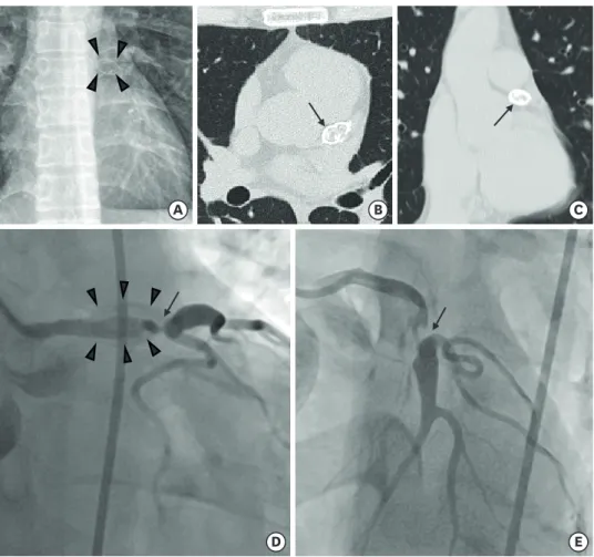
A 34-year-old male without past medical history was referred to the clinic with an ongoing chest pain. The pain was elicited by daytime activities, which was located on substernal area with a sense of tightness. An ovoid mass was found beside the left coronary cusp of the aorta on chest X-ray (Figure 1A). Chest computed tomography showed a 20 mm-sized ovoid calcified mass at distal portion of the left main coronary artery (LMCA) (Figure 1B and 1C).

The patient underwent coronary angiography, revealing a severe concentric stenosis at distal portion of the LMCA and the ostium of the left anterior descending artery (LADA) with a condensed calcified lesion (Figure 1D and 1E). LADA forming collaterals from the distal right coronary artery was also observed. The patient immediately underwent a coronary artery bypass graft (CABG) after the diagnosis because he complained of ongoing chest pain. For revascularization, parallel anastomoses of the left internal thoracic artery to the LADA and the right internal thoracic artery to a diagonal branch were done. Currently he was discharged from the clinic without complication.

Considerable portion of the coronary artery disease in young patients is driven from coronary aneurysm.

1)

Aneurysmal dilatation and stenosis sometimes cause sudden cardiac death in the young and is mainly associated with vascular sequelae from Kawasaki disease.

2)

The mass-like lesion on LMCA in this case was diagnosed as coronary aneurysm with condensed calcification. Surgical procedure seemed more beneficial and safer over percutaneous coronary intervention (PCI) in this patient, considering his need for long-term follow up.

The efficacy of CABG over PCI was proved on Korean study in Kawasaki disease patients with giant aneurysm (>8 mm), highlighting on good patency of graft function.

3)

REFERENCES

1. Aggarwal A, Srivastava S, Velmurugan M. Newer perspectives of coronary artery disease in young. World J Cardiol 2016;8:728-34.

PUBMED | CROSSREF

2. Beiser AS, Takahashi M, Baker AL, Sundel RP, Newburger JW. A predictive instrument for coronary artery aneurysms in Kawasaki disease. US Multicenter Kawasaki Disease Study Group. Am J Cardiol 1998;81:1116-20.

PUBMED | CROSSREF

3. Bang JS, Kim GB, Kwon BS, et al. Long-term prognosis for patients with Kawasaki disease complicated by large coronary aneurysm (diameter ≥6 mm). Korean Circ J 2017;47:516-22.

PUBMED | CROSSREF

Korean Circ J. 2019 Jan;49(1):113-114

https://doi.org/10.4070/kcj.2018.0243 pISSN 1738-5520·eISSN 1738-5555

Received: Jul 23, 2018 Revised: Aug 31, 2018 Accepted: Oct 23, 2018 Correspondence to Seung-Young Roh, MD

Division of Cardiology, Department of Internal Medicine, Dongguk University College of Medicine and Dongguk University Medical Center, 27 Dongguk-ro, Goyang 10326, Korea.

E-mail: [email protected]

Copyright © 2019. The Korean Society of Cardiology

This is an Open Access article distributed under the terms of the Creative Commons Attribution Non-Commercial License (https://

creativecommons.org/licenses/by-nc/4.0) which permits unrestricted noncommercial use, distribution, and reproduction in any medium, provided the original work is properly cited.

ORCID iDs Yuna Chung

https://orcid.org/0000-0002-7769-9484 Seung-Young Roh

https://orcid.org/0000-0002-0854-9079 Conflict of Interest

The authors have no financial conflicts of interest.

Author Contributions

Conceptualization: Roh SY; Investigation:

Chung Y; Supervision: Roh SY; Validation: Roh SY; Writing - original draft: Chung Y; Writing - review & editing: Roh SY.

Yuna Chung , MD, and Seung-Young Roh , MD

Division of Cardiology, Department of Internal Medicine, Dongguk University College of Medicine and Dongguk University Medical Center, Goyang, Korea

Ovoid-shaped Left Main Coronary Calcified Aneurysm Leading to

Unstable Angina Requiring Surgical Intervention

Images in

Cardiovascular Medicine

(2)

114 https://e-kcj.org https://doi.org/10.4070/kcj.2018.0243

Left Main Calcified Aneurysm

A B

D E

C

Figure 1. An ovoid mass near left side of the aorta in chest X-ray (arrow heads) (A). A condensed calcified mass located on LMCA (arrow) in horizontal section (B) and coronal section (C) of chest CT. Diagnostic CAG demonstrated a severe stenosis of distal portion of LMCA (arrows) surrounded by the calcified mass (arrow heads) on RAO (D) and LAO (E) view.

LMCA = left main coronary artery; CT = computed tomography; CAG = coronary angiography; RAO = right anterior

oblique; LAO = left anterior oblique.

수치

Figure 1. An ovoid mass near left side of the aorta in chest X-ray (arrow heads) (A). A condensed calcified  mass located on LMCA (arrow) in horizontal section (B) and coronal section (C) of chest CT

참조

관련 문서