230
A Case of Huge Coronary Aneurysm
After Implantation of a Sirolimus-Eluting Stent
Kyoung-Woo Seo, MD, Seung-Jea Tahk, MD, So-Yeon Choi, MD, Myeong-Ho Yoon, MD, Byoung-Joo Choi, MD, Hong-Seok Lim, MD, Joon-Han Shin, MD, Gyo-Seung Hwang, MD, Soo-Jin Kang, MD, Jin-Woo Kim, MD, Nam-Kyu Lim, MD and Myoung-Hee Lee, MD
Department of Cardiology, School of Medicine, Ajou University, Suwon, KoreaABSTRACT
This report describes the case of a 62-year-old woman who was previously diagnosed with stable angina. Coronary angiography revealed clinically significant stenosis in the middle of the left anterior descending (LAD) artery, the first diagonal branch, the distal left circumflex (LCX) artery and the proximal posterior descending artery (PDA).
After administering aspirin and clopidogrel, the patient underwent implantation of sirolimus-eluting stents in the middle LAD artery and the first diagonal branch. Bare-metal stents were implanted in the distal LCX artery and the proximal PDA. Nineteen months later, follow-up coronary angiography revealed aneurysmal dilation at the middle LAD artery and the first diagonal branch. Forty-six months after implantation of the sirolimus-eluting stents, the size of the coronary aneurysm had increased to 12.4 mm; however, no sign of aneurysmal dilatation was observed at the bare-metal stent sites. This suggested that the implantation of the sirolimus-eluting stent was partially responsible for causing the coronary aneurysm.
(Korean Circ J 2008;38:230-234)KEY WORDS:
Coronary aneurysm; Sirolimus; Drug-eluting stent.
Introduction
A coronary artery aneurysm (CAA) is a dilatation that exceeds 1.5 times the diameter of the normal ad- jacent segment of the coronary artery,
1)and its forma- tion is a rare complication of stenting with bare-metal stents. Experimental studies suggest that drug-eluting stents may induce toxic effects on the vessel wall because of incomplete stent apposition, aneurysm formation, and the potential for stent thrombosis or vessel rupture.
2)3)Coronary aneurysms have been described after the use of drug-eluting stents.
2-7)Only one such case report has been published for Koreans.
6)This report presents the first Korean case of a huge coronary aneurysm that was analyzed with using intravascular ultrasound (IVUS).
We describe here a 62-year-old woman who developed a huge coronary aneurysm in the middle left anterior descending (LAD) artery and the first diagonal branch after receiving a sirolimus-eluting stent (CYPHER; Cor-
dis, Miami Lakes, FL, USA). In contrast to the previous reports, she also had bare-metal stent sites with no sign of aneurysmal dilatation.
Case
A non-hypertensive, non-diabetic, non-smoking 62- year-old woman was admitted to our coronary care unit, and she had been experiencing intermittent chest pain for 3 years. The chest pain was exercise-induced and it lasted for 1 min. On physical examination, she had an arterial pressure of 140/100 mmHg, a heart rate of 76 beats/min and a respiratory rate of 18 breaths/min. Car- diopulmonary auscultation was normal. The initial elec- trocardiogram revealed no abnormality and the cardiac enzymes were all within their normal ranges. The echo- cardiogram showed a good global left ventricle (LV) ejection fraction (EF) of 75%, with no regional wall mo- tion abnormality of the left ventricle. Diagnostic coro- nary angiography revealed three-vessel disease, including a diffuse eccentric 72% diameter stenosis of the middle LAD artery, a diffuse eccentric 85% diameter stenosis of the first diagonal branch, a tubular eccentric 89%
stenosis of the distal left circumflex (LCX) artery and a tubular eccentric 72% stenosis of the proximal poste- rior descending artery (PDA). The middle LAD lesion
Received: October 23, 2007 Accepted: November 16, 2007
Correspondence: Seung-Jea Tahk, MD,Department of Cardiology, School of Medicine, Ajou University, San 5, Woncheon-dong, Yeongtong-gu, Suwon 443-721, Korea
Tel: 82-31-219-5723, Fax: 82-31-219-5708 E-mail: sjtahk@ajou.ac.kr
was predilated with an undersized balloon (2.5 mm), and a 3.0×23 mm sirolimus-eluting stent (CYPHER) was implanted with using an additional high-pressure balloon (2.75 mm). The first diagonal branch lesion was predilated with an undersized balloon (2.5 mm), and a 2.75×23 mm sirolimus-eluting stent (CYPHER) was implanted with using an additional high-pressure balloon (2.75 mm). The final kissing balloon inflation for the middle LAD and the first diagonal lesions was performed. We implanted a 2.5×20 mm bare-metal stent (Arthos; AMG, Westphalia, Germany) at the dis- tal LCX lesion and a 2.75×16 mm cobalt-chromium stent (Arthos Pico) at the proximal PDA lesion (Fig. 1).
The hospital course was unremarkable and she was dis- charged 3 days after the angiography procedure with aspirin 100 mg/day, clopidogrel 75 mg/day, simvastatin (a beta-blocker) and an angiotensin-converting enzyme inhibitor.
Six months later, she was admitted for routine co-
ronary angiography after the previous implantation of multiple stents. The coronary angiogram was normal, with no sign of in-stent changes. She presented with a new episode of precordial pain 19 months after the pre- vious implantation of the multiple stents. The subse- quent coronary angiogram revealed a new 70% diameter stenosis lesion at the middle LAD, an aneurysmal dila- tion at the middle LAD stent lesion and in-stent res- tenosis at the ostium of the first diagonal branch with aneurysmal dilation. A 2.75×16 mm cobalt-chromium stent (Arthos Pico) was implanted at the new middle LAD lesion. The ostial lesion of the first diagonal branch was dilated with a 3.0×20 mm balloon.
Twenty-four months later, the coronary angiogram revealed progressive dilatation of the aneurysm (4 mm in depth) at the middle LAD stent lesion, with no sign of in-stent changes.
Forty-six months later, she presented to the emer- gency room with chest pain. The coronary angiogram
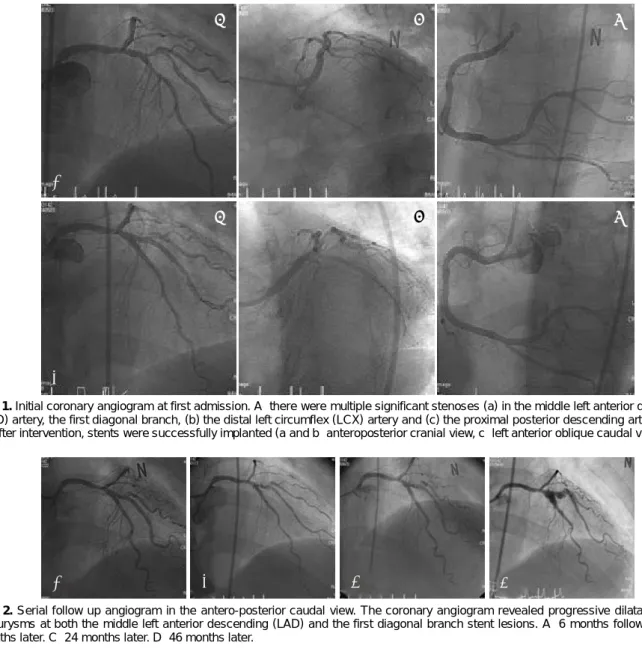
Fig. 1. Initial coronary angiogram at first admission. A: there were multiple significant stenoses (a) in the middle left anterior descending (LAD) artery, the first diagonal branch, (b) the distal left circumflex (LCX) artery and (c) the proximal posterior descending artery (PDA).
B: after intervention, stents were successfully implanted (a and b: anteroposterior cranial view, c: left anterior oblique caudal view).
A
B
a b c
a
b
cFig. 2. Serial follow up angiogram in the antero-posterior caudal view. The coronary angiogram revealed progressive dilatation of the aneurysms at both the middle left anterior descending (LAD) and the first diagonal branch stent lesions. A: 6 months follow-up. B: 19 months later. C: 24 months later. D: 46 months later.
A B C D
revealed progressive dilatation of the aneurysms at both the middle LAD and the first diagonal branch stent lesions with no sign of in-stent changes (Fig. 2). The stent localization and aneurysmatic segment were an- alyzed using IVUS (Galaxy
TM; Boston Scientific, Read- ing, PA, USA), which revealed aneurysmal dilatation of the vessel outside the well expanded stent with a ma- ximum aneurysm diameter of 12.4 mm (Figs. 3 and 4).
No major neointimal thickening was observed. Consi- dering her progressive disease state and the huge size of the aneurysm, she was advised to undergo further surgery.
Discussion
Randomized studies on the use of drug-eluting stents have demonstrated the inhibition of neointimal hyper- plasia in the majority of patients.
8)9)Moreover, gather- ing information on their long-term effect is extremely important with the increasing use of these stents.
CAAs are uncommon in patients undergoing angio- graphy, with a reported incidence of 0.3 to 4.9%. An atherosclerotic etiology is the most common. Other eti- ologies include Kawasaki syndrome and congenital an- eurysms, and aneurysms are now increasingly more com- mon as the sequelae of coronary intervention.
10)11)The pathogenesis of coronary aneurysm formation has not been fully elucidated. The several possible me- chanisms for coronary aneurysm formation include
coronary dissection,
12)13)the anti-inflammatory effects of the active drug,
14)incomplete stent apposition,
15)lo- calized hypersensitivity vasculitis
3)and the toxic effects of the carrier polymer.
4)Coronary aneurysms induced by balloon angiopla- sty may be caused by the use of oversized balloons or excessively high pressure. High-pressure balloon infla- tion is commonly used to optimize the expansion of coronary stents, and deep arterial injury is thought to be the main cause of aneurysm formation after coro- nary intervention. Injury or resection of the media may lead to gradual dilatation and thinning of the vessel wall. The reduced wall thickness and increased stress ultimately cause formation of the aneurysm. Dissec- tion of the coronary artery during percutaneous balloon angioplasty is the main factor related to the occurrence of a CAA.
12)13)IVUS following stent placement may have the ability to detect the incomplete coverage or incomplete appo- sition of the stent to the arterial wall, and this may not be detected by plain angiography alone.
Rab et al.
14)reported that aneurysm formation pro- bably increased with the concomitant use of anti-inflam- matory agents. Sirolimus acts specifically at the G1 phase of the cell cycle, and due to its early action on the cell cycle, sirolimus can block cell proliferation and so it has the potential to induce late vascular sequelae.
9)In addition, sirolimus stimulates apoptosis and reduces
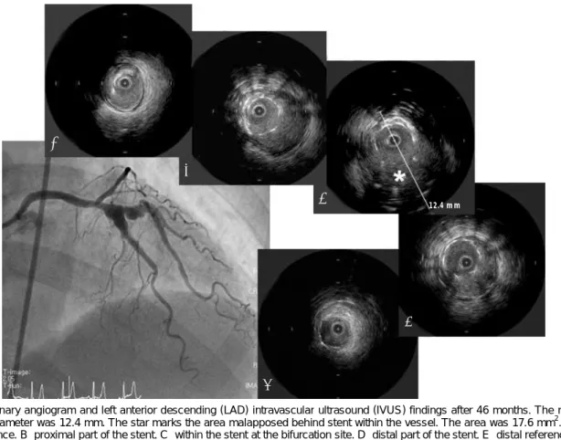
Fig. 3. Coronary angiogram and left anterior descending (LAD) intravascular ultrasound (IVUS) findings after 46 months. The maximal aneurysm diameter was 12.4 mm. The star marks the area malapposed behind stent within the vessel. The area was 17.6 mm2. A: pro- ximal reference. B: proximal part of the stent. C: within the stent at the bifurcation site. D: distal part of the stent. E: distal reference.
A
B
C
D
E
12.4 mm
inflammation. Therefore, drug-eluting stents can lead to the formation of coronary aneurysms.
Among the late postimplantation findings, incom- plete apposition, which is defined as separation of the stent filaments from the intima, could be the mecha- nism responsible for aneurysms.
15)Various hypotheses have been formulated to explain their origin, such as positive regional vascular remodeling, plaque regression, the late dissolution of the thrombotic material captured by the stent filaments, cell necrosis, apoptosis and an allergic reaction to sirolimus.
16)It was recently reported that late stent malapposition occurred more commonly after drug-eluting stent implantation than after bare- metal stent implantation.
17)18)A case of fatal acute myocardial infarction due to late thrombosis of a CYPHER stent deployed 18 months earlier has been reported.
3)The autopsy showed that the wall of the stented artery was dilated aneurysmally with an extensive inflammatory infiltrate involving the intima, media and adventitia. Based on the pathologi- cal evidence, local hypersensitivity vasculitis in response
to the polymer of the CYPHER stent was regarded as the most likely mechanism.
3)Another concern is the possible toxic effects of the carrier polymer that is used as the drug platform. Nume- rous synthetic polymers have been applied to the me- tallic surface to serve as a matrix for the active drug.
4)Concern about the fate of coronary aneurysm for- mation after drug-eluting stent implantation has been raised because of the substantial complications such as stent thrombosis, acute myocardial infarction,
19)vessel rupture and sudden death. Unfortunately, the optimal management for this condition remains controversial.
In our case, other causes may have been involved in addition to stenting with a drug-eluting stent. Consi- dering the patient’s progressive disease state and the huge size of the aneurysm, the patient was advised to undergo surgery.
In summary, we described here a case with delayed development of a giant coronary aneurysm after stent placement, and this occurred despite the excellent an- giographic result.
A
B
C
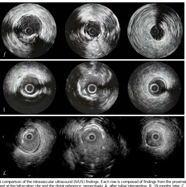
Fig. 4. Serial comparison of the intravascular ultrasound (IVUS) findings. Each row is composed of findings from the proximal reference, within the stent at the bifurcation site and the distal reference, respectively. A: after initial intervention. B: 19 months later. C: 46 months later.
REFERENCES
1) Swaye PS, Fisher LD, Litwin P, et al. Aneurysmal coronary artery disease. Circulation 1983;67:134-8.
2) Degertekin M, Serruys PW, Tanabe K, et al. Long-term follow- up of incomplete stent apposition in patients who received siroli- mus-eluting stent for de novo coronary lesions: an intravascular ultrasound analysis. Circulation 2003;108:2747-50.
3) Virmani R, Guagliumi G, Farb A, et al. Localized hypersensi- tivity and coronary thrombosis secondary to a sirolimus-eluting stent: should we be more cautious? Circulation 2004;109:701-5.
4) Vik-Mo H, Wiseth R, Hegbom K. Coronary aneurysm after im- plantation of a paclitaxel-eluting stent. Scand Cardiovasc J 2004;
38:349-52.
5) Zhang F, Qian JY, Ge JB. Rapid development of late stent mal- apposition and coronary aneurysm following implantation of a paclitaxel-eluting coronary stent. Chin Med J 2007;120:614-6.
6) Park KS, Cha ST, Sung JH, Kim IJ, Lim SW, Cha DH. A case of coronary artery aneurysm after sirolimus-eluting stent implanta- tion. Korean Circ J 2007;37:127-9.
7) Abreu L, Meireles GC, Forte AA, Sumita M, Hayashi J, Solano J.
Coronary artery aneurysm one year and five months after siroli- mus-eluting stent placement. Arq Bras Cardiol 2005;85:340-2.
8) Morice MC, Serruys PW, Souza JE, et al. A randomized com- parison of a sirolimus-eluting stent with a standard stent for coronary revascularization. N Engl J Med 2002;346:1773-80.
9) Moses JW, Leon M, Popma JJ, et al. Sirolimus-eluting stents versus standard stents in patients with stenosis in a native co- ronary artery. N Engl J Med 2003;349:1315-23.
10) Briguori C, Sarais C, Sivieri G, Takagi T, Di Mario C, Colombo A. Polytetrafluoroethylene-covered stents and coronary artery aneurysms. Catheter Cardiovasc Interv 2002;55:326-30.
11) Gercken U, Lansky A, Buellesfeld L, et al. Results of the Jostent coronary stent graft implantation in various clinical settings:
procedural and follow-up results. Catheter Cardiovasc Interv 2002;56:353-60.
12) Bal ET, Thijs Plokker HW, van den Berg EM, et al. Predictabi- lity and prognosis of PTCA-induced coronary artery aneurysms.
Cathet Cardiovasc Diagn 1991;22:85-8.
13) Vassanelli C, Turri M, Morando G, Menegatti G, Zardini P. Co- ronary arterial aneurysms after percutaneous transluminal co- ronary angioplasty: a not uncommon finding at selective follow up angiography. Int J Cardiol 1989;22:151-6.
14) Rab ST, King SB 3rd, Roubin GS, Carlin S, Hearns JA, Douglas JS Jr. Coronary aneurysms after stent placement: a suggestion of altered vessel wall healing in the presence of anti-inflammatory agents. J Am Coll Cardiol 1991;18:1524-8.
15) Stabile E, Escolar E, Weigold G, et al. Marked malapposition and aneurysm formation after sirolimus-eluting coronary stent implantation. Circulation 2004;110:e47-8.
16) Mintz GS, Shah VM, Weissman NJ. Regional remodeling as the cause of late malapposition. Circulation 2003;107:2660-3.
17) Ako J, Morino Y, Honda Y, et al. Late incomplete stent apposi- tion after sirolimus-eluting stent implantation: a serial intravas- cular ultrasound analysis. J Am Coll Cardiol 2005;46:1002-5.
18) Hong MK, Mintz GS, Lee CW, et al. Late stent malapposition after drug-eluting stent implantation: an intravascular ultra- sound analysis with long-term follow-up. Circulation 2006;113:
414-9.
19) Yun HJ, Kim KS, Hur SH, Park NH, Cho YW. A case report of a huge coronary artery aneurysm with acute myocardial infarction.
Korean Circ J 2002;32:720-4.