• 검색 결과가 없습니다.

An Unusual Form of Coronary Artery Fistula: A Small Aneurysm of Vieussens’ Arterial Ring Communicating with the Pulmonary Artery

N/A
N/A
Protected

Academic year: 2021

Share "An Unusual Form of Coronary Artery Fistula: A Small Aneurysm of Vieussens’ Arterial Ring Communicating with the Pulmonary Artery"

Copied!
3
0
0

로드 중.... (전체 텍스트 보기)

전체 글

(1)

Korean J Thorac Cardiovasc Surg 2014;47:152-154 □ Case Report □ http://dx.doi.org/10.5090/kjtcs.2014.47.2.152 ISSN: 2233-601X (Print) ISSN: 2093-6516 (Online)

− 152 −

Department of Thoracic and Cardiovascular Surgery, Kosin University Gospel Hospital, Kosin University College of Medicine Received: December 4, 2013, Revised: January 6, 2014, Accepted: January 7, 2014

Corresponding author: Seong Ho Cho, Department of Thoracic and Cardiovascular Surgery, Kosin University Gospel Hospital, Kosin University College of Medicine, 262 Gamcheon-ro, Seo-gu, Busan 602-702, Korea

(Tel) 82-51-990-6466 (Fax) 82-51-990-3066 (E-mail) aorta007@naver.com

C

The Korean Society for Thoracic and Cardiovascular Surgery. 2014. All right reserved.

CC
This is an open access article distributed under the terms of the Creative Commons Attribution Non-Commercial License (http://creative- commons.org/licenses/by-nc/3.0) which permits unrestricted non-commercial use, distribution, and reproduction in any medium, provided the original work is properly cited.

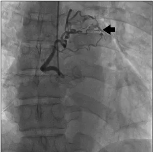
Fig. 1. Coronary angiography. Left coronary angiography demon- strated an anomalous coronary fistula from the proximal left ante- rior descending coronary artery to the main pulmonary artery (arrow).

An Unusual Form of Coronary Artery Fistula: A Small Aneurysm of Vieussens’ Arterial Ring Communicating with the Pulmonary Artery

Hae Young Lee, M.D., Seong Ho Cho, M.D.

Vieussens’ arterial ring (VAR) is the connection between the conus branch of the right coronary artery and the proximal right ventricular branch of the left anterior descending coronary artery. VARs are found in 48% of the population; however, pathologic VAR is rare. We experienced a case of pathologic VAR that involved a fistula con- necting to the main pulmonary artery.

Key words: 1. Coronary vessel anomalies 2. Fistula

CASE REPORT

A 53-year-old male presented chest discomfort and palpitations. Electrocardiography performed on admission re- vealed sinus tachycardia with no ischemic change. His blood pressure was 143/80 mmHg and pulse rate was 95 beats/min.

Laboratory results including complete blood count and cardiac

enzymes were within the normal range. There was no car-

diomegaly or evidence of active lung lesion on chest radi-

ography. Transthoracic echocardiography revealed normal eje-

ction fraction, chamber size, and valve function, and regional

wall motion abnormality was not present. However, there was

abnormal blood flow into the pulmonary artery on the para-

sternal short axis view, suggesting a coronary artery fistula

(CAF). The patient underwent coronary angiography, which

revealed a coronary fistula between the proximal left anterior

descending coronary artery (LAD) and the pulmonary artery

(Fig. 1). The right coronary artery (RCA) also had a con-

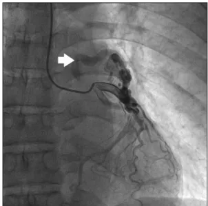
genital anomaly. The right conal artery, originating from a

separate orifice of the right coronary sinus and draining into

(2)

A Coronary Artery Fistula of Vieussens’ Arterial Ring

− 153 − Fig. 2. Coronary angiography. Right coronary angiography demon- strated the conus branch from a separate coronary ostium. The right conal branch has a fistula to the pulmonary artery (arrow).

Fig. 3. Three-dimensional reconstruction of computed tomography coronary angiography. The right conal branch had a separated coronary ostium and was connected to the proximal left anterior descending coronary artery via Vieussens’ arterial ring.

Fig. 4. Three-dimensional reconstruction of computed tomography coronary angiography. The right conal branch and the proximal left anterior descending coronary artery formed Vieussens’ arterial ring (arrow), which showed aneurismal dilatation prior to entering the main pulmonary artery.

the pulmonary artery, indicated the presence of a CAF be- tween the right conal artery and the pulmonary artery (Fig. 2).

Contrast-enhanced electrocardiogram-gated multidetector com- puted tomography (MDCT) was performed to evaluate the coronary fistulae and to reveal any other abnormalities.

MDCT showed an abnormal connection between the right conal artery and the LAD draining to the pulmonary artery via a 6.2-mm aneurysm. In other words, the right conal artery originating from a separate orifice of the right coronary sinus had an abnormal connection with the RCA via a short vessel just distal to its origin. It passed up and over the right ven- tricular outflow tract to provide flow to the aneurysm, which was connected to the LAD via a highly tortuous artery. There was also a fistula between the aneurysm and the main pulmo- nary artery (Figs. 3, 4). These features were compatible with the diagnosis of an aneurysm of VAR. The patient underwent an echocardiographic stress test and treadmill test to inves- tigate cardiac ischemia; these tests showed no ischemic change. Surgical resection was offered as a treatment option;

however, the patient refused treatment for personal reasons and is being followed closely.

DISCUSSION

CAFs are abnormal connections between coronary arteries

and cardiac chambers or great vessels, which have an in-

cidence of approximately 0.87% [1]. Most fistulae originate

(3)

Hae Young Lee and Seong Ho Cho

− 154 − from the right coronary or the left anterior descending artery, and both coronary arteries are the site of the CAF in approx- imately 5% of the cases. In the present case, the fistula, which showed aneurismal change, was fed by both the right conal artery and the LAD, and drained into the main pulmo- nary artery. These findings are compatible with the diagnosis of an aneurysm of VAR with a fistula to the pulmonary artery. Many studies have investigated CAFs and have re- ported that they have several possible origins; however, re- ports of CAFs of VAR are rare. VAR was first described in 1706 by Raymond de Vieussens [2] and is described as a connection between the conus branch of the RCA and the proximal right ventricular branch of the LAD. It is thought to be a remnant of the embryonic conotruncal ring and is found in 48% of the population [3]. However, the primary patho- logical features of VAR are rare. When significant stenosis occurs in the LAD or RCA, VAR will dilate and provide collateral blood flow to the left or right arterial system [4]. In our case, however, although there was no significant stenosis in the coronary artery system, hypoplastic RCA was observed. Atallah et al. [5] reported a case of VAR with a congenitally hypoplastic left coronary arterial system. In the case considered here, they demonstrated that VAR could not be the product of collateral circulation due to obstructive le- sions but was the result of congenitally hypoplastic left arte- rial circulation. In consideration of these features, in our case, VAR was also deemed to be unrelated to collateral circu- lation due to obstructive lesions; rather, it was the result of a congenitally hypoplastic right coronary system. There have

been previous reports of an aneurysm of VAR; however, re- ports of a fistula between VAR and the main pulmonary ar- tery are rare. The management of CAFs involving the pulmo- nary artery via VAR is no different from that of a typical CAF, although this remains controversal. However, a majority of these opinions advocate surgical management for sympto- matic patients because percutaneous intervention can cause a secondary ischemic event due to the accompanying atherosclerosis. Further, the results of numerous surgical mo- dalities are reported to be excellent.

CONFLICT OF INTEREST

No potential conflict of interest relevant to this article was reported.

REFERENCES

1. Angelini P, Velasco JA, Flamm S. Coronary anomalies: in- cidence, pathophysiology, and clinical relevance. Circulation 2002;105:2449-54.

2. Loukas M, Clarke P, Tubbs RS, Kapos T. Raymond de Vieussens. Anat Sci Int 2007;82:233-6.

3. Germing A, Mugge A. Images in cardiology: Vieussens’ ring.

Clin Cardiol 2003;26:441.

4. O’Leary EL, Garza L, Williams M, McCall D. Images in cardiovascular medicine. Vieussens’ ring. Circulation 1998;

98:487-8.

5. Atallah PC, Atallah PC, Spears JR. Vieussens’ ring with

congenitally hypoplastic left coronary arterial system. Int J

Cardiol 2011;152:e31-2.

수치

Fig. 1. Coronary angiography. Left coronary angiography demon- demon-strated an anomalous coronary fistula from the proximal left  ante-rior descending coronary artery to the main pulmonary artery  (arrow).
Fig. 4. Three-dimensional reconstruction of computed tomography  coronary angiography

참조

관련 문서

– 외경동맥(바깥목동맥 external carotid artery)의 안면 동맥(얼굴동맥 facial artery) 및 상인두동맥(오름인두 동맥 ascending pharyngeal

The “Asset Allocation” portfolio assumes the following weights: 25% in the S&P 500, 10% in the Russell 2000, 15% in the MSCI EAFE, 5% in the MSCI EME, 25% in the

1 John Owen, Justification by Faith Alone, in The Works of John Owen, ed. John Bolt, trans. Scott Clark, "Do This and Live: Christ's Active Obedience as the

Cerebral Cerebral Cerebral angiography Cerebral angiography angiography angiography shows shows shows shows feeding feeding feeding artery feeding artery

The locations of aneurysms were middle cerebral artery in 15 patients, cerebral artery in 15 patients, cerebral artery in 15 patients, cerebral artery in

These results of the histologic distribution of lamina propria, adipose tissue, and glandular tissue of the hard palatal mucosa and the topography of the greater palatine

• The supply and demand curves cross at the equilibrium price and quantity.. • You can read off approximate equilibrium values

_____ culture appears to be attractive (도시의) to the