69
CASE REPORT
Korean Circ J 2008;38:69-71
Print ISSN 1738-5520 / On-line ISSN 1738-5555 Copyright ⓒ 2008 The Korean Society of Cardiology
A Sirolimus-Eluting Stent Fracture Combined
with a Coronary Artery Aneurysm
Seung-Yul Lee, MD, Eui Im, MD, Woo-In Yang, MD, Jung-Sun Kim, MD, Yun-Hyeong Cho, MD and Won-Heum Shim, MD Division of Cardiology, Cardiovascular Center, Yonsei University College of Medicine, Seoul, Korea
ABSTRACT
A stent fracture combined with a coronary artery aneurysm is a rare event. As these events can lead to a harmful outcome, such as the development of myocardial ischemia by in-stent restenosis or thrombosis, repeated coronary intervention may be required. We report a case of a stent fracture combined with a coronary artery aneurysm. The fracture was thought to have developed by mechanical stress produced from a change of regional wall motion after an anteroseptal myocardial infarction. As detected by the use of intravascular ultrasound, neither in-stent restenosis nor a thrombus in the fractured stent was present. A cardiac magnetic resonance image showed that no viable myocardium in the anteroseptal wall was present. Therefore, the patient underwent medical treatment without intervention of the fractured stent. (Korean Circ J 2008;38:69-71)
KEY WORDS: Stents; Complications; Fracture; Aneurysm.
Introduction
The use of drug-eluting stents (DES) has reduced in-stent restenosis by inhibiting neointimal hyperplasia. The use of DES has been applied widely as an effective interventional therapeutic modality in coronary artery disease. However, complications such as restenosis or a stent thrombosis remain in this era of using DES. Re-cently, stent fractures and coronary artery aneurysms after the implantation of a drug-eluting stent have emerged as novel complications as these occurrences may cause restenosis or a thrombosis. We report here a case of stent fracture combined with coronary aneurysm after percutaneous coronary intervention (PCI) with a drug-eluting stent.
Case
A 70 year-old woman was admitted to the hospital with effort angina. Coronary angiography revealed dif-fuse and significant stenosis in the proximal and mid portion of the left anterior descending artery (LAD) (Fig. 1A). The LAD lesion was treated with two
over-lapping 3.5×23 mm and 3.0×33 mm sirolimus-eluting stents (SES; Cypher®, Cordis, Miami Lakes, FL
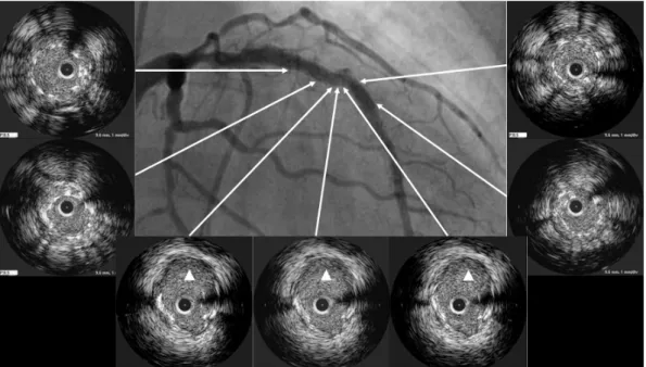
USA). Final angiography showed the presence of well-deployed stents without residual stenosis or dissection. However, the big septal artery was jailed with throm-bolysis in myocardial infarction (TIMI) 0 distal flow (Fig. 1B). Although we attempted to wire the jailed artery, wiring failed as the artery was below the over-lapping site and the lesion was tight. The day after the PCI, the level of a cardiac biomarker, the creatinine kinase MB fraction, increased to 114 ng/mL and echo-cardiography revealed akinetic regional wall motion abnormality at the anteroseptum from the mid to the apex. Sixteen months later, the patient suffered from resting chest discomfort again and underwent coronary angiography. There was complete stent fracture combin-ed with a coronary artery aneurysm, but no signi-ficant in-stent restenosis (Fig. 1C and D). Intravascular ultrasound (IVUS) confirmed the discontinuation of the stent strut and an aneurysmal change (Fig. 2). A cardiac magnetic resonance image (MRI) showed the presence of myocardial thinning with transmural delayed hyperenhancement and akinetic motion on the anteroseptal segement (Fig. 3). We concluded that the symptom was not associated with the stent fracture or the coronary artery aneurysm as previously placed stents were patent and the anteroseptal myocardial wall was not viable. Therefore, we decided to continue clinical surveillance. Six months later, the patient is receiving follow-up and has been without symptoms.
Received: August 16, 2007
Revision Received: September 27, 2007 Accepted: October 23, 2007
Correspondence: Won-Heum Shim, MD,Division of Cardiology, Cardiovas-cular Center, Yonsei University College of Medicine, 250 Seongsanno, Seodaemun-gu, Seoul 120-752, Korea
Tel: 82-2-2228-8460, Fax: 82-2-393-2041 E-mail: whshim@yuhs.ac
70·Stent Fracture with Coronary Aneurysm
Discussion
DES have been used widely for the effectiveness about restenosis. However, recently a few cases concerning the fracture of DES and stent related aneurysms have been reported. These occurrences may be considerable complications because of the association with restenosis or stent thrombosis.1-6)
Although the cause and pathophysiology of both complications are not known, most cases have occurred when there are higher radial forces, usage of longer stents or the use of overlapping stents.7) These events
typically occur in a hypermobile and tortuous vessel, especially the right coronary artery.8) Overexpansion of
the stent can be another risk factor as it may weaken struts and promote the fracture. Coronary artery aneu-rysms might result from local hypersensitivity vasculitis
in response to the polymer or coating drug on the DES.9)
In our case, the big septal artery was occluded totally after sirolimus-eluting stent implantation, resulting in akinetic motion on the anteroseptal wall. The change of regional wall motion might have developed a new hinge in the overlapping long stents during the cardiac cycle. Thus, mechanical stress on overlapping stents could provoke the fracture of the sirolimus-eluting stents. The coronary artery aneurysm of this case was ob-served at a fractured segment in the overlapping stents. Mechanical irritation of fractured struts may have caused aneurysmal dilation of the coronary vessel, and a local hypersensitivity reaction may be attributable to the coronary aneurysm.
Unfortunately, the natural history of DES fracture and coronary artery aneurysm has not yet been de-Fig. 1. Serial findings of coronary angiogram. A: initial coronary angiography showed diffuse stenosis in the proximal and mid portions of the LAD. B: post PCI angiography revealed well-deployed overlapping SESs, but the big septal artery (arrow) was totally occluded. C: fluoroscopy at follow-up showed a complete SES fracture. D: coronary angiography showed coronary artery aneurysm (arrowhead) at the fractured site. LAD: left anterior descending artery, PCI: percutaneous coronary intervention, SES: sirolimus-eluting stent.
C D
Seung-Yul Lee, et al.·71
fined. Moreover, the best treatment for the problems is unknown. In our case, there was restenosis or throm-bosis was not present at the fractured stent. Cardiac MRI was performed to formulate the management plan because the anteroseptal wall was already akinetic as determined by previous echocardiography. Consider-ing the absence of a viable portion in the anteroseptal wall as seen on cardiac MRI and the relative small sized coronary aneurysm, we decided to continue clinical follow-up instead of performing an interventional
pro-cedure on the LAD lesion.
Studies about DES fractures and coronary aneurysms are still very limited, but physicians should be con-cerned as these complications can be associated with restenosis or a thrombosis. A further investigation is needed to define the clinical significance of a DES frac-ture and coronary artery aneurysm.
REFERENCES
1) Brilakis ES, Maniu C, Wahl M, Barsness G. Unstable angina due to stent fracture. J Invasive Cardiol 2004;16:545.
2) Sianos G, Hofma S, Ligthart JM, et al. Stent fracture and res-enosis in the drug-eluting stent era. Catheter Cardiovasc Interv 2004;61:111-6.
3) Min PK, Yoon YW, Kwon HM. Delayed strut fracture of siro-limus-eluting stent: a significant problem or an occasional obser-vation? Int J Cardiol 2006;106:404-6.
4) Surmely JF, Kinoshita Y, Dash D, et al. Stent strut fracture-induced restenosis in a bifurcation lesion treated with the crush stenting technique. Circ J 2006;70:936-8.
5) Farb A, Burke AP, Kolodgie FD, Virmani R. Pathological me-hanisms of fatal late coronary stent thrombosis in humans. Cir-ulation 2003;108:1701-6.
6) Bae JH, Hyun DW, Kim KY, Yoon Hj, Nakamura S. Drug-eluting stent strut fracture as a cause of restenosis. Korean Circ J 2005;35:787-9.
7) Kim JS, Yoon YW, Hong BK, et al. Delayed stent fracture after successful sirolimus-eluting stent (Cypher®) implantation. Korean Circ J 2006;36:443-9.
8) Kim EJ, Rha SW, Wani SP, et al. Coronary stent fracture and restenosis in the drug-eluting stent era: do we have clues of ma-agement? Int J Cardiol 2007;120:417-9.
9) Joner M, Finn AV, Farb A, et al. Pathology of drug-eluting stents in humans: delayed healing and late thrombotic risk. J Am Coll Cardiol 2006;48:193-202.
Fig. 2. IVUS at follow-up angiography confirmed a complete stent fracture with coronary artery aneurysm (arrow heads). IVUS: intravas-cular ultrasound.
Fig. 3. Cardiac MRI revealed myocardial thinning with transmural delayed hyperenhancement (arrow) on the anteroseptal area.