세개의
4
0
0
전체 글
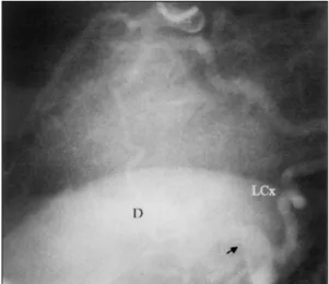
(2) 문헌고찰과 함께 보고하는 바이다.. 증. 례. 환 자:임○금, 여자, 70세. 주 소:노작성 호흡곤란. 현병력:내원 7년 전부터 본태성 고혈압으로 본원에 서 항고혈압제로 치료받아오던 환자이며 수개월 전부터 점차 심해지는 노작성 호흡곤란을 주소로 이에 대한 검 사 및 치료위해 입원하였다. 과거력:고혈압 외에는 특이사항 없었다. 가족력:특이사항 없었다. 이학적 검사:내원 당시 혈압은 130/80 mmHg, 맥 박수는 85/분, 호흡수 22회 / 분, 체온 36.7℃였다. 흉 부 청진상 강도 3/6의 지속성 이완기성 심잡음이 심첨부 에서 청진되고 다른 특이소견은 관찰되지 않았다. 검사실 소견:말초혈액검사에서 백혈구 7500/mm3, 혈색소 13.4 g/dL, 혈소판 347,000/mm3이었고, 혈청. Fig. 1. Selective left coronary angiogram. On the left lateral projection, the left circumflex coronary artery and the first big diagonal branch of the left anterior descending artery are very large and dilated, which directly communicate with left ventricle via common channel (arrow). D:the first big diagonal branch of the left anterior descending artery, LCx:left circumflex coronary artery.. 생화학 검사상에서 BUN 15.2 mg/dL, 크레아틴 0.7 mg/dL, AST 24 IU/L, ALT 26 IU/L이고 기타 생화학 검사 및 요 검사는 정상이었다. 심전도 및 흉부 X-선 검사:휴식기 심전도상 ST분 절 및 T파의 허혈소견은 없었으며, 흉부 X-선 검사상 심비대 및 폐부종등의 소견은 관찰되지 않았다. 경흉부 심초음파도 소견:비대칭적 심실중격 비후가 관찰되나 좌심실 유출로의 수축기 협착 소견은 없는 비 후성 심근증의 소견을 보이고 있었다. 심방 및 심실의 비대는 없었으며, 판막도 정상이었다. 관동맥 조영술 소견:선택적 좌관동맥 조영술상 큰 심 정맥(great cardiac vein)이 관정맥동으로 유입되기 직 전으로 추정되는 좌심실의 후측벽부위에서, 좌회선동맥 과 제 1 거대 대각선동맥이 공통경로를 이루어 좌심실 로 직접 유출되고 있었으며, 그 근위부는 심하게 확장된 소견을 보였다(Fig. 1). 선택적 우관동맥 조영술상에서. Fig. 2. Selective right coronary angiogram. On the left lateral projection, the right coronary artery is communicated directly with the left ventricle via common channel (arrow), with faint visualization of the left circumflex coronary artery and the first big diagonal branch of the left anterior descending artery.. 도 우관동맥이 좌회선동맥과 제 1 거대 대각선동맥과 같은 공통경로를 통하여 좌심실로 유출되고 있었으며. 되어 현재 외래에서 경과관찰중이다.. 혈류가 좌회선동맥과 제 1 거대 대각선동맥으로도 소 량의 역류를 보이고 있어 하나의 공통경로임을 확인하. 고. 찰. 였다(Fig. 2). 치료 및 경과:환자는 칼슘 통로 길항제와 안지오텐. 관동맥 조영술을 통하여 진단되는 선천성 관동맥루의. 신 II 수용체 길항제로 치료를 시작하였으며 증상 호전. 빈도는 대략 0.08%에서 0.3%로 보고1)되고 있으며 이. 272. Korean Circulation J 2002; 32(3):271-274.
(3) 중에서 관동맥-좌심실루의 빈도는 3%이내로 보고2)되. 그 외에 질산염제재12)와 최근에는 안지오텐신 전환효소. 고 있다. Nawa 등3)이 관동맥-좌심실루를 가진 73예를. 억제재3)가 시도되고 있다. 다발성 관동맥루를 가진 환자. 비교한 결과는 관상동맥을 침범한 수에 따라 각각 한 개. 에서는 관동맥루의 크기에 상관없이 감염성 심내막염에. 가 51예(69%), 두개가 12예(16.4%)였고 특히 본 예. 대한 예방이 필요하며,13) 지속적인 좌심실 이완기 용적. 와 같이 세개의 관동맥이나 그 분지를 모두 침범하는 경. 부하로 인한 심부전의 발현에 대해서도 지속적인 관찰이. 우는 10예(13.7%)였는데, 문헌상으로는 전세계적으로. 요구된다.. 약 30예만이 보고된 극히 드문 질환이다.. 요. 관동맥 조영술상 관동맥-심실루는 관동맥과 심실사이. 약. 에서 소혈관들이 다발을 이루고 뚜렷이 중첩되어 나타난 후 다발성 유출누공을 통해 심실로 유출되어 희미하게. 선천성 관동맥-좌심실루는 매우 드문 질환이며 세개. 좌심실이 조영되는 소견과 단일 관동맥이 직접 심실로 유. 의 주요 관동맥을 침범하는 다발성인 경우는 전 세계적. 출되어 심실이 바로 조영되는 소견으로 분류된다.4) 전자. 으로 그 보고가 매우 드물다. 특히 주요 관동맥이 공통. 의 경우 Thebesian vessels의 이상에 기인하는 것으로. 경로를 이루어 좌심실로 유출되는 특이한 관동맥 조영. 5). 보여지나 후자의 경우는 다른 병태생리학적 기전이 관. 술 소견을 보이는 관동맥-좌심실루는 문헌고찰상 그 보. 련되어질 것으로 생각된다. 본 증례는 후자에 해당하겠. 고를 찾기가 어려웠다. 이에 저자들이 경험한 공통경로. 다. 그러나, 본 증례와 같이 세개의 주요 관동맥이 하나. 를 갖는 세개의 주요 관동맥-좌심실루 1예를 문헌고찰. 의 공통경로를 이룬 후 함께 좌심실로 직접 유출되는 형. 과 함께 보고하는 바이다.. 태는 문헌 고찰상 찾아볼 수 없었으며, 본 증례가 최초 보고라고 생각된다.. 중심 단어:관상 혈관 이상;누공.. 다발성 관동맥-좌심실루를 가진 환자는 주로 협심증 의 증상을 호소한다.3) 죽상경화성 관동맥질환이나, 좌심. REFERENCES 1) Yamanaka O, Hobbs RE. Coronary artery anomalies in. 실비후가 없이도 심근의 허혈을 일으킬 수 있는데 이는 심근보다 낮은 저항의 루로 혈액단락에 의한 관도혈류현. 2). 상(coronary steal syndrome)이 주된 기전으로 이해 되고 있다.6) 특히 무증상인 환자에서 심근경색으로 발. 3). 현한 예7)가 있어 환자의 주관적 증상이나 이학적 검사 상 특이사항이 없더라도 심근허혈을 배제하여서는 안된 다. 이를 위해 휴식기 및 운동부하 심전도검사와 48시간 활동 중 심전도 검사,8) 심근관류 신티그라피,9) 심도자 후. 4). 관정맥동내 젖산 측정(coronary sinus lactate measurement),2) 및 도플러 심도자법을 이용한 관동맥 혈류. 5). 속도의 측정10)과 같은 적극적인 방법이 시도되고 있다. 다발성 관동맥-좌심실루에 의한 또 다른 중요한 혈역학. 6). 적 변화로 좌좌단락에 의한 이완기 용적 부하가 일어나 이에 의해 대동맥판 폐쇄부전증과 같은 임상양상이 나. 7). 타날 수 있다.11) 이 질환의 치료와 질환의 예후에 관해서는 그 경험이. 8). 부족하고 치료효과에 대해서도 논란이 많은 실정이다. 수 술적 치료나 중재적 시술에 의한 치료보다는 주로 증상 조절을 위한 내과적 치료가 시도되고 있는데 심근 허혈 증상이 있는 경우 베타 차단제8)나 칼슘 통로 길항제,5). 9). 126,595 patients undergoing coronary arteriography. Cathet Cardiovasc Diagn 1990;21:28-40. Levin DC, Fellows KE, Abrams HL. Hemodynamically significant primary anomalies of the coronary arteries: angiographic aspects. Circulation 1978;58:25-34. Nawa S, Miyachi Y, Toshino N, Shiba T, Hayashi K, Tamesue K, Yamamoto H, Shimizu N. Three major coronary artery-to-left ventricular shunts: report of three cases and review of literature. Cardiovasc Intervent Radiol 1997; 20:300-4. Elian D, Zahav YH, Agranat O, Rath S, Di Segni E. Coronary arterioluminal communications in routine angiography. Cathet Cardiovasc Diagn 1998;43:29-32. Black IW, Loo CK, Allan RM. Multiple coronary arteryleft ventricular fistulae: clinical, angiographic, and pathologic findings. Cathet Cardiovasc Diagn 1991;23:133-5. Stierle U, Giannitsis E, Sheikhzadeh A, Potratz J. Myocardial ischemia in generalized coronary artery-left ventricular microfistulae. Int J Cardiol 1998;63:47-52. McLellan BA, Pelikan PC. Myocardial infarction due to multiple coronary-ventricular fistulas. Cathet Cardiovasc Diagn 1989;16:247-9. Koh KK, Cho SK, Kim SS. Left and right coronary artery to left ventricular fistula: demonstration of myocardial ischemia by treadmill test and Holter monitoring: a case report. Angiology 1993;44:977-80. Frustaci A, Caldarulo M, Pagliari G, Adragna L. Coronary angiodysplasia causing left ventricular shunt and myocardial ischemia. Am Heart J 1993;125:889-91.. 273.
(4) 10) Meissner A, Lins M, Herrmann G, Simon R. Multiple. coronary artery-left ventricular fistulae: haemodynamic quantification by intracoronary Doppler ultrasound. Heart 1997;78:91-3. 11) Amin H, Solankhi N, Uzun O. Coronary arterial-left ventricular fistulae. Heart 2001;85:648. 12) Duckworth F, Mukharji J, Vetrovec GW. Diffuse coron-. 274. ary artery to left ventricular communications: an unusual cause of demonstrable ischemia. Cathet Cardiovasc Diagn 1987;13:133-7. 13) Wolf A, Rockson SG. Myocardial ischemia and infarction due to multiple coronary-cameral fistulae: two case reports and review of the literature. Cathet Cardiovasc Diagn 1998;43:179-83.. Korean Circulation J 2002; 32(3):271-274.
(5)
수치
관련 문서
(a) A twisted nematic cell, The LC molecules are aligned horizontally on the left window and vertically on the right window.. They gradually twist (plane upon plane) from one
• The supply and demand curves cross at the equilibrium price and quantity.. • You can read off approximate equilibrium values
systemic circulation, in the right ventricle and oxygenated blood from the lungs, or pulmonary circulation, in the left ventricle, as in birds and mammals.. Two vessels,
If the volume of the system is increased at constant temperature, there should be no change in internal energy: since temperature remains constant, the kinetic
Chest X-ray: Posterio-anterior (PA) View - Left Anterior Oblique
1 John Owen, Justification by Faith Alone, in The Works of John Owen, ed. John Bolt, trans. Scott Clark, "Do This and Live: Christ's Active Obedience as the
Measurement of proximal contact tightness between the left first molar and second molar(dental implant) in the mandible... Clinical dental anatomy, histology,
The average position of the posterior superior alveolar artery, the wall thickness of the lateral wall, and the average volume of the maxillary sinus will
