54
Case Report
Intraocular Involvement of a Nasal Natural Killer T-Cell Lymphoma:
A Case Report
Jae Ho Yoo, Soo Young Kim, Kyu Bong Jung, Jung Joo Lee, Sang Joon Lee
Department of Ophthalmology, Kosin University College of Medicine, Busan, Korea pISSN: 1011-8942 eISSN: 2092-9382
Korean J Ophthalmol 2012;26(1):54-57 http://dx.doi.org/10.3341/kjo.2012.26.1.54
Malignant lymphomas that develop in the orbit and ocu- lar adnexa account for 8% of all extranodal lymphomas [1,2]. Most ocular and orbital lymphomas are non-Hodg- kin’s B-cell lymphoma [1]. At these sites, with the excep- tion of B-cell lymphoma, lymphoblastic diseases are rare and occur in only 1% to 3% of cases [2]. Ocular and ocular adnexa T-cell lymphomas are rare. There are few cases of nasal natural killer T-cell lymphoma (NKTL) reported in the medical literature [1-4]. Additionally, pathologically confirmed intraocular involvement of nasal NKTL, such as the case reported here, is very rare [3].
Case Report
A 57-year-old woman presented with a three-day his- tory of photophobia and diplopia in the left eye. She was diagnosed with histologically confirmed nasal NKTL of the right nasal cavity one month prior. The Snellen visual
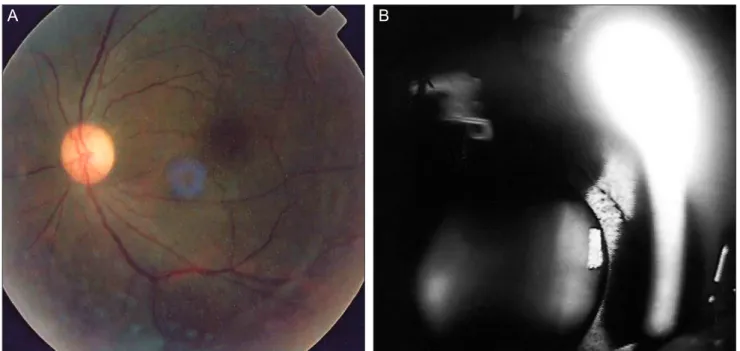
acuity of the affected eye with correction was 1.0 / 0.63, and the intraocular pressure in the right and left eyes was 17 and 18 mmHg, respectively. The patient had mild left eye conjunctival injection, ptosis, and swelling of the eye- lid. The left pupil was fully dilated with no pupillary light reflex. Ocular movement on left-upper gaze was limited in the left eye. Five days later, the left corrected visual acuity deteriorated and ptosis worsened. Slit lamp examination revealed anterior uveitis refractory to topical steroid treat- ment. Opthalmoplegia, vitreous opacity, and an iris mass developed in the left eye (Fig. 1A and 1B). Orbital magnet- ic resonance imaging and cerebrospinal fluid investigations were unremarkable. Aqueous humor aspiration from the anterior chamber was performed with a 26-gauge needle.
Thirty-five percent of the cells obtained from the aspira- tion were morphologically atypical lymphocytes.
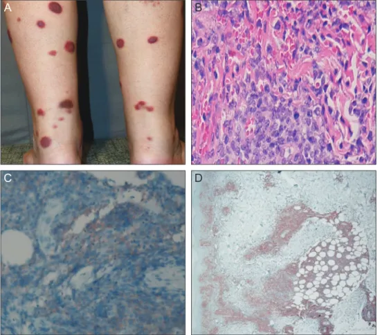
The patient was diagnosed with intraocular dissemina- tion of the lymphoma. Treatment with intravitreal triam- cinolone injections, radiotherapy, and chemotherapy was administered. Skin manifestations of the lymphoma were noted 40 days post-chemotherapy (Fig. 2A). Skin biopsy confirmed neoplastic infiltration of small- to medium- sized pleomorphic lymphocytes with irregular nuclei, inconspicuous nucleoli, and scant cytoplasm (Fig. 2B). Im-
© 2012 The Korean Ophthalmological Society
This is an Open Access article distributed under the terms of the Creative Commons Attribution Non-Commercial License (http://creativecommons.org/licenses /by-nc/3.0/) which permits unrestricted non-commercial use, distribution, and reproduction in any medium, provided the original work is properly cited.
Herein, we report a case of nasal natural killer T-cell lymphoma (NKTL) with intraocular involvement. A 57-year- old woman was referred due to a three-day history of photophobia and diplopia in the left eye. One-month previously, she was diagnosed with nasal NKTL of the right nasal cavity. Ophthalmic examination revealed conjunctival injection and ptosis. The left pupil was fully dilated and non-reactive to light. Ocular motion was restricted on left-upper gaze. Five days later, anterior uveitis developed and persisted despite topical steroid treatment. An orbital magnetic resonance imaging was without specific findings, however, ophthalmoplegia, vitreous opacity, and an iris mass were observed. A diagnostic anterior chamber aspiration was performed.
Aqueous humor aspiration revealed 35% morphologically atypical lymphocytes. After an intravitreal triamcinolone injection, radiotherapy and chemotherapy were administered; this resolved the uveitis and iris mass. When refractory uveitis or orbital pseudotumor occurs in patients with nasal NKTL, ocular and orbital involvement of the NKTL should be considered.
Key Words: Extranodal NK-T-cell lymphoma
Received: December 6, 2010 Accepted: February 9, 2011
Corresponding Author: Sang Joon Lee, MD. Department of Ophthal- mology, Gospel Hospital, Kosin University College of Medicine, #34 Amnam-dong, Seo-gu, Busan 602-702, Korea. Tel: 82-51-990-6140, Fax:
82-51-990-3026, E-mail: [email protected]
munohistochemical staining was positive for CD56 (Fig.
2C) and CD3 (Fig. 2D). The patient was diagnosed with nasal NKTL with ocular involvement. After receiving ra- diotherapy at 900 cGy, her visual acuity improved and her
55 JH Yoo, et al. Intraocular Nasal NK/T-Cell Lymphoma
Fig. 1. (A) Fundoscopy revealed vitreous haziness and opacity. (B) Slit lamp exam revealed a nodular, depigmented, mass-like appear- ance of the iris (black arrow) and a pinkish lump in the iris (white arrow).
A
Fig. 2. (A) This photograph demonstrates multiple ery- thematous and violaceous, well- defined, coin-sized plaques and nodules in the lower limbs. (B) The neoplastic infiltrate was composed of small- to medium- sized pleomorphic lymphocytes with irregular nuclei, incon- spicuous nucleoli, and scant cy- toplasm. (C) Immunotype was CD56+. (D) Immunotype was CD3+.
A B
C D
B
56
Korean J Ophthalmol Vol.26, No.1, 2012
anterior uveitis, vitreous opacity, and iris mass resolved (Fig. 3A and 3B). However, the patient’s condition contin- ued to deteriorate despite radiotherapy and chemotherapy, and her ptosis and ophthalmoplegia gradually progressed.
She died of sepsis three months after diagnosis.
Discussion
Extranodal NKTL, including nasal NKTL, previously known as lethal midline granuloma, is a definitive diag- nostic entity according to the World Health Organization lymphoma classification [1,4,5]. The nasal cavity is the most common site of involvement. However, histopatho- logically identical tumors may be identified at other ex- tranodal sites, including the skin, muscle, gastrointestinal tract, liver, kidney, and retroperitoneal space [6,7]. Orbital and adnexal involvement has been infrequently reported in patients with this disorder [6]. There are few reported cases of nasal NKTL involving the orbit and/or ocular ad- nexa [2,6]. Coupland et al. [2] and Woog et al. [6] reported the formation of chronic uveitis and vitritis as intraocular manifestations of NKTL. Cimino et al. [3] did report a case of histologically confirmed nasal NKTL with intraocular involvement, however, in most cases intraocular involve- ment has not been confirmed histologically. The presence of intraocular involvement raises the possibility of lepto- meningeal or central nervous system dissemination, and prompts consideration for external-beam radiotherapy to the eye and orbit in addition to systemic chemotherapy [1,6,8]. NKTL involving the ocular adnexa is generally a rapidly progressing disease, with a short survival from
time of diagnosis, despite standard therapy [6,9,10]. This was true in the current case as well; the patient died three months after diagnosis. Since NKTL with ocular and or- bital involvement is very rare and is characterized by rapid disease progression and a poor prognosis, NKTL should be considered in the differential diagnosis of uveitis or orbital pseudotumor refractory to therapy.
Conflict of Interest
No potential conflict of interest relevant to this article was reported.
References
1. Choi KH, Lee SJ, Suh YL, Kim YD. Nasal-type natural killer/T-cell lymphoma of the orbit. J Korean Ophthalmol Soc 2004;45:2145-50.
2. Coupland SE, Krause L, Delecluse HJ, et al. Lymphoprolif- erative lesions of the ocular adnexa. Analysis of 112 cases.
Ophthalmology 1998;105:1430-41.
3. Cimino L, Chan CC, Shen D, et al. Ocular involvement in nasal natural killer T-cell lymphoma. Int Ophthalmol 2009;29:275-9.
4. Hon C, Kwok AK, Shek TW, et al. Vision-threatening complications of nasal T/NK lymphoma. Am J Ophthalmol 2002;134:406-10.
5. Al-Hakeem DA, Fedele S, Carlos R, Porter S. Extranodal NK/T-cell lymphoma, nasal type. Oral Oncol 2007;43:4-14.
6. Woog JJ, Kim YD, Yeatts RP, et al. Natural killer/T-cell lymphoma with ocular and adnexal involvement. Ophthal- mology 2006;113:140-7.
7. Nakamura S, Suchi T, Koshikawa T, et al. Clinicopatho-
A B
Fig. 3. (A) Vitreous opacity resolved after radiotherapy at 900 cGy. (B) The iris masses resolved after radiotherapy at 900 cGy.
57 JH Yoo, et al. Intraocular Nasal NK/T-Cell Lymphoma
logic study of CD56 (NCAM)-positive angiocentric lym- phoma occurring in sites other than the upper and lower respiratory tract. Am J Surg Pathol 1995;19:284-96.
8. Cheung MM, Chan JK, Lau WH, et al. Primary non- Hodgkin’s lymphoma of the nose and nasopharynx: clinical features, tumor immunophenotype, and treatment outcome in 113 patients. J Clin Oncol 1998;16:70-7.
9. Chan JK, Sin VC, Wong KF, et al. Nonnasal lymphoma expressing the natural killer cell marker CD56: a clinico- pathologic study of 49 cases of an uncommon aggressive neoplasm. Blood 1997;89:4501-13.
10. Chan JK. Natural killer cell neoplasms. Anat Pathol 1998;3:77-145.