Peripheral T-cell lymphoma (PTCL) is a subtype of non-Hodgkin’s lymphoma (NHL) and PTCL is quite rare in western countries: this accounting for 10 to 15% of all the case of NHL in Europe (1). Although PTCL may in- volve many organs, including the sino-nasal cavity and airway, intestinal tract, skin, lymph nodes and liver, it rarely involves the lung, and there are few radiological descriptions to guide making the proper diagnosis (2).
We report here on a case of PTCL, unspecified (PTCL- U) that which did involve the lung parenchyma and me- diastinal lymph nodes, and we present the clinical mani- festations and pathological findings.
Case Report
A 39-year-old man was admitted with a mild fever and
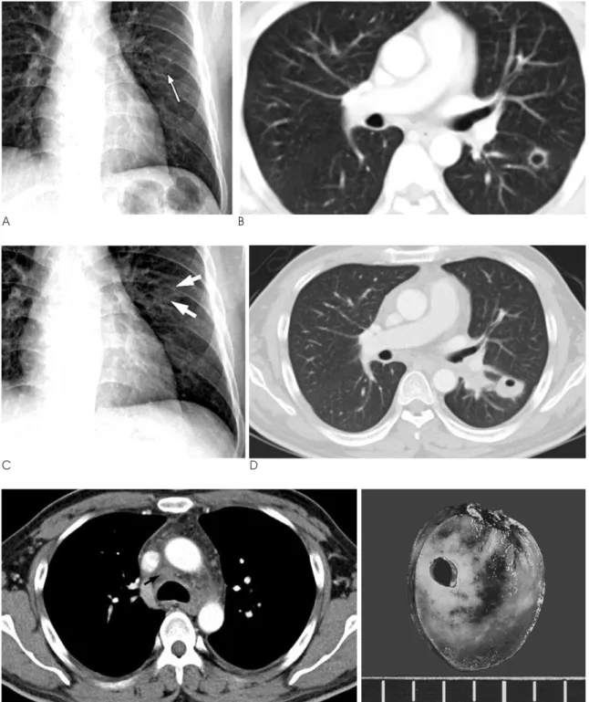
cough of 3 days duration. The laboratory findings were an erythrocyte sedimentation rate (ESR) of 24 mm/hr, a C-reactive protein (CRP) level of 77 mg/L, the cytoplas- mic antineutrophil cytoplasmic antibody (cANCA) was negative and the perinuclear anti-neutrophilic cytoplas- mic antibody (pANCA) was negative. The chest radiog- raphy on admission revealed a cavitary lesion in the left middle lung zone (Fig. 1A). The chest CT image showed a thin-walled cavitary lesion in the superior segment of the left lower lobe (Fig. 1B). The upper level of the chest CT image, through the carina, revealed multiple lym- phadenopathies in the mediastinum. The patient was treated with antibiotics and then he was discharged af- ter the fever and cough had subsided.
About 2 months later, the patient was readmitted to our hospital after 2 days of high fever. The laboratory findings were a white blood cell count of 14, 500/uL, a CRP level of 103 mg/L, the C-ANCA and P-ANCA were negative and the sputum acid-fast bacilli was negative.
The antibody titer for Epstein-Barr virus was not elevat- ed. Chest radiography showed an ill-defined consolida- tion in the left middle lung field and a prominent left hilum (Fig. 1C). The follow-up chest CT images showed a thick-walled cavity in the left lower lobe that was
J Korean Soc Radiol 2010;62:37-40
─ 37 ─
Pulmonary Involvement of Peripheral T-cell Lymphoma, Unspecified: A Case Report1
Jin Chung, M.D., Hyo Sub Shim, M.D.2, Seok Jin Ham, M.D.3, Tae Hoon Kim, M.D., Sang Jin Kim, M.D.
Peripheral T-cell lymphoma is a rare type of lymphoma that’s derived from post- thymic lymphoid T cells. Pulmonary involvement of peripheral T-cell lymphoma of the unspecified type is very rare and the imaging findings of this illness have rarely been reported. We present here a case of peripheral T-cell lymphoma of the unspeci- fied type with a cavitary lesion in the lung parenchyma, and we pathologically con- firmed this illness by performing video-assisted thoracoscopic surgery.
Index words :Lymphoma, T-Cell, peripheral Tomography, X-Ray Computed Lung
1Department of Radiology, Gangnam Severance Hospital, Yonsei University College of Medicine, Seoul, Korea
2Department of Pathology, Gangnam Severance Hospital, Yonsei University College of Medicine, Seoul, Korea
3Department of Thoracic Surgery, Gangnam Severance Hospital, Yonsei University College of Medicine, Seoul, Korea
Received April 13, 2009 ; Accepted July 21, 2009
Address reprint requests to : Tae Hoon Kim, M.D., Department of Radiology, Gangnam Severance Hospital, 146-92 Dogok-dong, Gangnam- gu, Seoul 135-720, Korea.
Tel. 82-2-2019-3517 Fax. 82-2-3462-5472 E-mail: [email protected]
more enlarged than that seen on the previous chest CT scan (Fig. 1D). The mediastinal window images of the chest CT scan revealed multiple lymphadenopathies with poorly defined margins in the mediastinum and an
irregular diffuse infiltration in the mediastinal fat (Fig.
1E).
The consolidation with the cavitary lesion was re- moved by performing video-assisted thoracoscopic
Jin Chung, et al: Pulmonary Involvement of Peripheral T-cell Lymphoma, Unspecified
─ 38 ─
A B
C D
F E
Fig. 1. A. The initial chest X-ray of a 39-years-old male shows a thin-walled cavitary lesion (arrow) in the left middle lung zone.
B. The chest CT images show a thin-walled cavitary lesion with a speculated margin in the superior segment of the left lower lobe.
C. The chest X-ray after 2 months shows consolidation (arrows) in the left middle lung zone.
D, E. The follow-up CT scan shows a thick-walled cavitary lesion, which has enlarged as compared to that seen on the previous chest CT scan in Figure 1B. An irregular diffuse infiltration in the mediastinal fat is visualized with a poorly defined lymph node (arrow) (E).
F. Grossly, the cut specimen shows parenchymal necrosis and hemorrhage with a cavitary lesion.
surgery. Macroscopically, the cut specimen showed parenchymal hemorrhage and necrosis with a cavitary lesion (Fig. 1F). Microscopically, the specimen showed a cavitary lesion and dilated vessels with surrounding he- morrhagic necrosis that showed angiocentric, pleomor- phic, atypical lymphoid cells on hematoxylin and eosin staining (Fig. 1G). Immunohistochemical staining show- ed positive reactions for cluster of differentiation 3 (CD 3), which is a T cell marker, and for T cell intracytoplas- mic antigen (TIA), which is a cytotoxic granule-associat- ed protein, in the pleomorphic atypical lymphoid cells.
Staining for CD56, a natural killer/T (NK/T) cell marker, and CD 20, a B cell marker, was negative (Fig. 1H).
Discussion
Peripheral T-cell lymphoma of the unspecified type accounts for 4 to 11% of all non-Hodgkin lymphomas, and pulmonary involvement occurs in 6 to 10% of all patients with PTCL-U (3, 4). The median age of onset for PTCL-U is reported to be in the seventh decade (4, 5).
This lymphoma usually takes an aggressive course, with relapses being more common than for B-cell lym- phomas. Most patients with PTCL-U present with nodal involvement, but the disease can also involve extranodal sites, including the liver, bone marrow, spleen, gastroin- testinal tract and skin (2).
On CT scans, the pulmonary manifestations of lym- phoma generally appear as multiple nodules or mass- like areas of consolidation with pleural effusion and
lymph node enlargement being visible in 42% and 35%
of patients, respectively, and without ground glass opac- ities or reticular opacities (6). Yet pulmonary involve- ment of PTCL-U has rarely been reported. Lee et al. (2) reported that generalized lymphadenopathies were the most common findings for this lymphoma. The CT find- ings of lymph node involvement in the neck of PTCL patients were reported as central necrosis, an ill-defined margin and heterogeneous enhancement (7). In our case, the lymph nodes lost their defined margins and there was an irregular diffuse infiltration in the medi- astinal fat. Mavi et al. (8) reported on the pulmonary in- volvement of PTCL with multiple cavitary lesions in both lungs. Our case also showed a thick-walled cavi- tary lesion on the CT images. This cavitary lesion in the lung pathologically represented necrosis, which is a rare finding of lymphoma (9). However, AIDS-related pul- monary lymphoma usually showed multiple nodules that may be cavitated (10).
Pathologically, the peripheral T-cell lymphomas, un- specified (PTCL-U) that do not match one of the defined entities of PTCL show a variety of cellular morpholo- gies, including medium-sized cells, mixed medium and large cells, large cells and lymphoepithelioid cells (2, 3).
Immunohistochemical staining of PTCL-U produces positive reactions for CD 3, a T cell marker, and TIA, a cytotoxic granule-associated protein in the pleomorphic atypical lymphoid cells, but there are negative reactions for CD56, a marker for NK/T cells, and CD 20, a B cell marker. Peripheral T-cell lymphoma, unspecified, also
J Korean Soc Radiol 2010;62:37-40
─ 39 ─
G H
Fig. 1. G. The microscopic image shows a cavitary lesion and dilated vessels with surrounding hemorrhagic necrosis (hematoxylin and eosin staining, ×40).
H. Immunohistochemical staining for CD 3 (T cell marker) gives a positive reaction.
gives a negative reaction for Epstein-Barr virus (EBV), and extranodal NK/T cell marker (3, 10).
In summary, we reported here on a case of peripheral T-cell lymphoma, unspecified, with pulmonary parenchymal involvement that presented as a thick- walled cavitary lesion. Lymphadenopathies with poorly defined margins and a surrounding infiltration in the mediastinal fat may also reflect the severity of the un- derlying disease, as was noted in this case of peripheral T cell lymphoma, unspecified.
References
1. Salar A, Fernandez de Sevilla A, Romagosa V, Domingo-Claros A, Gonzalez-Barca E, de Sanjose S, et al. Distribution and incidence rates of lymphoid neoplasms according to the REAL classification in a single institution. A prospective study of 940 cases. Eur J Haematol 1997;59:231-237
2. Lee HJ, Im JG, Goo JM, Kim KW, Choi BI, Chang KH, et al.
Peripheral T-cell lymphoma: spectrum of imaging findings with clinical and pathologic features. Radiographic 2003;23:7-28 3. Lee Y, Uhm JE, Lee HY, Park MJ, Kim H, Oh SJ, et al. Clinical fea-
tures and prognostic factors of patients with “peripheral T-cell lymphoma, unspecified”. Ann Hematol 2009;88:111-119
4. Montalban C, Obeso G, Gallego A, Castrillo JM, Bellas C, Rivas C.
Peripheral T-cell lymphoma: a clinicopathological study of 41 cas- es and evaluation of the prognostic significance of the updated kiel classification. Histopathology 1993;22:303-310
5. Savage KJ, Chhanabhai M, Gascoyne RD, Connors JM.
Characterization of peripheral T-cell lymphomas in a single North American institution by the WHO classification. Ann Oncol 2004;15:1467-1475
6. Lewis ER, Caskey CI, Fishman EK. Lymphoma of the lung: CT findings in 31 patients. AJR Am J Roentgenol 1991;156:711-714 7. Choi JW, Kim SS, Kim EY, Heran M. Peripheral T-cell lymphoma
in the neck: CT findings of lymph node involvement. AJNR Am J Neuroradiol 2006;27:1079-1082
8. Mavi A, Dhuriraj T, Cermik TF, Urhan M, Wasik M, Basu S, et al.
Central photopenic lesions on FDG-PET scan in a patient with pe- ripheral T-cell lymphoma. Ann Nucl Med 2008;22:629-633 9. Gadkowski LB, Stout JE. Cavitary pulmonary disease. Clin
Microbiol Rev 2008;21:305-333
10. Lopez-Guillermo A, Cid J, Salar A, Lopez A, Montalban C, Castrillo JM, et al. Peripheral T-cell lymphomas: initial features, natural history, and prognostic factors in a series of 174 patients di- agnosed according to the R.E.A.L. Classification. Ann Oncol 1998;
9:849-855
Jin Chung, et al: Pulmonary Involvement of Peripheral T-cell Lymphoma, Unspecified
─ 40 ─
대한영상의학회지 2010;62:37-40
미분류된 말초성 T세포림프종의 폐의 병발: 증례 보고1
1연세의대 강남세브란스병원 영상의학과
2연세의대 강남세브란스병원 병리과
3연세의대 강남세브란스병원 흉부외과
정 진∙심효섭2∙함석진3∙김태훈∙김상진
말초성 T세포림프종은 드문 림프종의 형태이다. 그 중에서도 분류되지 않은 말초성 T세포림프종이 폐에 생긴 경 우는 매우 드문 경우로 영상적 소견은 거의 보고된 것이 없다. 저자들은 비디오 보조 흉강경 수술 (video-assisted thoracoscopic surgery, 이하 VATS)로 병리적으로 확진 된 폐 실질의 미분류된 말초성 T세포림프종의 영상적 소견을 보고하고자 한다.