ISSN 0378-6471 (Print)⋅ISSN 2092-9374 (Online)
https://doi.org/10.3341/jkos.2019.60.6.594
Case Report
원발 안구 내 T세포 림프종
Primary Intraocular T-cell Lymphoma
신용일1⋅김주미1⋅이종주1⋅김정열1⋅김진만2⋅조영준1
Yong-Il Shin, MD1, Ju Mi Kim, MD1, Jong-Joo Lee, MD, PhD1, Jung-Yeul Kim, MD, PhD1, Jinman Kim, MD, PhD2, Young-Joon Jo, MD, PhD1
충남대학교 의과대학 안과학교실1, 충남대학교 의과대학 병리학교실2
Department of Ophthalmology, Chungnam National University College of Medicine1, Daejeon, Korea Department of Pathology, Chungnam National University College of Medicine2, Daejeon, Korea
Purpose: Intraocular lymphoma can be divided into primary and secondary usually involving B-cell lymphoma. Intraocular T-cell lymphoma is mostly secondary lymphoma while primary intraocular T-cell lymphoma is extremely rare. We report a case of pri- mary T-cell lymphoma.
Case summary: A 62-year-old male without any systemic disease presented with a floater in the right eye. A fundus examination showed multiple whitish retinal infiltrations in the right eye. Intraocular lymphoma was suspected, and systemic examination was performed, but all results were normal. During steroid treatment, previous lesions were enlarged, new lesions developed, and a diagnosis of primary T-cell lymphoma was made by diagnostic vitrectomy. Consecutive intravitreal injections of methotrexate were performed. After eight injections, the vitreous and retinal lesions improved but we decided to terminate the injections due to corneal epitheliopathy. The corneal epitheliopathy was recovered and the patient is currently undergoing periodic follow-ups without progression of the lesion.
Conclusions: Although intraocular T-cell lymphoma is a rare condition, this primary T-cell type should be considered when an in- traocular lymphoma lesion is suspected.
J Korean Ophthalmol Soc 2019;60(6):594-599
Keywords: Corneal epitheliopathy, Methotrexate, Primary intraocular T-cell lymphoma
■Received: 2018. 9. 20. ■ Revised: 2018. 11. 6.
■Accepted: 2019. 5. 16.
■Address reprint requests to Young-Joon Jo, MD, PhD
Department of Ophthalmology, Chungnam National University Hospital, #282 Munhwa-ro, Jung-gu, Daejeon 35015, Korea Tel: 82-42-280-7607, Fax: 82-42-255-3745
E-mail: [email protected]
* This work was supported by 2015 research fund of Chungnam National University.
ⓒ2019 The Korean Ophthalmological Society
This is an Open Access article distributed under the terms of the Creative Commons Attribution Non-Commercial License (http://creativecommons.org/licenses/by-nc/3.0/) which permits unrestricted non-commercial use, distribution, and reproduction in any medium, provided the original work is properly cited.
원발 안구 내 림프종은 뇌, 척수, 뇌척수막 또는 눈에서 발생하는 원발 중추신경계 림프종의 아형이다. 중추신경계 를 침범하는 비호지킨 림프종이 가장 흔하며 주로 광범위 큰 B세포로 구성되어 있고, T세포 림프종은 매우 드물다.1,2
안구 내 T세포 림프종의 대부분은 전신 림프종이 안구 내 로 전이된 이차성 림프종이다.3-5 원발성으로 밝혀진 안구 내 T세포 림프종은 전 세계적으로 매우 드물게 보고되었으 며,6,7 국내에서는 현재까지 이에 대한 보고된 바 없다. 저자 들은 단안에 발생한 원발 T세포 림프종 1예를 보고하고자 한다.
증례보고
기저질환이 없는 62세 남자 환자가 3개월 전부터 우안 부유물을 호소하여 타 병원에서 경구 스테로이드 및 스테 로이드 점안액으로 포도막염에 대한 치료를 하다가 호전이 없어 내원하였다. 양안 최대교정시력은 1.0이었고, 전안부
A B
C D
Figure 1. Fundus findings of a 62-year-old man with primary intraocular T-cell lymphoma. At the initial visit, fundus photography
shows a vitreous opacity and retinal infiltrations (A, white arrow). Fluorescein angiography (FA) shows multiple hyperfluorescent dots (B, yellow arrows). Three months later, the lesions were aggravated. Previous lesions were enlarged, and the number of yellow- ish retinal infiltrations was increased (C, white arrows; D, yellow arrows). A new lesion developed in the inferotemporal area (C, red arrow). Inferotemporal lesion shows a leopard spot pattern on FA (D, green arrow).에 특이 소견은 없었다. 안저검사에서 우안의 황반부 주변 에 흰색의 두 개의 둥근 망막침윤과 아래쪽 적도 부위에 다 수의 둥근 흰색 망막 침윤이 있었으며, 상이측 유수신경섬
유층이 보였고, +1 정도의 유리체 혼탁이 있었다. 형광안저 혈관조영술에서 침윤 부위에 여러 개의 과형광 반점이 관 찰되었다(Fig. 1A, B). 안구 내 림프종 의증하에 혈액종양
50 μm 50 μm
A B
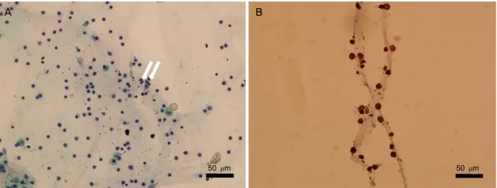
Figure 2. Microscopic features of vitreous tap. Hematoxylin and eosin stain reveals scattered small T-lymphocytes (arrows) with
macrophages (A, ×400). Immunohistochemical stain of vitreous shows positive for CD3 (B, ×400).A B C
Figure 3. Consecutive findings of toxic keratopathy followed by multiple intravitreal methotrexate (MTX) injections for intraocular
T-cell lymphoma. After 8 times intravitreal MTX injection, the patient demonstrated a severe corneal epitheliopathy (A, B). There were severe conjunctival injection, cystic change and limbal haziness causing the loss of palisades of Vogt (A). A month after treat- ment, the patient had improvement of his corneal epitheliopathy (C).내과에 의뢰하였다. 두경부 자기공명영상검사와 뇌척수액 검사 결과는 정상이었다. 혈액검사에서는 톡소카라증 양성 소견을 보였으나 위양성으로 판단하였다. 전신검사에서 정 상이었으며, 초기에 특이적인 망막 소견을 보이지 않아, 우 선 스테로이드 경구 복용을 시작하였다. 스테로이드 치료 중 부유물이 감소하는 양상을 보였으나, 초진 3개월 후 이 전 병변의 크기가 커지고, 하이측에 새로운 병변이 생겼으 며, 새로운 병변은 형광안저혈관조영술에서 leopard spot 형태를 보였다(Fig. 1C, D). 이에 스테로이드 치료를 중단 하고 유리체 천자를 시행하였다. 유리체의 세포검사 결과 T세포가 산재되어 있었고, 면역조직화학염색 결과 CD20 음성, CD3 양성이었다(Fig. 2). 진단적 유리체절제술을 시 행하였고, 면역조직화학염색 결과는 이전과 동일하였다. 원 발 안구 내 T세포 림프종을 진단하고 유리체 내 메토트렉 세이트(Methotrexate [MTX] 주사[0.4 mg/0.1 mL])를 시작
하였다. 1주에 2번씩, 4주간 총 8차례의 MTX 주입술 후 안 저병변의 크기와 개수는 감소하였고, 색이 옅어졌으나 결 막충혈, 표층점모양상피병증, 각막의 낭성 변화 및 윤부의 흐릿함이 관찰되어 독성각막병증 진단하에 MTX 주사를 중단하였다(Fig. 3A, B). 인공누액과 자가혈청 점안 및 엽 산 복용 1개월 후 각막은 회복되었고(Fig. 3C), 안저 병변은 호전된 상태로 유지되었다. 유리체절제술 6개월 후 우안 백 내장이 진행하여, 우안 수정체유화술 및 인공수정체삽입술 을 시행하였으며, 술 후 최대교정시력은 1.0이었다. 8차례 의 주사 치료 이후 이전의 병변 부위는 위축성, 색소성 변 화가 관찰되고, 형광안저혈관조영술에서 leopard spot 형태 를 동반한 창문형광을 보였다(Fig. 4). 병변의 악화 소견이 없어 유리체 내 주사를 시행하지 않고, 안저 병변의 변화 및 전신 전이 여부에 대하여 주기적인 경과관찰 중이다.
A B
C
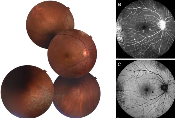
Figure 4. Fundus findings at 10 months after multiple intravitreal methotrexate injections. Fundus photograph shows atrophic and
pigmentary changes corresponding to the previous infiltrates lesions (A). Fluorescein angiography (FA) shows a window defect with leopard spot pattern, and mild disk leakage in the late phase (B). Fundus autofluorescence (FAF) shows hypoautofluorescent dot and granular appearance. Hypoautofluorescent spots on FAF are hyperfluorescent on FA (C).고 찰
안구 내 림프종은 전체 안구 내 악성 종양 중 1.86%를 차지하는 매우 드문 질환으로,8 원인에 따라 원발성과 이차 성으로 분류한다. 원발 안구 내 림프종(primary intraocular lymphoma)은 원발 중추신경계 비호치킨 림프종(primary central nervous system lymphoma)의 아형으로서 주로 유리 체와 망막을 침범하기 때문에 원발 유리체망막 림프종 (primary vitreoretinal lymphoma)으로도 불린다.9 이차성 안 구 내 림프종은 전신을 침범하는 악성 림프종의 전이 또는 이차적 발현으로 나타난다. 안구 내 림프종의 대부분은 B세 포에서 기원하며, 상대적으로 T세포 림프종은 매우 드문 것으로 알려져 있다. Hoffman et al10의보고에 의하면 중추 신경계 침범이 없고 안구 내에만 국한된 원발 안구 내 T세 포 림프종은 23예 중 2예(9%)로 드물었고, 대부분이 균상 식육종(mycosis fungoides)이나 전신 림프종과 연관된 이차 성 안구 내 림프종이었다.
안구 내 림프종은 비특이적인 양상으로 인해 포도막염으 로 오인되고 진단이 늦어지는 경우가 많다.11 기존 연구들 에서 안과적 증상의 발현 후 안구 내 림프종 확진에 걸리는
시간이 평균 10.2개월로 보고되었다.10 본 증례에서도 타 병 원에서 경구 스테로이드 및 스테로이드 점안액으로 포도막 염에 대한 치료를 3개월간 하다가 호전이 없어 본원에 내 원하였고, 증상이 발생한 시점으로부터 안구 내 T세포 림 프종을 확진받기까지 약 6개월이 소요되었다. 검사가 까다 로운 점 역시 안구 내 림프종의 진단을 어렵게 한다. 임상 소견상 안구 내 림프종이 의심되나 전신에 이상 소견이 없 는 경우 원발 안구 내 림프종에 대한 침습적인 조직검사가 필요하다. 유리체 또는 망막 조직을 채취해야 하며 그 검체 의 양은 매우 제한적이다. 또한 유리체는 많은 반응성 T림 프구, 괴사된 세포 및 잔해, 피브린을 포함하고 있어 악성 세포를 검출하는 데 혼란을 줄 수 있다.12 림프종 세포는 매 우 약하므로 검체 채취 즉시 검사실로 보내져야 하며, 검체 를 처리하는 과정에서 종양 세포가 소실되어 검사의 민감 도가 매우 낮다. 특히 T세포 림프종은 종양 세포 크기가 다 양하여 악성 종양 세포와 비감염성 염증 반응을 감별해야 하는 어려움이 따른다. 조직검사 전 스테로이드 치료를 받 은 림프종 환자들은 종양 세포가 손상되고 이로 인해 위음 성을 나타낼 가능성이 높아진다는 것도 염두에 두어야 한 다.11 중합효소 연쇄반응검사(polymerase chain reaction)를
통한 T세포 수용체 유전자의 재배열 확인, 유세포 분석 (flow cytometry), 효소결합 면역흡착검사(enzyme-linked im- munosorbent assay)로 알아본 사이토카인 interleukin (IL)-10 과 IL-6의 비율 등 추가적 검사 결과가 진단의 정확도를 높 이는 데 도움이 될 수 있다.
본 증례에서는 환자에게 초진 시와 다른 새로운 망막침 윤이 발생하여 스테로이드 치료를 중단하고 유리체 천자를 시행하였다. 유리체의 세포검사 결과 T세포가 산재되어 있 었고, 면역조직화학염색 결과 CD20 음성, CD3 양성이었 다. 진단적 유리체절제술을 통한 세포학적 검사에서 역시 동일한 결과를 보였다. 두경부 자기공명영상검사와 뇌척수 액검사 결과는 정상으로 원발 T세포 림프종을 진단하였다.
중추 신경계에 침범하지 않은 원발 안구 내 림프종의 일 차적 치료로 유리체 내 MTX 주사를 시행할 수 있다.13,14 0.4 mg/0.1 mL의 MTX를 2회/주, 총 4주간 유도 요법, 1회/주, 8주 동안 강화 요법, 1회/월, 총 9개월간 주입하는 유지 요 법 프로토콜을 보고하였다.13 유리체 내 MTX 주사는 전신 항암 치료 및 방사선 치료보다 합병증과 재발이 적다고 알 려져 있다. Frenkel et al14은 평균 6.4회의 MTX 주사 이후 임상적 관해 상태가 되었고, 10년의 경과관찰 동안 안구 내 림프종의 재발은 없었다고 보고하였다. 본 증례에서도 1주에 2번씩, 4주간 총 8차례의 유리체 내 MTX 주사 이후 안저 의 병변은 호전되었지만, 심한 독성각막병증이 발생하였다.
유리체 내 MTX 주사는 백내장, 각막상피병증, 전방 내 신 생 혈관 생성 등의 합병증이 발생할 수 있다.13 각막상피병 증이 발생한 환자에서 MTX 주사의 중단 및 인공누액, 자 가혈청 및 경구 엽산의 복용15을 통해 각막 합병증은 호전 되었다.
원발 안구 내 림프종 환자에서는 주기적인 경과관찰이 필요하다. Chaput et al3의 보고에 의하면 원발 안구 내 T세 포 림프종 환자 두 명에서 각각 진단 8개월, 36개월 후에 중추신경계와 피부 전이가 발생하였다. 따라서 안과적인 치료뿐만 아니라 전신 전이 여부에 대한 혈액종양내과 의 사와의 협진을 통한 검사 및 주기적인 추적관찰도 매우 중 요하다.
저자들은 단안의 유리체 침윤을 보인 환자에서 전신검사 결과 원발 부위는 찾을 수 없었고, 진단적 유리체절제술을 통해 원발 안구 내 T세포 림프종이 진단된 1예를 경험하였 으며, 유리체 내 MTX 주사를 통하여 안구 내 병변의 호전 을 보였기에 이를 문헌 고찰과 함께 보고하는 바이다. 본
증례에서 유리체의 면역조직화학염색검사 시 B세포 림프 종에 국한된 검사만 시행했다면 진단 및 치료에 더 많은 시 간이 소요되었을 것이다. 임상에서 안구 내 림프종이 의심 되는 환자에서 드물지만 T세포 림프종이 있을 수 있음을 고려해야 한다.
REFERENCES
1) Hong JT, Chae JB, Lee JY, et al. Ocular involvement in patients with primary CNS lymphoma. J Neurooncol 2011;102:139-45.
2) Suh MH, Yu HG. Clinical manifestations of intraocular lymphoma.
J Korean Ophthalmol Soc 2009;50:78-84.
3) Chaput F, Amer R, Baglivo E, et al. Intraocular T-cell lymphoma:
clinical presentation, diagnosis, treatment, and outcome. Ocul Immunol Inflamm 2017;25:639-48.
4) Hunyor AP, Harper CA, O'Day J, McKelvie PA. Ocular-central nervous system lymphoma mimicking posterior scleritis with exu- dative retinal detachment. Ophthalmology 2000;107:1955-9.
5) Saga T, Ohno S, Matsuda H, et al. Ocular involvement by a periph- eral T-cell lymphoma. Arch Ophthalmol 1984;102:399-402.
6) Lobo A, Larkin G, Clark BJ, et al. Pseudo‐hypopyon as the present- ing feature in B‐cell and T‐cell intraocular lymphoma. Clin Exp Ophthalmol 2003;31:155-8.
7) Char DH, Ljung BM, Deschênes J, Miller TR. Intraocular lympho- ma: immunological and cytological analysis. Br J Ophthalmol 1988;72:905-11.
8) Reddy EK, Bhatia P, Evans RG. Primary orbital lymphomas. Int J Radiat Oncol Biol Phys 1988;15:1239-41.
9) Turaka K, Bryan JS, De Souza S, et al. Vitreoretinal lymphoma:
changing trends in diagnosis and local treatment modalities at a single institution. Clin Lymphoma Myeloma Leuk 2012;12:412-7.
10) Hoffman PM, Mckelvie P, Hall AJ, et al. Intraocular lymphoma: a series of 14 patients with clinicopathological features and treat- ment outcomes. Eye (Lond) 2003;17:513-21.
11) Whitcup SM, de Smet MD, Rubin BI, et al. Intraocular lymphoma.
Clinical and histopathologic diagnosis. Ophthalmology 1993;100:
1399-406.
12) Sagoo MS, Mehta H, Swampillai AJ, et al. Primary intraocular lymphoma. Surv Ophthalmol 2014;59:503-16.
13) Smith JR, Rosenbaum JT, Wilson DJ, et al. Role of intravitreal me- thotrexate in the management of primary central nervous system lymphoma with ocular involvement. Ophthalmology 2002;109:
1709-16.
14) Frenkel S, Hendler K, Siegal T, et al. Intravitreal methotrexate for treating vitreoretinal lymphoma: 10 years of experience. Br J Ophthalmol 2008;92:383-8.
15) Gorovoy I, Prechanond T, Abia M, et al. Toxic corneal epitheliop- athy after intravitreal methotrexate and its treatment with oral folic acid. Cornea 2013;32:1171-3.
= 국문초록 =
원발 안구 내 T세포 림프종
목적: 안구 내 림프종은 원발성과 이차성으로 나눌 수 있으며, 대부분이 B세포 림프종이다. 안구 내 T세포 림프종은 대부분 이차성 림프종이며, 원발 안구 내 T세포 림프종은 극히 드물다. 저자들은 국내에서 아직 보고된 바가 없는 원발 T세포 림프종 1예를 보고하고 자 한다.
증례요약: 기저질환 없는 62세 남자 환자가 우안의 부유물을 주소로 내원하였다. 우안에 다수의 유리체 망막 침윤이 있어 안구 내 림프종 의증하에 전신검사를 시행하였으나, 모두 정상이었다. 스테로이드 치료를 시행한 후 이전의 병변이 커지고, 새로운 병변이 생겨 진단적 유리체절제술을 시행하여, 원발 안구 내 T세포 림프종으로 최종 진단하였다. 유리체 내 메토트렉세이트 주사를 시행하였 다. 8회의 주사 후 망막 및 유리체 병변은 호전되었으나 합병증으로 나타난 각막상피병증이 악화되어 치료를 중단하였다. 1달 뒤 각막 상태는 호전되었고 망막 및 유리체 병변은 호전된 채 유지되었다. 현재 전신 전이 여부 및 안저 소견 악화 여부에 대해 주기적인 경과관찰 중이다.
결론: 흔하지는 않지만 안구 내 림프종이 의심되는 환자에서 원발 안구 내 T세포 림프종이 있을 수 있음을 고려하여야 한다.
<대한안과학회지 2019;60(6):594-599>
신용일 / Yong-Il Shin
충남대학교 의과대학 안과학교실 Department of Ophthalmology, Chungnam
National University College of Medicine