A Case of Primary Intraocular Lymphoma Treated by Intravitreal Methotrexate
5
0
0
전체 글
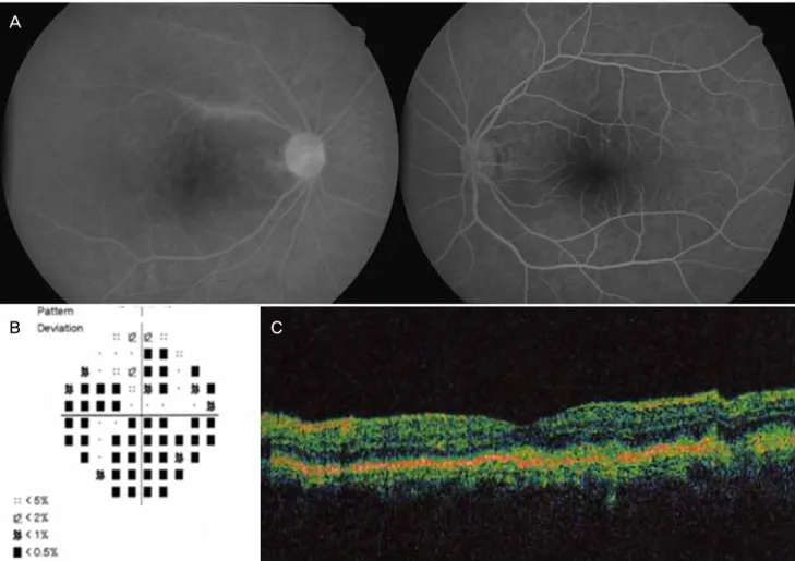
(2) E Kim, et al. A CASE OF PIOL TREATED BY INTRAVITREAL MTX. A. B. C. Fig. 1. (A) Fluorescein angiography; some perivascular dye leakage, disc hyperfluorescence and granular patterns of scattered hypofluorescent and hyperfluorescent spots are seen in the right eye. (B) Visual field of the right eye; considerable peripheral constriction is seen in pattern deviation. (C) OCT of the right eye; small RPE humps are seen.. mined by pattern visual evoked potentials (VEP). Prominent suppression of the central signal was noted in multifocal ERG of the right eye compared with that of the left eye (Fig. 2). After three months of failure to follow-up, the patient’s BCVA decreased to no light perception (NLP) OD and 20/200 OS, and active panuveitis was observed in the right eye. Shallow retinal detachment, vitreous debris and chorioretinal thickening were detected by ultrasonography. The left eye had mild uveitis and multiple vitreous seedings. It showed a granular pattern and leakage in angiography, small RPE humps in OCT, and considerable visual field constriction (Fig. 3). A thorough systemic evaluation was performed, but there was no evidence of intracranial or visceral involvement, and the hematologic exam was nonspecific except for an elevated erythrocyte sedimentation rate. Mega-dose intravenous steroid therapy was carried out, followed by diagnostic vitrectomy with retinal biopsy of the right eye. Diffuse large malignant B-cells with strong immunoreactivities in CD20 immunostaining were seen in the preretinal membrane biopsy specimen otherwise, no abnormal cells were obtained from the vitreous biopsy (Fig. 4). Intravitreal injections of MTX (800 μg/0.1 ml in the right eye, 400 μg/0.05 ml. in the left eye) were performed twice weekly for one month, once weekly for the next month, once every two weeks for the following month, followed by nine monthly injections. During the treatment, punctuate epithelial erosions and vortex keratopathy developed, but they subsided completely with topical instillation of 0.003% leucovorin eye drops and lubricants (Fig. 5). The final BCVA was NLP OD, 20/20 OS, and both eyes were free from malignant cells in vitreous biopsy six months later. The granular pattern on fluorescein angiography was persistent but there was no dye leakage (Fig. 6). In the left eye, the visual field constriction was slightly improved, and the small RPE humps in OCT had disappeared. Although P1 latency was markedly delayed in the right eye compared with that of the left eye, the P1 amplitudes showed a minimal difference in flash VEP. The EOG Arden ratio was 0.9604 OD and 1.231 OS. The amplitude of b waves was markedly decreased in a scotopic ERG of the left eye the photopic ERG response, oscillatory potentials and 30 Hz flicker response were relatively well preserved, with ERG waves being flat in the right eye. The patient was free of intracranial and ocular recurrence until six months later. No ocular complications except minimal opacities of the crystalline lenses were noted in both eyes. 211.
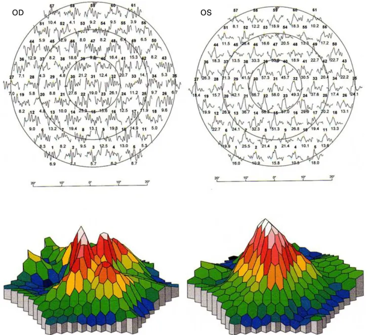
(3) Korean J Ophthalmol Vol.23, No.3, 2009. OD. OS. Fig. 2. Multifocal ERG; trace arrays and 3D-topography ofthe response density in the right eye shows prominent suppression of the central signal compared with that of the left eye.. D iscussion Of ocular masquerade syndromes, PIOL is reported to be the 14 4 most common. PIOL typically presents as a posterior uveitis non-specific findings of vitreous debris, chorioretinal thickening, widening of the optic nerve, elevated chorioretinal lesions, 15 and retinal detachment can be seen in ultrasonography. Subretinal and sub-RPE infiltrations of PIOL are reported to be common, and creamy-yellow lesions on a fundal exam might 16 be associated with PED, which was demonstrated in our OCT images. On fluorescein angiography, small sub-RPE infiltrations may appear as blocked hypofluorescence, and as 16 the tumor regresses, it can become an RPE window defect. In our case, vasculitis and disc leakage disappeared as the tumor 212. regressed; the granular pattern which might have been due to clumps of pigment epithelial cells, RPE atrophy and subretinal fibrosis, were persistent. Vitreous biopsy can be a useful tool to diagnose PIOL, but prior steroid therapy might suppress the number of vitreous cells, including lymphoma cells, which may 17 result in a negative vitreous cytology. Electrophysiological findings obtained from eyes with PIOL 18 were commented on in a brief report by Wang et al. which showed subnormal rod and cone responses. In our case, the b wave amplitude of the scotopic ERG of the left eye was markedly decreased; other ERG waves might be subnormal but were relatively well preserved. The EOG Arden ratio was significantly reduced in both eyes and could represent abnormal outer retinal and RPE functions..
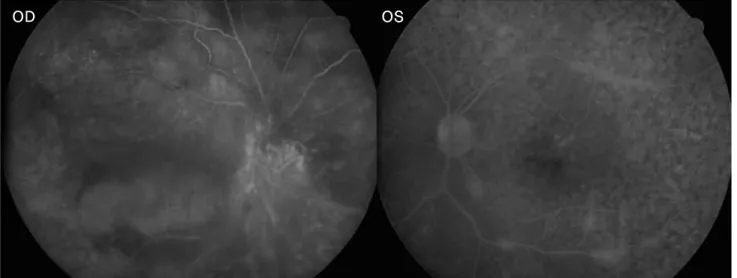
(4) E Kim, et al. A CASE OF PIOL TREATED BY INTRAVITREAL MTX. OD. OS. Fig. 3. Fluorescein angiography shows a granular pattern and leakage from vessels in the left eye.. A. B. Fig. 4. (A) H-E stain of the preretinal membrane shows a diffuse infiltration of atypically large lymphoid cells (×400). (B) CD20 immunostaining shows diffuse strong immunoreactivities in the tumor cells (×400).. OD. OS. Fig. 5. Toxic epithelial keratopathy is seen in both eyes.. 213.
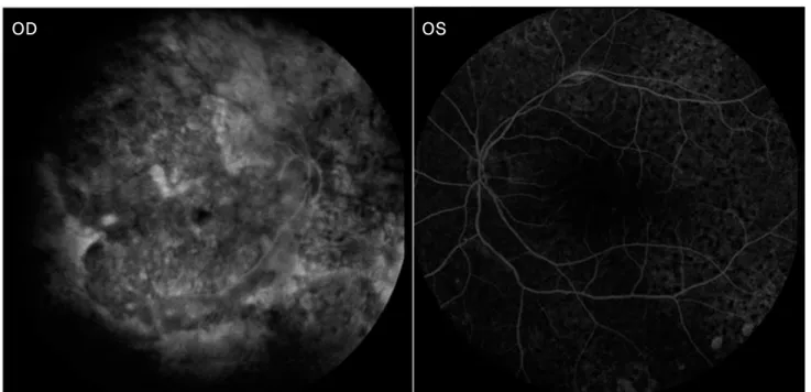
(5) Korean J Ophthalmol Vol.23, No.3, 2009. OD. OS. Fig. 6. Fluorescein angiography shows a persistent granular pattern but no dye leakage.. Intravitreal chemotherapy has been reported to have few complications,11,12 and because MTX is well tolerated by intraocular tissues, repeated injections are feasible without retinal toxicity.19 Toxic keratopathy can also be easily managed with topical leucovorin eye drops and lubricants. We modified the chemotherapy protocol of a previous report12 by adding two additional once-every-two-week injections for consolidation because the patient had useful vision in only one eye and any other treatment choice was not readily available. We also used a double dose regimen in the right eye where tumor involvement of the vitreous and retina was massive, no useful vision remained, and, being a vitrectomized eye, a shorter half-life of MTX was expected. Intravitreal chemotherapy was effective even though the patient had a history of systemic chemotherapy failure for PCNSL. In summary, primary intravitreal chemotherapy can be effectively used to treat PIOL and had minimal ocular complications in this patient.. References 1. Chan CC, Buggage RR, Nussenblatt RB. Intraocular lymphoma. Curr Opin Ophthalmol 2002;13:411-8. 2. Peterson K, Gordon KB, Heinemann MH, DeAngelis LM. The clinical spectrum of ocular lymphoma. Cancer 1993;72:843-9. 3. Davis JL. Diagnosis of intraocular lymphoma. Ocul Immunol Inflamm 2004;12:7-16. 4. Nussenblatt RB, Chan CC, Wilson WH, et al. International Central Nervous System and Ocular Lymphoma Workshop: recommendations for the future. Ocul Immunol Inflamm 2006;14:139-44. 5. Hochberg FH, Miller DC. Primary central nervous system lymphoma. J Neurosurg 1988;68:835-53. 6. Lee SH, Kim DJ, Kim IT. A Case of Primary Central Nervous System Lymphoma with Ocular Involvement. J Korean Ophthalmol. 214. Soc 2005;46:565-71. 7. Nelson DF, Martz KL, Bonner H, et al. Non-Hodgkin’s lymphoma of the brain: can high dose, large volume radiation therapy improve survival? Report on a prospective trial by the Radiation Therapy Oncology Group (RTOG): RTOG 8315. Int J Radiat Oncol Biol Phys 1992;23:9-17. 8. Batchelor TT, Kolak G, Ciordia R, et al. High-dose methotrexate for intraocular lymphoma. Clin Cancer Res 2003;9:711-5. 9. Berenbom A, Davila RM, Lin HS, Harbour JW. Treatment outcomes for primary intraocular lymphoma: implications for external beam radiotherapy. Eye 2007;21:1198-201. 10. Margolis L, Fraser R, Lichter A, Char DH. The role of radiation therapy in the management of ocular reticulum cell sarcoma. Cancer1980;45:688-92. 11. Sou R, Ohguro N, Maeda T, et al. Treatment of primary intraocular lymphoma with intravitreal methotrexate. Jpn J Ophthalmol 2008; 52:167-74. 12. Frenkel S, Hendler K, Siegal T, et al. Intravitreal methotrexate for treating vitreoretinal lymphoma: 10 years of experience. Br J Ophthalmol 2008;92:383-8. 13. Behin A, Hoang-Xuan K, Carpentier AF, Delattre JY. Primary brain tumours in adults. Lancet 2003;361:323-31. 14. Rothova A, Ooijman F, Kerkhoff F, et al. Uveitis masquerade syndromes. Ophthalmology 2001;108:386-99. 15. Ursea R, Heinemann MH, Silverman RH, et al. Ophthalmic, ultrasonographic findings in primary central nervous system lymphoma with ocular involvement. Retina 1997;17:118-23. 16. Corriveau C, Easterbrook M, Payne D. Lymphoma simulating uveitis (masquerade syndrome). Can J Ophthalmol 1986;21:144-9. 17. Whitcup SM, de Smet MD, Rubin BI, et al. Intraocular lymphoma. Clinical and histopathologic diagnosis. Ophthalmology 1993;100: 1399-406. 18. Wang JK, Yang CM, Lin CP, et al. An Asian patient with intraocular lymphoma treated by intravitreal methotrexate. Jpn J Ophthalmol 2006;50:474-8. 19. Velez G, Yuan P, Sung C, et al. Pharmacokinetics and toxicity of intravitreal chemotherapy for primary intraocular lymphoma. Arch Ophthalmol 2001;119:1518-24..
(6)
수치
관련 문서
버킷림프종(Burkitt lymphoma) 3) 명명에 –oma 가 포함되나 임상적으로 악성인 종양. 예) Glioma,
It considers the energy use of the different components that are involved in the distribution and viewing of video content: data centres and content delivery networks
After first field tests, we expect electric passenger drones or eVTOL aircraft (short for electric vertical take-off and landing) to start providing commercial mobility
• Phase II clinical trials of PI3Kδ inhibitor (idelalisib) showed dramatic response in patients with previously treated indolent non-hodgkin’s
1 John Owen, Justification by Faith Alone, in The Works of John Owen, ed. John Bolt, trans. Scott Clark, "Do This and Live: Christ's Active Obedience as the
In addition to the problem of this bias, the problem caused by using the time variable with a weighted wage rate may be a multicollinearity between time value (=time x
Anoxic
Median overall survival of 28 multiple myeloma patients treated with Cyclophosphamide-Prednisone combination regimens as a primary therapy was 115 weeks.. Median
