Veterinary Science
Transplantation of canine umbilical cord blood-derived mesenchymal stem cells in experimentally induced spinal cord injured dogs
Ji-Hey Lim
1, Ye-Eun Byeon
1, Hak-Hyun Ryu
1, Yun-Hyeok Jeong
2, Young-Won Lee
3, Wan Hee Kim
1, Kyung-Sun Kang
2,*, Oh-Kyeong Kweon
1,*
1
Department of Veterinary Surgery,
2Laboratory of Stem Cell and Tumor Biology, Department of Veterinary Public Health, College of Veterinary Medicine, Seoul National University, Seoul 151-742, Korea
3
College of Veterinary Medicine, Research Institute of Veterinary Medicine, Chungnam National University, Daejeon 305-764, Korea
This study was to determine the effects of allogenic umbilical cord blood (UCB)-derived mesenchymal stem cells (MSCs) and recombinant methionyl human granulocyte colony-stimulating factor (rmhGCSF) on a canine spinal cord injury model after balloon compression at the first lumbar vertebra. Twenty-five adult mongrel dogs were assigned to five groups according to treatment after a spinal cord injury: no treatment (CN); saline treatment (CP); rmhGCSF treatment (G); UCB-MSCs treatment (UCB-MSC); co-treatment (UCBG). The UCB- MSCs isolated from cord blood of canine fetuses were prepared as 10
6cells/150
µl saline. The UCB-MSCs were directly injected into the injured site of the spinal cord and rmhGCSF was administered subcutaneously 1 week after the induction of spinal cord injury. The Olby score, magnetic resonance imaging, somatosensory evoked potentials and histopathological examinations were used to evaluate the functional recovery after transplantation. The Olby scores of all groups were zero at the 0-week evaluation.
At 2 week after the transplantation, the Olby scores in the groups with the UCB-MSC and UCBG were significantly higher than in the CN and CP groups. However, there were no significant differences between the UCB-MSC and UCBG groups, and between the CN and CP groups. These comparisons remained stable at 4 and 8 week after transplantation. There was significant improvement in the nerve conduction velocity based on the somatosensory evoked potentials. In addition, a distinct structural consistency of the nerve cell bodies was noted in the lesion of the spinal cord of the UCB-MSC and UCBG groups. These results suggest that transplantation of the UCB-MSCs resulted in recovery of nerve function in dogs with a spinal cord injury and may be considered as a therapeutic modality for spinal cord injury.
Key words: dog, spinal cord injury, stem cell, transplantation, umbilical cord blood
Introduction
Regeneration of the central nervous system is limited after injuries related to ischemia, trauma and degenerative disease [33]. Recently, cell transplantation therapy has been considered for treatment in the field of regenerative medicine [3,27]. At the early stages of these studies, neural stem cells were obtained mainly from fetal tissue to explore their use for regeneration [5]. However, due to their limited availability alternative sources of cells for neural transplantation, such as embryonic, bone marrow, adipose and umbilical cord blood (UCB) stem cells are being evaluated [27,30,41]. The cells obtained from these additional sources can survive, proliferate, migrate, and differentiate into neuronal phenotypes in the damaged brain and spinal cord [1,21,34,40].
UCB stem cells, used for cell therapy, have advantages over the use of other sources of stem cells [4]. UCB cells have more mesenchymal progenitor cells per volume, and are more pluripotent and genetically flexible than bone marrow-derived mesenchymal stem cell [4,7]. In addition, it has been suggested that they are not as mature as other adult stem cells so that they may not elicit alloreactive responses that modulate the immune system [2,13,16,37]. Furthermore, they have been shown to have lower carcinogenic potential than embryonic stem cells [15].
Recently, UCB stem cells have been shown to differentiate into neural cells in vitro [7,8]. In addition, functional recovery after transplantation of UCB stem cells into an injured area of the spinal cord in vivo [40] and the efficacy of intravenous administration of UCB stem cells in traumatic brain injury have been demonstrated [20]. Furthermore, there has been a report on recovery after transplantation of UCB stem cells with neurotrophic factors [15]. The treatment with granulocyte colony-stimulating factor (GCSF) has been
*Corresponding author
Tel: +82-2-880-1248; Fax: +82-2-888-2866
E-mail: [email protected], [email protected]
shown to stimulate bone marrow stem cell proliferation and mobilization [25].
Although canine bone marrow-derived mesenchymal stem cells have been isolated and characterized in many studies, there are only a few reports on the isolation of mesenchymal stem cells from canine cord blood (UCB- MSCs) [22,34]. In this study, using a previously developed modified canine acute spinal cord injury model [19], we examined the effectiveness of canine UCB-MSCs and recombinant methionyl human GCSF (rmhGCSF) on the improvement of neurological function.
Materials and Methods
Animals
Twenty-five healthy adult mongrel dogs that weighed 3.77 ± 0.59 kg, were used for the experimental spinal cord injury study. This investigation was performed according to the guidelines of the ‘Guide for the Care and Use of Laboratory Animals’ of Seoul National University. The dogs were assigned, without bias, to five groups according to treatment after spinal cord injury. The control group with no treatment (CN: n = 5); the control group with media- injection (CP: n = 5); the group with subcutaneous injection of rmhGCSF (G: n = 5); the group with transplantation of canine UCB-MSCs into the spinal cord injury site (UCB- MSC: n = 5); the groups with transplantation of canine UCB-MSCs and subcutaneous injection of rmhGCSF (UCBG: n = 5).
Induction of spinal cord injury
The spinal cord injury (SCI) was performed using balloon compression methods as described previously [19]. Briefly, the dogs were anesthetized with intravenous administration of diazepam (Melode; Dong Wha Pharm, Korea) at a dose of 0.3 mg/kg and propofol (Anepol; Ha Na Pharm, Korea) at 6 mg/kg with atropine sulfate (Atropine; Je Il Pharm, Korea) at 0.05 mg/kg subcutaneously. Anesthesia was maintained by inhalation of 2% isoflurane (Aerane; Ilisung, Korea).
Datex-Ohmeda (Microvtec Display, UK) was used for monitoring of physiologic measures including rectal temperature, oxygen saturation and pulse rate during anesthesia. The dogs were placed in a ventral recumbent position. The hemilaminectomy was performed by a left paramedian approach at L
4. A three to five millimeter hole was made in the left vertebral arch at L
4using a high-speed pneumatic burr. A three-French embolectomy catheter (SORIN Biomedica, Italy) was inserted into the hole made at the L
4vertebral arch. The balloon was advanced, under fluoroscopic guidance, until the tip of the catheter was placed at the cranial margin of the L
1vertebral body. The balloon was then inflated 150 µl by injection of a contrast agent (Omnipaque; Amersham Health, Ireland) diluted 50 : 50 with saline. The soft tissues and skin were closed as per standard methods. The balloon
was fixed with a Chinese finger type suture, and then removed after 12 h. After the operation, the dogs were monitored in an intensive care unit, and if needed, manual bladder expression was performed at least three times daily until voluntary urination was established.
Preparations of canine UCB-MSCs
Cell collection: UCB was obtained during the Caesarian section of a 58 kg mongrel dog at Seoul National University Veterinary Medical Teaching Hospital. Eight ml of blood was collected using 10 ml plastic syringes that contained 2 ml of citrate-phosphate-dextrose anticoagulant.
Isolation and culture of UCB-MSCs: The low-density mononuclear cells were isolated using Ficoll-Plaque Plus (Amersham Biosciences, Sweden). Then, the cells were cultured in growth medium [Dulbeco’s Modified Eagle media-low glucose with the addition of 10% fetal bovine serum (Gibco-BRL, USA) with 2mmol/l L-glutamine (Gibco- BRL, USA)] and 0.3% penicillin-streptomycin (Gibco- BRL, USA) at 37
oC and 5% CO
2concentration [26]. The UCB-MSCs were cultured and the mononucleated cells that proliferated from the cord blood were characterized by FACS analysis [11]. The cells were prepared as 1 × 10
6in 150 µ l of saline solution for the injection.
Transplantation of canine UCB-MSCs and injection of rmhGCSF: Transplantation of canine UCB-MSCs was performed a week after the SCI. The dogs were anesthetized using the same methods described above. The CN group did not have any cells or medium transplanted after the injury. In the CP group, a laminectomy was performed and the injured site was exposed by a durotomy. One hundred and fifty micro liters of saline was injected into the spinal cord at three locations (center of the injury, 1.0 mm proximal and 1.0 mm distal to the injury, 3.0 mm in depth) using a 30 gauge needle. The incision region of the dura mater was sutured with 8.0 absorbable materials. The remaining lesion was closed routinely. In the UCB-MSCs group, 1 × 10
6of cells suspended in 150 µ l of saline solution were injected at the SCI site. In the UCBG group, 1 × 10
6of cells suspended in 150 µ l of saline solution were injected at the SCI site and 100 µ l of rmhGCSF solution (GCSF; Dong Wha Pharm, Korea) was injected subcutaneously for 7 days, once a day.
The rmhGCSF was prepared with 5 µ g/animal/day and diluted with sterilized saline. In group G, 100 µ l of rmhGCSF solution was injected using the same method as in the UCBG group.
Evaluations
Behavioral assessment: Behavioral assessment was
performed to evaluate the functional recovery of the hind
limbs. Each dog was videotaped for a minimum of 10 steps
from both sides and behind when walking on the floor. Dogs
with non-weight bearing on their hind limbs were also videotaped, supported by holding the base of their tail.
Using a 15-point scoring system (Olby score), the dogs’ gait was independently scored from the videotapes by two individuals unaware of the experimental conditions [24]. A mean score at 0, 2, 4, and 8 weeks after the SCI was calculated.
Somatosensory evoked potential assessment: Somato- sensory evoked potentials (SEP) were measured using the Neuropack 2 (Nihon Kohden, Japan) and two subdermal channels at 0, 4 and 8 weeks after the cell transplantation.
Channel 1 was located in the subdermal region at the midline between the sixth and seventh lumbar vertebra and channel 2 was positioned between the tenth and eleventh thoracic vertebra using platinum needle Grass stimulating electrodes (Astro-Med, USA). The posterior tibial nerve was stimulated for 0.2 msec, with 2 Hz and 3 mA [39]. The latency response was converted into the velocity as a measure of spinal cord dysfunction with the evoked potentials. Spinal conduction velocity (CV) from L
6~L
7to T
10~T
11was calculated by the following equation [39]:
Conduction velocity (m/sec) = distance (cm) of two points/
latency (msec) difference × 10.
Magnetic resonance image: Magnetic resonance imaging (MRI) was performed with a 0.2 Tesla Magnet scanner (Esaote, Italy) at a week after the SCI and at 4 and 8 weeks after the cell injection. The majority of the images obtained were interleaved 5.0 mm with a slice thickness of 5.0 mm.
The repetition time (TR) and time to echo (TE) were adjusted. T1-weighted (T1W) (TR/TE = 540/26 msec) and T2-weighted (T2W) echo (TR/TE = 380/90 msec) images were obtained. All dogs in each of the groups were examined and the spinal cord injury lesions expressed in T2W sagittal planes at 0, 4 and 8 weeks after the injury.
Histopathological assessment: To assess the histopathological changes, all dogs were euthanized at 8 weeks after the cell treatment. The spinal cords from T
10to L
4of all dogs were sampled and fixed in 10% buffered neutral formalin. And then the tissues were rountinely processed, embedded and sectioned at 4 µ m. These sections were mounted on silane- coated slide glass. The slides were stained with hematoxylin and eosin (H&E) to detect vacuolar formation and Luxol fast blue to identify myelin [6]. The volumes of the cavities in the damaged spinal cord were calculated from images of the transverse sections using image analyzer software (ImageJ version 1.37; National Institutes of Health, USA). The myelinated area was analyzed using the same software.
These two analyses were performed at the epicenter of the damaged spinal cord.
Statistical analysis: Data was expressed as the mean ±
SD. Statistical analysis was carried out using SPSS 12.0 software (SPSS, USA). The statistical significance of the differences among group means was assessed using a one- way ANOVA. A p < 0.05 was considered significant.
Results
Behavioral outcomes
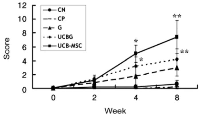
The Olby scores for the degree of nerve function, in all groups, was zero at 0 week. The Olby scores in the groups UCB-MSC, G and UCBG were increased 1.2, 0.8 and 1.4 at 2 weeks after treatment and 7.4, 3.0 and 4.2 at 8 weeks, respectively (Fig. 1). However, the increase in the Olby scores in the CN and CP groups was below 1.0 until 8 weeks. The Olby scores in the UCB-MSC and UCBG groups were significantly higher than in the CN and CP groups from 2 weeks onward after treatment. However, there were no significant differences between the UCB- MSC and UCBG groups and between the CN and CP groups. Group G did not show any significant difference from the other groups at 2 weeks. However, group G had significantly lower Olby scores than the UCB-MSC and UCBG groups at 4 and 8 weeks, although there were no differences between the UCB-MSC and UCBG groups at this time.
Somatosensory evoked potentials
The somatosensory evoked potentials of the posterior tibial nerves at the thoracic level revealed no response in all groups at 0 week (Table 1). At 4 weeks, it was possible to measure the evoked potentials in the G, UCB-MSC and UCBG groups. However, the CP and CN showed no responses until 8 weeks. The conduction velocities in G, UCB-MSC and UCBG at 8 weeks were improved up to 43.92 ± 37.77 m/s, 48.92 ± 26.13 m/s and 39.64 ± 30.99 m/
s, respectively.
Fig. 1.
Olby scores during 8 weeks. The groups of UCB-MSC
and UCBG improved more as shown with their functional scores
compared with the groups of CN and CP at 2 week after
transplantation (
p< 0.05). The UCBG and UCB-MSC groups
were significantly improved as compared with all other groups at
8 week after transplantation (
p< 0.05).
MRI results
Most dogs in all of the groups showed clear hyperintense signals in the T2W sagittal plane at the L
1lesion at 1 week after the SCI (Fig. 2). The CN and CP groups had a similar
appearance at the injured site that became a clearer hyperintense area, in the parenchyma at the L
1level, until 9 weeks after the SCI. The hyperintense area in the T2W sagittal plane at the L
1level was decreased in two dogs in the group G at 8 weeks. One of the dogs in the UCBG group had a slightly decreased hyperintense lesion in the T2W sagittal plane at the L
1level. In the UCB-MSC group, one dog showed a normointense intramedullar lesion but it was narrower than the near normal appearing areas.
Histopathological findings
At 8 weeks after treatment, the margins of normal grey and white matter were not identified in all of the dogs studied. There was a generalized infiltration of fibrous tissue and adhesions in the dura mater. Most of the dogs in the CN and CP groups showed damaged tissues and severe vacuolar formation. Cavity formation and Luxol fast blue positive area were very small in the CP group (Fig. 3A & B). The G
Table 1.
Mean conduction velocities calculated from the somatosensory evoked potentials at week 0, 4 and 8
Groups Weeks
0 4 8
CN NE NE NE
CP NE NE NE
G NE *22.24 ± 9.70* 43.92 ± 37.77 UCB- MSC NE 26.54 ± 9.48 48.92 ± 26.13 UCBG NE 025.44 ± 16.98 39.64 ± 30.99
*m/sec, CN: no treatment, CP: saline treatment, G: rmhGCSF treatment, UCB-MSC: umbilical cord blood-derived mesenchymal stem cell treatment, UCBG: co-treatment, NE: no effects. All values are mean ± SD.
Fig. 2.
Magnetic resonance images of the spinal cord in T2W sagittal view of the group CN (A & B), CP (C & D), G (E & F), UCB-
MSC (G & H) and UCBG (I & J). Most of the dogs were shown the clear localization of the spinal cord injury lesion (circle). A, C, E, G
and I: Before cell transplantation, 1 week after spinal cord injury. B, D, F, H and J: 8 weeks after cell transplantation.
group showed severe crushing damage in both the white and grey matters as well as cavity formation (Fig. 3C & D).
Although the UCB-MSC group showed similar histological findings with the other groups, neuronal cell like structures in a small area were observed (Fig. 3E-G). The mean percentage of cavities in the CN, CP, G, UCB-MSC and UCBG groups were 41.06± 10.39, 2.64± 4.55, 21.37 ±17.13, 6.55 ± 4.69, and 16.30 ± 11.46, respectively ( p < 0.05) (Table 2). The CN group showed a significantly higher percentage of cavity formation. The mean percentages of areas stained with Luxol fast blue in the CN, CP, G, UCB- MSC, and UCBG groups were 13.70 ± 9.27, 8.39 ± 9.63, 30.39 ± 13.65, 46.33 ± 7.02, and 33.87 ± 25.33, respectively ( p < 0.05).
Discussion
The Olby scoring system was used for quantitative evaluation of functional outcomes in this study. Olby et al . modified the BBB open field scoring system for dogs based on the pelvic limbs [24]. Several investigators have confirmed
the reliability of the Olby scoring system [23,38]. The spinal cord injury model in the present study had more than 75% of the spinal canal occluded during a 12 h period; the resulting lesions showed histopathologically severe hemorrhage and vacuolar formation. The dogs had paraplegia and were not expected to regain a normal gait without treatment [6] as demonstrated in the control groups, CN and CP, with or without saline injection. In the CN and CP groups, the dogs
Fig. 3.
Histopathological findings at 8 weeks after cell transplantation. (A) The epicenter of injured spinal cord of a dog in the group CP, which showed small cavity formation with a little Luxol fast blue positive area. Luxol fast blue stain. Counterstain with cresyl violet.
×12.5. (B) High magnification of A. ×40. (C) The epicenter of injured spinal cord of a dog in the group G, which was crushed and damaged in both white and grey matters with cavity formation. H&E stain. ×12.5. (D) Same lesion as C. It showed small amount of remained myelin (arrow). Luxol fast blue stain. Counterstain with cresyl violet. ×12.5. (E) The epicenter of injured spinal cord of a dog in the group UCB-MSC, which revealed abnormal structures, however it showed structural consistency with nerve cell body (circle).
H&E stain. ×12.5. (F) High magnification of E (circle). H&E stain. ×400. (G) Nerve cell body in the same lesion of F (arrows). Luxol fast blue stain. Counterstain with cresyl violet. ×400.
Table 2.
Percentages of cavity formation and Luxol fast blue staining positive area in transverse section of the epicenter of injured spinal cord
Groups Cavity formation (%) Positive area (%) CN 41.06 ± 10.39
a13.70 ± 9.27
a0
CP 2.64 ± 4.55
b8.39 ± 9.63
aG
b21.37 ± 17.13
ab30.39 ± 13.65
bUCB-MSC 6.55 ± 4.69
b46.33 ± 7.02
b0 UCBG
b16.30 ± 11.46
ab33.87 ± 25.33
ba,b