INTRODUCTION
Studies of diverse kinds of mesenchymal stem cells (MSCs) have recently been extended to include MSCs from many dif- ferent types of tissues. Although teeth are hard tissues, they contain stem cells originating from dental pulp, periodontal ligaments, apical papilla, and exfoliated deciduous teeth.
1-3MSCs modulate inflammatory reactions and suppress T- cell proliferation.
4One of the mechanisms of immune modu- lation by MSCs is the induction of regulatory T-cells (Tregs).
5-8pISSN: 0513-5796 · eISSN: 1976-2437
Received: May 12, 2017 Revised: June 20, 2017 Accepted: June 27, 2017
Corresponding author: Dr. Jong Won Hong, Department Plastic & Reconstructive Surgery, Severance Hospital, College of Medicine, Yonsei University, 50-1 Yonsei-ro, Seodaemun-gu, Seoul 03722, Korea.
Tel: 82-2-2228-2210, Fax: 82-2-393-6947, E-mail: [email protected]
•The authors have no financial conflicts of interest.
© Copyright: Yonsei University College of Medicine 2017
This is an Open Access article distributed under the terms of the Creative Com- mons Attribution Non-Commercial License (http://creativecommons.org/licenses/
by-nc/4.0) which permits unrestricted non-commercial use, distribution, and repro- duction in any medium, provided the original work is properly cited.
Yonsei Med J 2017 Sep;58(5):1031-1039 https://doi.org/10.3349/ymj.2017.58.5.1031
Immune Tolerance of Human Dental Pulp-Derived Mesenchymal Stem Cells Mediated by CD4 + CD25 + FoxP3 + Regulatory T-Cells and Induced
by TGF- β1 and IL-10
Jong Won Hong
1,2, Jung Hyun Lim
1,2, Chooryung J. Chung
3, Tae Jo Kang
1,4, Tae Yeon Kim
3, Young Seok Kim
1,2, Tae Suk Roh
1,2, and Dae Hyun Lew
1,21
Department of Plastic & Reconstructive Surgery, College of Medicine, Yonsei University, Seoul;
2
Institute for Human Tissue Restoration, College of Medicine, Yonsei University, Seoul;
3
Department of Orthodontics, College of Dentistry, Yonsei University, Seoul;
4
Yujin Plastic Surgery, Seoul, Korea.
Purpose: Most studies on immune tolerance of mesenchymal stem cells (MSCs) have been performed using MSCs derived from bone marrow, cord blood, or adipose tissue. MSCs also exist in the craniofacial area, specifically in teeth. The purpose of this study was to evaluate the mechanisms of immune tolerance of dental pulp-derived MSC (DP-MSC) in vitro and in vivo.
Materials and Methods: We isolated DP-MSCs from human dental pulp and co-cultured them with CD4
+T-cells. To evaluate the role of cytokines, we blocked TGF-β and IL-10, separately and together, in co-cultured DP-MSCs and CD4
+T-cells. We analyzed CD25 and FoxP3 to identify regulatory T-cells (Tregs) by fluorescence-activated cell sorting (FACS) and real-time PCR. We per- formed alloskin grafts with and without DP-MSC injection in mice. We performed mixed lymphocyte reactions (MLRs) to check immune tolerance.
Results: Co-culture of CD4
+T-cells with DP-MSCs increased the number of CD4
+CD25
+FoxP3
+Tregs (p<0.01). TGF-β or/and IL- 10 blocking suppressed Treg induction in co-cultured cells (p<0.05). TGF-β1 mRNA levels were higher in co-cultured DP-MSCs and in co-cultured CD4
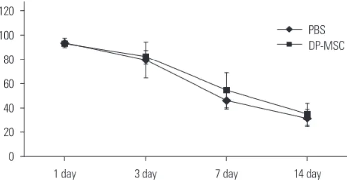
+T-cells than in the respective monocultured cells. However, IL-10 mRNA levels were not different. There was no difference in alloskin graft survival rate and area between the DP-MSC injection group and the non-injection group. None- theless, MLR was reduced in the DP-MSC injected group (p<0.05).
Conclusion: DP-MSCs can modulate immune tolerance by increasing CD4
+CD25
+FoxP3
+Tregs. TGF-β1 and IL-10 are factors in the immune-tolerance mechanism. Pure DP-MSC therapy may not be an effective treatment for rejection, although it may module immune tolerance in vivo.
Key Words: Dental pulp, mesenchymal stem cell, immune tolerance
In addition, soluble factors secreted from MSCs increase Tregs.
7Early studies of MSCs with diverse functions were performed mainly using MSCs derived from bone marrow (BM-MSCs).
5-7Because many kinds of MSCs have been found in different sources, MSC studies have expanded to study MSC character- istics depending on the specific source of the MSCs.
9-11Tolerance studies have indicated increased importance not only for solid organ transplantation but also for vascularized composite allotransplantation (VCA). Based on the concept of
“like with like,” the numbers of hand transplantations and fa- cial transplantations have been increasing, and satisfactory re- sults have been reported.
12,13Immunosuppressive agents should be used throughout a patient’s life after VCA; however, side ef- fects can include infection, malignancy, and diabetes. There- fore, studies of the immune tolerance of MSCs associated with VCA have been performed to reduce the requirement for im- munosuppressants.
14MSCs of dental origin are known to modulate immune resp- onses,
3,15-18although it is not yet known whether stem cells of various dental origins have the same mechanisms of immune tolerance as other stem cells. We investigated the mechanism of immune tolerance by dental pulp-derived MSC (DP-MSC).
We analyzed CD4
+CD25
+FoxP3
+Tregs to evaluate the effect of DP-MSCs on immune tolerance. We also investigated the ef- fects of the soluble factors TGF-β and IL-10, and we examined the immune tolerance effects in vivo.
MATERIALS AND METHODS
DP-MSC isolation and culture
We obtained normal human premolars extracted for orthodon- tic purposes and isolated DP-MSCs from the extracted teeth according to a previously published method.
19-22The process was performed with the approval of our Institutional Review Board (Yonsei University, Gangnam Severance Hospital, IRB#
3-2012-0082). We obtained teeth from a total of 6 patients. We cleaned the surface of the teeth with sterile normal saline. We then made a crack on the teeth by dental fissure burr in the ver- tical direction and cut the teeth using a pin cutter. After that, we extracted the dental pulp. We cut the pulp tissues to a size of 2×2×1 mm and cultured the pulp cells to produce outgrowths from the fragments.
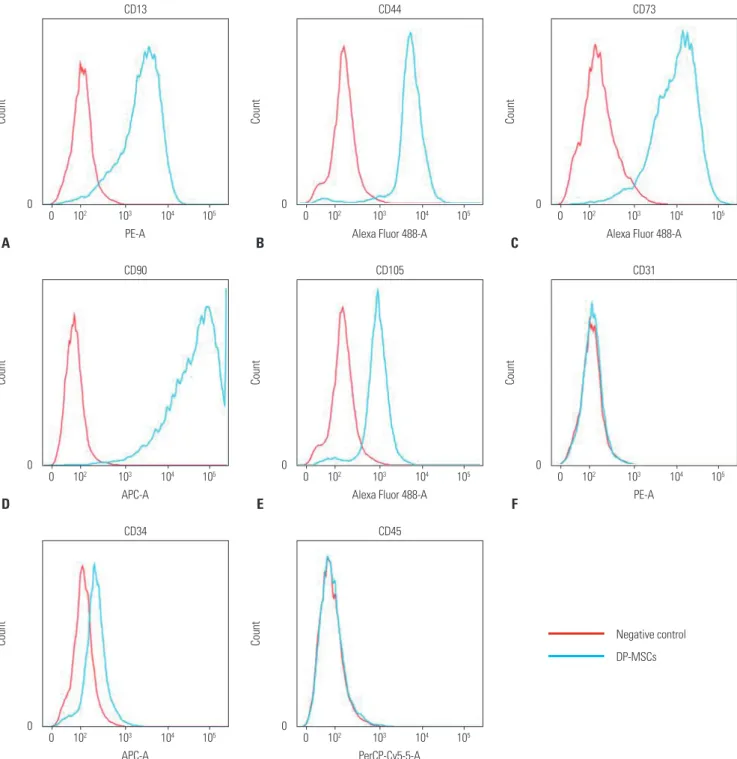
19,21Mesenchymal stem cell markers
We analyzed the membrane-associated molecules of the hu- man DP-MSCs by fluorescence activated cell sorting (FACS).
We analyzed CD13 (anti-human PE, eBioscience, San Diego, CA, USA), CD44 (anti-human Alexa Fluor 488, Molecular Probes, Rockford, IL, USA), CD73 (anti-human Alexa Fluor 488, Molecular Probes), CD90 (anti-human APC, Molecular Probes), CD105 (anti-human Alexa Fluor 488, Molecular Probes), CD31 (anti-human PE, BD Pharmingen, San Jose, CA, USA), CD34
(anti-human APC, Molecular Probes), and CD45 (anti-human PreCP-Cy5-5-A, Molecular Probes).
PBMC isolation & CD4
+T-cell preparation
We isolated human peripheral blood mononuclear cells (PB- MCs) from a healthy individual by density gradient centrifuga- tion using the Ficoll-Hypaque. We isolated CD4
+T-cells from the PBMCs using CD4
+T-cell isolation kit (Miltenyi Biotec, Ber- gisch Gladbach, Germany).
7We first counted the PBMCs and resuspended 1.0× 10
7cells in 80 μL of MACS buffer [0.5% bo- vine serum albumin in phosphate-buffered saline (PBS)]. We then added 20 μL of CD4 microbeads (Miltenyi Biotec) to the aspirated cells. We incubated the cell suspension for 15 min at 4°C and then applied the prepared cells to an MS column (Milt- enyi Biotec) in the magnetic field of the MACS separator. We collected the labeled cells on the tube after removing the col- umn from the separator.
Co-culture with and without TGF- β and IL-10 blocking We seeded 1.0×10
5DP-MSCs per well on a six-well culture plate. We then added 3.0×10
5CD4
+T-cells per well and incu- bated the cultures for 72 h at 37°C. To determine the effects of TGF-β and IL-10 on immune tolerance of the DP-MSCs, we add- ed anti-TGF-β (10 μg/mL; R&D systems, Minneapolis, MN, USA) and/or anti-IL-10 (0.05 μg/mL; R&D systems) to the co- cultured DP-MSCs and CD4
+T-cells. The co-culture groups with blocking were TGF-β single block, IL-10 single block, and TGF-β and IL-10 double block. All of the cultures were grown for 72 h for FACS and for 24 h for real-time PCR.
CD25 and FoxP3 analysis by FACS
To identify Tregs, we labeled the CD4
+T-cells cultured for 72 h for surface CD4 and CD25 and for intracellular FoxP3. We used CD4-FITC (anti-human CD4, Miltenyi Biotec), CD25-PE (anti- human CD25, BD Pharmingen) antibodies, and FoxP3-APC
Table 1. List of Primer Pairs
Gene Sequence
GAPDH Forward 5'-GGA GCC AAA AGG GTC ATC AT-3' (20 mer) Reverse 5'-GTG ATG GCA TGG ACT GTG GT-3' (20 mer) CD25 Forward 5'-ATC TGG TGG AAG ATC TCC CC-3' (20 mer) Reverse 5'-CTG CTG AAA ACT GCC GTG AT-3' (20 mer) FoxP3 Forward 5'-GCC ATG GAA ACA GCA CAT TC-3' (20 mer)
Reverse 5'-CTC ATT GAG TGT CCG CTG CT-3' (20 mer) TGF- β 1 Forward 5'-GCG GCA GCT GTA CAT TGA CT-3' (20 mer)
Reverse 5'-CCA CGT AGT ACA CGA TGG GC-3' (20 mer) TGF- β 2 Forward 5'-GGG TAC CTT GAT GCC ATC CC-3' (20 mer)
Reverse 5'-GCA ATA GGC CGC ATC CAA AG-3' (20 mer)
TGF- β 3 Forward 5'-TGA GCA CAT TGC CAA ACA GC-3' (20 mer)
Reverse 5'-GAG GCA GAT GCT TCA GGG TT-3' (20 mer)
IL-10 Forward 5'-TCA CCT TCC AGT GTC TCG GA-3' (20 mer)
Reverse 5'-TAG CTG GGA TTA CAG GTG CG-3' (20 mer)
(anti-human Foxp3, eBioscience). The final cell concentration for FACS was 1.0×10
5cells/mL. We performed FACS using a BD FACS Canto II Flow Cytometer and the FACSDiva software (BD Biosciences, Piscataway, NJ, USA).
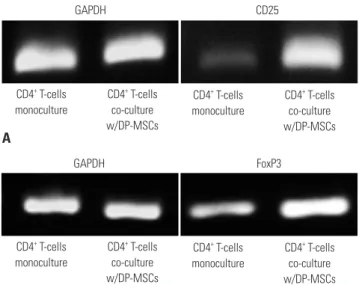
CD25, FoxP3, TGF- β 1, 2, 3, and IL-10 analysis by semi-quantitative PCR and real-time PCR
After culturing them for 24 h, we analyzed CD4
+T-cells for CD25
and FoxP3. We performed semi-quantitative PCR to analyze levels of CD25, FoxP3, IL-10, and TGF-β1, 2, and 3. We investi- gated the cytokine origin and variation according to the cells, CD4
+T-cells or DP-MSC. We performed different experiments:
DP-MSC monoculture, DP-MSC co-culture with CD4
+T-cells, CD4
+T-cell monoculture, and CD4
+T-cell co-culture with DP- MSCs. Because we did not observe any difference between mo- noculture and co-culture in TGF-β2, 3 levels, we performed re-
0
Count
PE-A
0 10
210
310
410
5CD13
A
0
Count
APC-A
0 10
210
310
410
5CD90
D
0
Count
APC-A
0 10
210
310
410
5CD34
G
0
Count
Alexa Fluor 488-A 0 10
210
310
410
5CD73
C
0
Count
PE-A
0 10
210
310
410
5CD31
F 0
Count
Alexa Fluor 488-A 0 10
210
310
410
5CD44
B
0
Count
Alexa Fluor 488-A 0 10
210
310
410
5CD105
E
0
Count
PerCP-Cy5-5-A 0 10
210
310
410
5CD45
H
Negative control DP-MSCs
Fig. 1. DP-MSC surface marker analysis using flow cytometry. (A) CD13 positive, (B) CD44 positive, (C) CD73 positive, (D) CD90 positive, and (E) CD105
positive. (F) CD31 negative, (G) CD34 negative, and (H) CD45 negative. These signals are shown as blue lines, while isotype matched control antibod-
ies are shown as red lines. DP-MSC, dental pulp-derived mesenchymal stem cell.
7.00 6.00 5.00 4.00 3.00 2.00 1.00 0.00
Relative change
CD4
+T-cells co-culture w/DP-MSCs CD4
+T-cells
monoculture
Non block Block TGF-β Block IL-10 Block TGF-β, IL-10
†
*
†
†