313 http://dx.doi.org/10.4196/kjpp.2011.15.5.313
ABBREVIATIONS: EMF, extremely low frequency electromagnetic fields; ROCC, receptor operated Ca2+ channels; CRAC, Ca2+-release activated Ca2+.
Received October 11, 2011, Revised October 15, 2011, Accepted October 21, 2011
Corresponding to: Sang Soo Sim, College of Pharmacy, Chung-Ang University, 221, Huksuk-dong, Dongjak-gu, Seoul 156-756, Korea.
(Tel) 82-2-820-5615, (Fax) 82-2-821-7680, (E-mail) [email protected]
*First two authors contributed equally to this article.
Intracellular Ca
2+Mobilization and Beta-hexosaminidase Release Are Not Influenced by 60 Hz-electromagnetic Fields (EMF) in RBL 2H3 Cells
Yeon Hee Hwang1,*, Ho Sun Song1,*, Hee Rae Kim1, Myoung Soo Ko1, Jae Min Jeong1, Yong Ho Kim1, Jeong Soo Ryu1, Uy Dong Sohn1, Yoon-Myoung Gimm2, Sung Ho Myung3, and Sang Soo Sim1
1College of Pharmacy, Chung-Ang University, Seoul 156-756, 2Korea EMF Safety, Dankook University, Yongin 448-701, 3Smart Grid Research Division, Korea Electrotechnology Research Institute, Changwon 641-120, Korea
The effects of extremely low frequency electromagnetic fields (EMF) on intracellular Ca2+ mobiliza- tion and cellular function in RBL 2H3 cells were investigated. Exposure to EMF (60 Hz, 0.1 or 1 mT) for 4 or 16 h did not produce any cytotoxic effects in RBL 2H3 cells. Melittin, ionomycin and thapsigargin each dose-dependently increased the intracellular Ca2+ concentration. The increase of intracellular Ca2+ induced by these three agents was not affected by exposure to EMF (60 Hz, 1 mT) for 4 or 16 h in RBL 2H3 cells. To investigate the effect of EMF on exocytosis, we measured beta- hexosaminidase release in RBL 2H3 cells. Basal release of beta-hexosaminidase was 12.3±2.3% in RBL 2H3 cells. Exposure to EMF (60 Hz, 0.1 or 1 mT) for 4 or 16 h did not affect the basal or 1μ M melittin-induced beta-hexosaminidase release in RBL 2H3 cells. This study suggests that exposure to EMF (60 Hz, 0.1 or 1 mT), which is the limit of occupational exposure, has no influence on intracellular Ca2+ mobilization and cellular function in RBL 2H3 cells.
Key Words: EMF, Ca2+ mobilization, Exocytosis, Beta-hexosaminidase
INTRODUCTION
Many epidemiologic studies suggested the possibility that exposure to extremely low frequency electromagnetic fields (EMF) may be related to the risk of acute lymphoblastic leukemia in children [1-3]. There is a public concern about the possible adverse health effects associated with exposure to EMF. However, the mechanism of the interaction be- tween EMF and cellular systems is still unclear.
We previously reported that there are no significant ch- anges in phospholipase activity such as PLA2, PLC and PLD in RAW 264.7 cells and RBL 2H3 cells exposed to EMF (60 Hz, 0.1 or 1 mT) for 4 or 16 h [4]. However, cellular function may be affected by a variety of signaling pathways in addi- tion to the phospholipase pathway. Considering that many cellular functions are closely related to the increase of intra- cellular Ca2+ concentration, it is plausible that EMF expo- sure may cause alterations in Ca2+ mobilization via both ex- tracellular Ca2+ influx and intracellular Ca2+ release.
Ca2+ is a universal messenger that controls a variety of cell functions, including secretion. The increase of intracellular Ca2+ is caused by the influx of extracellular Ca2+ via Ca2+
channel, intracellular Ca2+ release via inositol-1,4,5-tripho-
sphate (IP3) [5,6] and inhibition of the Ca2+ pump that lowers the intracellular Ca2+ concentration. Intracellular Ca2+ mobi- lization can be regulated by a variety of exogenous stimulants.
Melittin-induced increase of intracellular Ca2+ is related to PLC-mediated inositol triphosphate (IP3) accumulation with receptor operated Ca2+ channels (ROCC) [7]. Ionomycin is a well-known ionophore and increases intracellular Ca2+ con- centration through Ca2+-release activated Ca2+ (CRAC) chan- nels [8]. Thapsigargin also increases intracellular Ca2+ con- centration via the inhibition of sarco/endoplasmic reticulum Ca2+ ATPase [9]. In this study, we investigated the effect of EMF on intracellular Ca2+ mobilization stimulated by me- littin, ionomycin and thapsigargin and on beta-hexosamini- dase release in RBL 2H3 cells.
METHODS Materials
Melittin, ionomycin, thapsigargin and ρ-nitrophenyl-N- acetyl-β-glucosaminide were purchased from Sigma Che- mical Co. (St. Louis, MO, USA). Dulbecco's modified Eagle’s minimum essential medium (DMEM) and fetal bovine se- rum (FBS) were purchased from Invitrogen Co. (Grand Island, NY, USA). Fura-2/AM was purchased from Enzo Life Sciences (Plymouth, PA, USA). Other reagents were
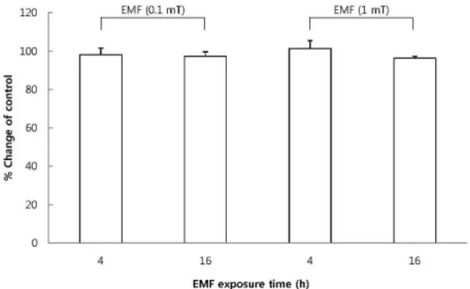
Fig. 1. The effect of EMF on cell viability of RBL 2H3 cells. The cells were exposed to EMF (60 Hz, 0.1 or 1 mT) for 4 or 16 h and viability was measured with MTT assay. Results are indicated in mean±S.D. from four separate experiments.
purchased from Sigma Chemical Co. (USA).
Cell culture
Rat basophilic leukemia (RBL 2H3) cells were grown in Dulbecco’s modified Eagle minimum essential medium (DMEM) supplemented with 10% fetal bovine serum (FBS) and antibiotic-antifungal mix (100 IU/ml penicillin G, 100μg/ml of streptomycin and 0.25μg/ml of amphotericin B) at 37oC in 5% CO2.
EMF exposure system
EMF generation equipment was designed and constructed by Korea Electrotechnology Research Institute (Korea).
Monitoring of magnetic field was conducted under observation of the current injected to the exposure system, because the magnetic field is proportional to the injected current. The field generator consisted of four square-shaped coils and one cage with three testing floors (top, middle and bottom floor).
The voltage fluctuation rate and the harmonic rate of the power quality using a power amp was under 1%. After fix- ing the magnetic field of the center of the middle floor to 1 mT, the fields at various points were measured. The spa- tial variation of the magnetic field was less than 3%. This strongly demonstrates that the field generator is well suit- ed for a small-scale in vitro study. Using a water-jet cooling system, the temperature in the incubator at 1 mT was maintained at 37±0.3oC. Also, a magnetic field shielding system using ferrite material was adopted to shield the strong magnetic field in the outer regions of the EMF ex- posure system. The coils were turned on for at least 30 min before use, and the cells were exposed to 0.1 mT and 1 mT in 60 Hz magnetic field for 4 h and 16 h. All experiments were conducted under the same environmental conditions.
MTT assay
Cell viability was performed with an MTT-based colori- metric assay [10]. Cells in 96-well plates (1×10⁵ cells/well) were exposed to EMF at 37oC for 4 or 16 h. 20μl of MTT solution (5 mg/ml in phosphate buffered saline) was added and further incubated for 3 h. After aspirating the super- natant from the wells, 100μl of dimethyl sulfoxide was added to dissolve the formazan crystals. The absorbance of each well was then read at 520 nm.
Measurement of intracellular Ca2+ mobilization The intracellular Ca2+ level was measured using Fura-2/
AM by monitoring a fluorospectrometer [11]. Briefly, cul- ture medium was replaced and cells were washed 3 times with PBS. After that, cells were detached by trypsin and suspended with 10 ml Krebs solution, then loaded with Fura-2/AM to a final concentration of 2μM and incubated at 37oC for 1 h. The loaded cells were washed twice with Krebs solution and centrifuged at 3,000×g for 10 min. The Fura-2 fluorescence was monitored on a Quanta Master (Qm4, Photon Technology International, NJ, USA.) at 37oC with excitation at 340 and 380 nm and emission at 500 nm. The ratio of F340/F380 was recorded, and the maximum fluorescence ratio (Rmax) was measured by using 0.1%
Triton X-100. The minimum fluorescence ratio (Rmin) was measured following depletion of external Ca2+ by addition of 5 mM EGTA/Tris pH8.5.
Measurement of beta-hexosaminidase release
To investigate the effect of EMF on exocytosis, we measured beta-hexosaminidase release in RBL 2H3 cells. RBL 2H3 cells exposed to EMF were washed with PIPES buffer 3 times and then suspended in PIPES buffer. The cells were stimulated with 0.5μM melittin for 30 min at 37oC [12]. The cell suspen- sion was centrifuged at 300×g for 10 min. 20μl of the super- natant was incubated with an equal volume of substrate sol- ution (2 mM ρ-nitrophenyl-N-acetyl-β-glucosaminide, 0.1 M citrate, pH 4.5) in duplicate in 96-well plates for 1 h at 37oC. The reaction was stopped by adding 200μl of 0.1 M sodium carbonate buffer, pH 10.0 [13]. The absorbance was measured at 405 nm with fluorospectrophotometer (FL600 Microplate Fluorescence Reader, Bio-tek).
Statistical analysis
Results are presented as mean±S.D. and were analyzed statistically by analysis of variance (ANOVA). Differences be- tween groups were determined with the Newman-Keul’s test.
The level of significance was set at less than 5% (p<0.05).
RESULTS
Effect of EMF on the viability of RBL 2H3 cells To exclude the possibility that EMF may affect cellular viability, we first confirmed the cytotoxicity of EMF using an MTT assay. The exposure to EMF (60 Hz, 0.1 or 1 mT) for 4 or 16 h did not produce any cytotoxic effects in RBL 2H3 cells (Fig. 1).
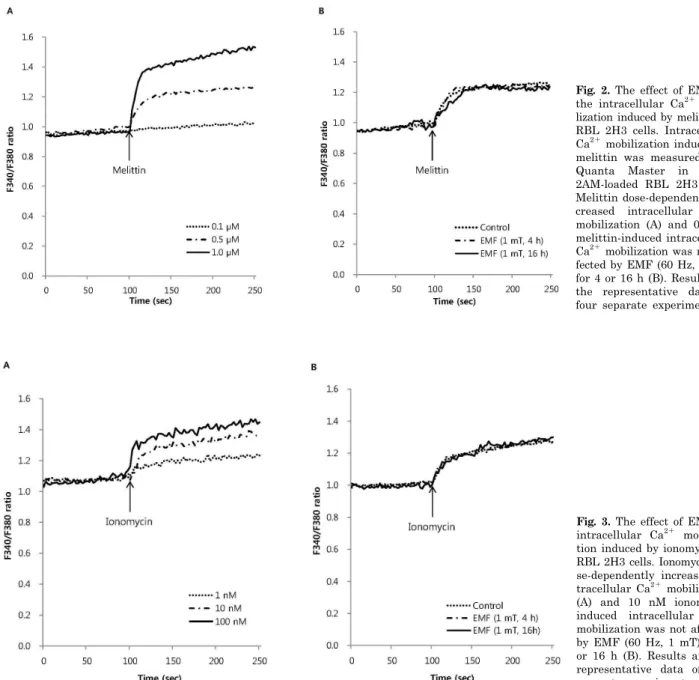
Effect of EMF on intracellular Ca2+ mobilization To investigate the effect of EMF on the influx of ex- tracellular Ca2+ via receptor operated Ca2+ channels (ROCC), we measured melittin-induced intracellular Ca2+
mobilization in RBL 2H3 cells exposed to EMF (60 Hz, 1 mT) for 4 or 16 h. Melittin dose-dependently increased the intracellular Ca2+ concentration (Fig. 2A). EMF did not af-
Fig. 2. The effect of EMF on the intracellular Ca2+ mobi- lization induced by melittin in RBL 2H3 cells. Intracellular Ca2+ mobilization induced by melittin was measured with Quanta Master in Fura- 2AM-loaded RBL 2H3 cells.
Melittin dose-dependently in- creased intracellular Ca2+
mobilization (A) and 0.5μM melittin-induced intracellular Ca2+ mobilization was not af- fected by EMF (60 Hz, 1 mT) for 4 or 16 h (B). Results are the representative data of four separate experiments.
Fig. 3. The effect of EMF on intracellular Ca2+ mobiliza- tion induced by ionomycin in RBL 2H3 cells. Ionomycin do- se-dependently increased in- tracellular Ca2+ mobilization (A) and 10 nM ionomycin- induced intracellular Ca2+
mobilization was not affected by EMF (60 Hz, 1 mT) for 4 or 16 h (B). Results are the representative data of four separate experiments.
fect the 0.5μM melittin-induced intracellular Ca2+ mobi- lization (Fig. 2B). As an ionophore, ionomycin increased in- tracellular Ca2+ concentration in a dose-dependent manner (Fig. 3A), while thapsigargin significantly increased the concentration via inhibition of sarco/endoplasmic reticulum Ca2+ ATPase in RBL 2H3 cells (Fig. 4A). EMF did not influ- ence the intracellular Ca2+ concentration induced by 10 nM ionomycin or 100 nM thapsigargin (Fig. 3B, 4B). EMF (60 Hz, 0.1 mT) for 4 or 16 h did not change the intracellular Ca2+ concentration induced by 0.5μM melittin, 10 nM ion- omycin or 100 nM thapsigargin (data not shown).
Effect of EMF on beta-hexosaminidase release in RBL 2H3 cells
To investigate the effect of EMF on exocytosis, we meas-
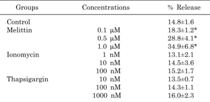
ured beta-hexosaminidase release in RBL 2H3 cells. Basal release of beta-hexosaminidase was 12.3±2.3% in RBL 2H3 cells. Melittin increased beta-hexosaminidase release in a dose-dependent manner, whereas ionomycin and thapsi- gargin did not cause beta-hexosaminidase release (Table 1).
Exposure to EMF (60 Hz, 0.1 or 1 mT) for 4 or 16 h did not affect the basal and 1μM melittin-induced beta-hex- osaminidase release in RBL 2H3 cells (Fig. 5).
DISCUSSION
The aim of this study was to investigate the effect of EMF on intracellular Ca2+ mobilization and exocytosis of be- ta-hexosaminidase in RBL 2H3 cells. Before measuring the exocytosis of beta-hexosaminidase, it was very important
Fig. 4. The effect of EMF on intracellular Ca2+ mobiliza- tion induced by thapsigargin in RBL 2H3 cells. Thapsi- gargin dose-dependently inc- reased intracellular Ca2+ mo- bilization (A) and 100 nM thapsigargin-induced intrac- ellular Ca2+ mobilization was not affected by EMF (60 Hz, 1 mT) for 4 or 16 h (B). Re- sults are the representative data of four separate experi- ments.
Table 1. Dose-response of beta-hexosaminidase release by melittin, ionomycin and thapsigargin in RBL 2H3 cells
Groups Concentrations % Release
Control 14.8±1.6
Melittin 0.1 μM 18.3±1.2*
0.5 μM 28.8±4.1*
1.0 μM 34.9±6.8*
Ionomycin 1 nM 13.1±2.1
10 nM 14.5±3.6
100 nM 15.2±1.7
Thapsigargin 10 nM 13.5±0.7
100 nM 14.3±1.1
1000 nM 16.0±2.3
*Significantly different from control (p<0.05).
Fig. 5. The effect of EMF on basal (A) and 1μM melittin-induced beta-hexosaminidase release (B) in RBL 2H3 cells. Both basal and 1μM melittin-induced beta-hexosaminidase release were not affected by exposure to EMF (60 Hz, 0.1 or 1 mT) for 4 or 16 h.
Results indicate mean±S.D. from four separate experiments.
to confirm the cytotoxicity of EMF. Exposure to EMF (60 Hz, 0.1 or 1 mT) for 4 or 16 h did not show any cytotoxic activity in RBL 2H3 cells. It has been reported that 0.5 mT EMF has no significant effect on proliferation of human periph- eral blood mononuclear cells [14], but 20 mT EMF for up to 23 days could inhibit the growth of human mesenchymal stem cells [15]. The cytotoxicity of EMF may be dependent on the intensity of EMF. The current data did not reveal any cytotoxic activity in RBL 2H3 cells, suggesting that exocytosis of beta-hexosaminidase may be independent on cell membrane rupture by EMF.
Ca2+ is a universal messenger that controls various cel- lular functions, including secretion, contraction, prolifera- tion and differentiation. Intracellular Ca2+ is increased by a variety of pathways, such as the influx of extracellular Ca2+, release of intracellular Ca2+ stores [5,6] and inhibi- tion of the Ca2+ pump that reduces intracellular Ca2+ con- centration. In this experiment, melittin, ionomycin and thapsigargin each dose-dependently increased intracellular Ca2+ concentration. The increase of intracellular Ca2+ in- duced by these three agents was not affected by exposure to EMF (60 Hz, 1 mT) for 4 or 16 h in RBL 2H3 cells. The melittin-induced increase of intracellular Ca2+ was related to PLC-mediated inositol triphosphate (IP3) accumulation
with receptor operated Ca2+ channels (ROCC) [7]. Ionomy- cin is a well-known ionophore and increases intracellular Ca2+ concentration through Ca2+-release activated Ca2+
(CRAC) channels [8]. Thapsigargin also increases intra-
cellular Ca2+ concentration via the inhibition of sarco/endo- plasmic reticulum Ca2+ ATPase [9]. These data suggest that EMF (60 Hz, 0.1 or 1 mT) for 4 or 16 h does not influ- ence intracellular Ca2+ mobilization via the influx of ex- tracellular Ca2+, the release of intracellular Ca2+ store or the Ca2+ pump in RBL 2H3 cells. However, there has been much debate regarding the effect of EMF on intracellular Ca2+ concentration: EMF has been shown to increase intra- cellular Ca2+ concentration [16,17], to decrease it [18] and to have no effect on it [19,20]. Such different effects of EMF on intracellular Ca2+ concentration may be due to the cell types used and the experimental conditions.
To investigate the effect of EMF on exocytosis, we meas- ured beta-hexosaminidase release in RBL 2H3 cells. Expo- sure to EMF (60 Hz, 0.1 or 1 mT) for 4 or 16 h did not affect the basal and 1μM melittin-induced beta-hex- osaminidase release in RBL 2H3 cells. This data agreed with the previous report that single exposure to EMF did not cause the degranulation of mast cells [21].
Guidelines on 50/60 Hz electromagnetic fields were is- sued by IRPA/INIRC in 1990. The basic hypothesis sug- gested that 50/60 Hz magnetic fields from external sources such as powerlines could be linked to an increased risk of childhood leukemia [1]. However, the mechanism under- lying the interaction between EMF and cellular functions remains elusive. It is therefore necessary to assess the changes of intracellular Ca2+ and cellular function caused by EMF in order to understand the possible adverse effects of EMF in leukocytes. In this study, we used a kind of leu- kocyte, RBL 2H3 cells (rat basophilic leukemia cells), and exposed the cells to EMF (60 Hz, 0.1 or 1 mT) for 4 or 16 h. It has been reported that the limit of EMF for general public exposure and occupational exposure are 0.2 mT and 1 mT, respectively [22]. The intensity of EMF used in this experiment is the limit of occupational exposure. Although further studies are necessary to identify the changes in in- tracellular Ca2+ mobilization and cellular function by EMF exposure, we suggest that EMF (60 Hz, 0.1 or 1 mT) for 4 or 16 h may have no influence on intracellular Ca2+ mobi- lization and cellular function in RBL 2H3 cells.
ACKNOWLEDGEMENTS
This work was supported by the Power Generation &
Electricity Delivery of the Korea Institute of Energy Technology and Planning (KETEP) grant funded by the Korean Government Ministry of Knowledge and Economy (No. 2009101030003E).
REFERENCES
1. Wertheimer N, Leeper E. Electrical wiring configurations and childhood cancer. Am J Epidemiol. 1979;109:273-284.
2. Floderus B, Persson T, Stenlund C, Wennberg A, Ost A, Knave B. Occupational exposure to electromagnetic fields in relation to leukemia and brain tumors: a case-control study in Sweden.
Cancer Causes Control. 1993;4:465-476.
3. Matanoski GM, Elliott EA, Breysse PN, Lynberg MC. Leukemia in telephone linemen. Am J Epidemiol. 1993;137:609-619.
4. Song HS, Kim HR, Ko MS, Jeong JM, Kim YH, Kim MC, Hwang YH, Sohn UD, Gimm YM, Myung SH, Sim SS. Effect of extremely low frequency electromagnetic fields (EMF) on phospholipase activity in the cultured cells. Korean J Physiol Pharmacol. 2010;14:427-433.
5. Berridge MJ, Irvine RF. Inositol trisphosphate, a novel second messenger in cellular signal transduction. Nature. 1984;312:
315-321.
6. Somlyo AV, Bond M, Somlyo AP, Scarpa A. Inositol tris- phosphate-induced calcium release and contraction in vascular smooth muscle. Proc Natl Acad Sci USA. 1985;82:5231-5235.
7. Nielsen OH, Bouchelouche PN, Berild D. Arachidonic acid and calcium metabolism in rnelittin stimulated neutrophils. Media- tors Inflamm. 1992;1:313-317.
8. Kahr H, Schindl R, Fritsch R, Heinze B, Hofbauer M, Hack ME, Mörtelmaier MA, Groschner K, Peng JB, Takanaga H, Hediger MA, Romanin C. CaT1 knock-down strategies fail to affect CRAC channels in mucosal-type mast cells. J Physiol.
2004;557:121-132.
9. Shukla N, Freeman N, Gadsdon P, Angelini GD, Jeremy JY.
Thapsigargin inhibits angiogenesis in the rat isolated aorta:
studies on the role of intracellular calcium pools. Cardiovasc Res. 2001;49:681-689.
10. Woerdenbag HJ, Merfort I, Passreiter CM, Schmidt TJ, Willuhn G, van Uden W, Pras N, Kampinga HH, Konings AW.
Cytotoxicity of flavonoids and sesquiterpene lactones from Arnica species against the GLC4 and the COLO 320 cell lines.
Planta Med. 1994;60:434-437.
11. Tseng PH, Lin HP, Hu H, Wang C, Zhu MX, Chen CS. The canonical transient receptor potential 6 channel as a putative phosphatidylinositol 3,4,5-trisphosphate-sensitive calcium entry system. Biochemistry. 2004;43:11701-11708.
12. Huang F, Yamaki K, Tong X, Fu L, Zhang R, Cai Y, Yanagisawa R, Inoue K, Takano H, Yoshino S. Inhibition of the antigen-induced activation of RBL-2H3 cells by sinomenine.
Int Immunopharmacol. 2008;8:502-507.
13. Hemmerling J, Nell S, Kipp A, Schumann S, Deubel S, Haack M, Brigelius-Flohé R. alpha-Tocopherol enhances degranulation in RBL-2H3 mast cells. Mol Nutr Food Res. 2010;54:652-660.
14. Ikeda K, Shinmura Y, Mizoe H, Yoshizawa H, Yoshida A, Kanao S, Sumitani H, Hasebe S, Motomura T, Yamakawa T, Mizuno F, Otaka Y, Hirose H. No effects of extremely low frequency magnetic fields found on cytotoxic activities and cytokine production of human peripheral blood mononuclear cells in vitro. Bioelectromagnetics. 2003;24:21-31.
15. Yan J, Dong L, Zhang B, Qi N. Effects of extremely low- frequency magnetic field on growth and differentiation of human mesen- chymal stem cells. Electromagn Biol Med. 2010;29:165-176.
16. Morabito C, Guarnieri S, Fanò G, Mariggiò MA. Effects of acute and chronic low frequency electromagnetic field exposure on PC12 cells during neuronal differentiation. Cell Physiol Bio- chem. 2010;26:947-958.
17. Gaetani R, Ledda M, Barile L, Chimenti I, De Carlo F, Forte E, Ionta V, Giuliani L, D'Emilia E, Frati G, Miraldi F, Pozzi D, Messina E, Grimaldi S, Giacomello A, Lisi A. Differentiation of human adult cardiac stem cells exposed to extremely low- fr- equency electromagnetic fields. Cardiovasc Res. 2009;82:411-420.
18. Bernabò N, Tettamanti E, Pistilli MG, Nardinocchi D, Berardinelli P, Mattioli M, Barboni B. Effects of 50 Hz extremely low frequency magnetic field on the morphology and function of boar spermatozoa capacitated in vitro. Therio- genology. 2007;67:801-815.
19. Pilger A, Ivancsits S, Diem E, Steffens M, Kolb HA, Rüdiger HW. No effects of intermittent 50 Hz EMF on cytoplasmic free calcium and on the mitochondrial membrane potential in human diploid fibroblasts. Radiat Environ Biophys. 2004;43:203-207.
20. Madec F, Billaudel B, Charlet de Sauvage R, Sartor P, Veyret B.
Effects of ELF and static magnetic fields on calcium oscillations in islets of Langerhans. Bioelectrochemistry. 2003;60:73-80.
21. Rajkovic V, Matavulj M, Johansson O. Combined exposure of peripubertal male rats to the endocrine-disrupting compound atrazine and power-frequency electromagnetic fields causes degranulation of cutaneous mast cells: a new toxic environmental hazard? Arch Environ Contam Toxicol. 2010;59:334-341.
22. International Commission on Non-Ionizing Radiation Protection.
Guidelines for limiting exposure to time-varying electric and magnetic fields (1 Hz to 100 kHz). Health Phys. 2010;99:818-836.