ISSN: 2233-601X (Print) ISSN: 2093-6516 (Online)
− 360 −
Received: April 9, 2018, Revised: May 14, 2018, Accepted: June 12, 2018, Published online: October 5, 2018
Corresponding author: Cheul Lee, Department of Thoracic and Cardiovascular Surgery, Seoul St. Mary’s Hospital, College of Medicine, The Catholic University of Korea, 222 Banpo-daero, Seocho-gu, Seoul 06591, Korea
(Tel) 82-2-2258-6131 (Fax) 82-2-594-8644 (E-mail) [email protected]
© The Korean Society for Thoracic and Cardiovascular Surgery. 2018. All right reserved.
This is an open access article distributed under the terms of the Creative Commons Attribution Non-Commercial License (http://creativecommons.org/
licenses/by-nc/4.0) which permits unrestricted non-commercial use, distribution, and reproduction in any medium, provided the original work is properly cited.
A Rare Case of Tetralogy of Fallot Associated with Pulmonary Artery Sling
Seha Ahn, M.D., Cheul Lee, M.D.
Department of Thoracic and Cardiovascular Surgery, Seoul St. Mary’s Hospital, College of Medicine, The Catholic University of Korea
Pulmonary artery sling is a rare congenital cardiac anomaly, in which the left pulmonary artery originates from the right pulmonary artery and courses leftward between the trachea and the esophagus. Tetralogy of Fallot associated with pulmonary artery sling is even rarer, and only a few cases have been reported in the literature. We present a case of tetralogy of Fallot associated with pulmonary artery sling that was repaired successfully.
Key words: 1. Tetralogy of Fallot 2. Vascular ring
Case report
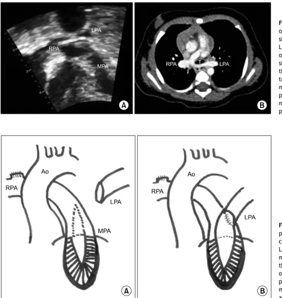
A 6-month-old girl from another country was ad- mitted to Seoul St. Mary’s Hospital for the manage- ment of congenital heart disease. Echocardiography performed in her country had revealed tetralogy of Fallot (TOF) with an invisible left pulmonary artery (LPA). At admission to our hospital, her arterial oxy- gen saturation by pulse oximetry was around 85%, and stridor was heard on auscultation without respi- ratory symptoms. Echocardiography confirmed TOF and a pulmonary artery (PA) sling (Fig. 1A). The di- ameter of the pulmonary valve annulus was 5 mm (Z-score of −5.7) and there was a small patent duc- tus arteriosus (PDA) connected to the LPA. The di- ameter of the LPA was 4.5 mm and the diameter of the right pulmonary artery (RPA) was 5 mm.
Computed tomography (CT) was performed to better delineate the anatomy of the PAs and to evaluate the airway (Fig. 1B). No tracheal stenosis associated with complete tracheal cartilage was found.
Surgery was performed under aorto-bicaval car- diopulmonary bypass. The main pulmonary artery (MPA) was hypoplastic and coursed rightward and posterior to the ascending aorta owing to the abnor- mal course of the LPA. The PDA was divided and the entire course of the LPA was carefully dissected to prevent injury to the trachea and the esophagus.
Closure of the ventricular septal defect (VSD) and re- section of the right ventricular outflow tract (RVOT) muscles were carried out through a right atrial incision. Then, the LPA was transected at its origin from the RPA and a cuff of the LPA was left on the RPA to prevent RPA stenosis and to shorten the LPA.
The defect of the RPA was repaired primarily with a 6-0 polypropylene running suture. A short longi- tudinal incision was made along the RVOT, and the pulmonary valve was found to be bicuspid and dys- plastic with a small opening. Transannular extension was carried out up to the distal MPA, and additional RVOT muscle resection was performed through the RVOT incision. The LPA was trimmed obliquely to
Korean J Thorac Cardiovasc Surg 2018;51:360-362 □ CASE REPORT □
https://doi.org/10.5090/kjtcs.2018.51.5.360
Tetralogy of Fallot with Pulmonary Artery Sling
− 361 −
Fig. 1. (A) Subcostal coronal view of a preoperative echocardiogram showing the hypoplastic MPA and LPA originating from RPA. (B) Pre- operative computed tomography showing the LPA originating from the RPA and coursing between the trachea and the esophagus. MPA, main pulmonary artery; LPA, left pulmonary artery; RPA, right pul- monary artery; T, trachea; E, eso- phagus.
Fig. 2. Illustration of the operative procedure. (A) The transannular in- cision and the obliquely trimmed LPA. (B) The end-to-side anasto- mosis between the posterior half of the LPA and the left lateral aspect of the MPA. Ao, aorta; MPA, main pulmonary artery; RPA, right pul- monary artery; LPA, left pulmonary artery.
Fig. 3. Enhanced postoperative computed tomography showing the well-reconstructed pulmonary arteries. Ao, aorta; MPA, main pulmonary artery; RPA, right pulmonary artery; LPA, left pulmo- nary artery.
obtain a natural geometry and to prevent kinking af- ter anastomosis to the MPA (Fig. 2A), and the poste- rior half of the LPA was anastomosed to the left lat- eral aspect of the MPA in an end-to-side fashion us- ing a 6-0 polypropylene running suture (Fig. 2B). The transannular incision and the anterior aspect of the LPA were covered with a bovine pericardial patch se- cured with a 6-0 polypropylene running suture. The cardiopulmonary bypass time was 172 minutes and the aortic cross-clamp time was 114 minutes.
Postoperative echocardiography showed no re- sidual VSD, no RVOT obstruction, severe pulmonary regurgitation, good flow in the branches of the PA, and good ventricular function. Postoperative CT also revealed well-reconstructed PAs (Fig. 3). The pa- tient’s recovery was uneventful and she was dis- charged on the ninth postoperative day.
Seha Ahn and Cheul Lee
− 362 −
Discussion
PA sling is a rare congenital cardiac anomaly in which an anomalous LPA arises from the RPA and courses between the trachea and the esophagus. PA sling can be associated with various cardiovascular anomalies, including VSD, atrial septal defect, and PDA. However, PA sling associated with TOF is very rare. Gikonyo et al. [1] found that among 130 cases of PA sling, only 3 (2.3%) were associated with TOF, and concomitant repair of TOF associated with PA sling has rarely been reported [2-6].
The clinical manifestations of PA sling vary and depend on the severity of airway compromise. Our patient had stridor without other respiratory symp- toms, and there was no tracheal stenosis. When asso- ciated tracheal stenosis is present, slide tracheoplasty can be a good surgical option [6]. Goldstein et al. [7]
reported that the incidence of PA stenosis was rela- tively common, occurring in 79% of patients after the repair of PA sling or anomalous origin of 1 PA from the ascending aorta. However, Yong et al. [5]
reported an incidence of PA stenosis of only 4.8% af- ter PA sling repair in 21 patients. Similar results re- garding the patency of the LPA were presented by Backer et al. [6]. Yong et al. [5] and Backer et al. [6]
credited cardiopulmonary bypass and the im- plantation of the LPA to the proximal MPA as the surgical techniques that have lowered the incidence of LPA stenosis. In this case, we left a cuff of the LPA on the RPA to shorten the LPA, because the LPA is often redundant. In addition, we trimmed the LPA obliquely to obtain a natural geometry and to pre- vent kinking after it was anastomosed to the MPA.
We chose to anastomose the posterior wall of the LPA to the MPA incision and to cover the anterior wall with a patch for 2 reasons. First, the diameter of the LPA was only 4.5 mm, and we expected that LPA stenosis would occur if the LPA was implanted to the separate MPA opening in an end-to-side
manner. Secondly, the hypoplastic MPA was not ide- ally suited for re-implantation of the LPA at the sep- arate MPA opening, and this drove our decision to make the MPA incision.
In conclusion, we successfully repaired a rare com- bination of TOF and PA sling. Although concomitant repair of these defects is technically straightforward, care should be taken to prevent post-repair LPA stenosis.
Conflict of interest
No potential conflict of interest relevant to this ar- ticle was reported.
References
1. Gikonyo BM, Jue KL, Edwards JE. Pulmonary vascular sling:
report of seven cases and review of the literature. Pediatr Cardiol 1989;10:81-9.
2. Hanafusa Y, Uemura H, Yagihara T, Kagisaki K, Takahashi M, Kitamura S. Surgical repair of pulmonary arterial sling associated with tetralogy of Fallot. Jpn J Thorac Cardiovasc Surg 2003;51:160-2.
3. Takeda Y, Asou T, Fakhri D, Rahayoe AU, Yoshimura H, Rachmat J. Pulmonary artery sling associated with tetral- ogy of fallot. Asian Cardiovasc Thorac Ann 2005;13:77-8.
4. Joshi A, Agarwal S, Aggarwal SK, Datt V, Sethi GR, Satsangi DK. Single stage repair of tetralogy of fallot associated with left pulmonary artery sling and tracheal stenosis. J Card Surg 2013;28:595-8.
5. Yong MS, d’Udekem Y, Brizard CP, et al. Surgical manage- ment of pulmonary artery sling in children. J Thorac Cardiovasc Surg 2013;145:1033-9.
6. Backer CL, Russell HM, Kaushal S, Rastatter JC, Rigsby CK, Holinger LD. Pulmonary artery sling: current results with cardiopulmonary bypass. J Thorac Cardiovasc Surg 2012;
143:144-51.
7. Goldstein BH, Bergersen L, Powell AJ, Graham DA, Bacha EA, Lang P. Long-term outcome of surgically repaired uni- lateral anomalous pulmonary artery origin. Pediatr Cardiol 2010;31:944-51.