ISSN: 2233-601X (Print) ISSN: 2093-6516 (Online)
− 280 −
Received: March 19, 2018, Revised: April 4, 2018, Accepted: April 11, 2018, Published online: August 5, 2018
Corresponding author: Jae-Ik Lee, Department of Thoracic and Cardiovascular Surgery, Gachon University Gil Medical Center, 21 Namdong-daero 774beon-gil, Namdong-gu, Incheon 21565, Korea
(Tel) 82-32-460-8922 (Fax) 82-32-460-3117 (E-mail) [email protected]
© The Korean Society for Thoracic and Cardiovascular Surgery. 2018. All right reserved.
This is an open access article distributed under the terms of the Creative Commons Attribution Non-Commercial License (http://creativecommons.org/
licenses/by-nc/4.0) which permits unrestricted non-commercial use, distribution, and reproduction in any medium, provided the original work is properly cited.
Isolated Unilateral Absence of Pulmonary Artery Associated with Contralateral Lung Cancer
Kun Woo Kim, M.D. 1 , Jae-Ik Lee, M.D., Ph.D. 1 , Kuk-Hui Son, M.D., Ph.D. 1 , Eun Young Kim, M.D. 2 , Kook-Yang Park, M.D., Ph.D. 1 , Chul-Hyun Park, M.D., Ph.D. 1
Departments of
1Thoracic and Cardiovascular Surgery and
2Radiology, Gachon University Gil Medical Center
Unilateral absence of a pulmonary artery (UAPA) is a rare congenital anomaly that may present with various symptoms, depending on the nature and severity of other cardiovascular anomalies. Furthermore, con- tralateral lung surgery in patients with UAPA is extremely rare, and clinical experience is limited. This report describes a case of surgical treatment of contralateral primary lung cancer in a patient with isolated UAPA.
A 56-year-old man was diagnosed with primary lung cancer accompanied by isolated UAPA on the con- tralateral side. He underwent meticulous cardiorespiratory function tests preoperatively. We performed a right lower lobectomy. Although in the immediate postoperative period, the patient suffered from a mild decline in his respiratory function, he recovered uneventfully. The present case shows that preoperative awareness of UAPA and meticulous perioperative management enable contralateral lung surgery to be performed safely.
Key words: 1. Unilateral absence of pulmonary artery 2. Lung neoplasms
3. Surgery
4. Perioperative care
Case report
A 56-year-old man presented with a mass shadow in the right lung field on a chest X-ray. Chest com- puted tomography (CT) revealed a mass measuring roughly 4 cm in the right lower lobe (Fig. 1A), and a 3-dimensional reconstructed CT image confirmed left unilateral absence of a pulmonary artery (UAPA) with hypertrophic bronchial arteries reflecting collat- eral circulation (Fig. 1B). After percutaneous needle biopsy, the tumor was identified as adenocarcinoma.
In room air, arterial blood gas analysis (ABGA) yield- ed results of 92.5 mm Hg for PO
2, 34.7 mm Hg for PCO
2, and 97.4% for oxygen saturation (SaO
2).
Pulmonary function testing revealed a forced vital ca-
pacity (FVC) of 3.1 L (70% of predicted), a forced expiratory volume in 1 second (FEV1) of 2.2 L (64%
of predicted), and a FEV1/FVC ratio of 0.71. Echocar- diography revealed no cardiac anomaly and the right ventricular systolic pressure (RVSP) was 23.7 mm Hg. Based on the results of these tests, we planned thoracoscopic lobectomy. During the operation, im- mediately after applying single-lung ventilation, SaO
2decreased rapidly. Because this event repeated, con- version to open thoracotomy was required, and the planned lobectomy was completed uneventfully un- der double-lung ventilation. Postoperatively, in the resting state, SaO
2was 97%–99% with an oxygen supply of 3 L/min via a nasal prong and 88%–92%
in room air. Even during simple daily activities, he
Korean J Thorac Cardiovasc Surg 2018;51:280-282 □ CASE REPORT □
https://doi.org/10.5090/kjtcs.2018.51.4.280
UAPA Associated with Contralateral Lung Cancer
− 281 −
Fig. 1. (A) Chest CT shows a mass larger than 4 cm in the right lower lobe (arrow). (B) Three-dimen- sional reconstructed CT image shows a normal RPA and absence of the left pulmonary artery, as well as compensatory hypertrophy of the collateral systemic arteries (arrows). Lung cancer is noted in the right lower lobe (asterisk). CT, computed tomography; RPA, right pulmonary artery.
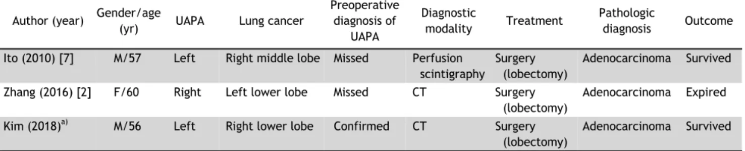
Table 1. Reports of unilateral absence of a pulmonary artery associated with contralateral lung cancer Author (year) Gender/age
(yr) UAPA Lung cancer
Preoperative diagnosis of
UAPA
Diagnostic
modality Treatment Pathologic
diagnosis Outcome Ito (2010) [7] M/57 Left Right middle lobe Missed Perfusion
scintigraphy
Surgery (lobectomy)
Adenocarcinoma Survived Zhang (2016) [2] F/60 Right Left lower lobe Missed CT Surgery
(lobectomy)
Adenocarcinoma Expired Kim (2018)
a)M/56 Left Right lower lobe Confirmed CT Surgery
(lobectomy)
Adenocarcinoma Survived
UAPA, unilateral absence of a pulmonary artery; M, male; F, female; CT, computed tomography.
a)