CASE REPORT pISSN 1225-7737/eISSN 2234-8042 http://dx.doi.org/10.12701/yujm.2014.31.2.109
Yeungnam Univ J Med 2014;31(2):109-112YUJM VOLUME 31, NUMBER 2, DECEMBER 2014 109
성인 무증상 폐동맥 슬링
정한희1, 백주열2, 이원익2, 장지혜1, 정민영1, 우기현1, 박성일1, 김일규1
1가톨릭대학교 의과대학 내과학교실, 2청주성모병원 내과
An adult asymptomatic pulmonary artery sling
Han Hee Chung
1, Ju Yeol Baek
2, Won Yik Lee
2, Ji Hye Jang
1, Min Young Jeong
1, Gi Hyeon Woo
1, Seong Il Park
1, Il Kyu Kim
11
Department of Internal Medicine, College of Medicine, The Catholic University of Korea, Seoul;
2
Department of Internal Medicine, Cheongju St. Mary’s Hospital, Cheongju, Korea
A pulmonary artery sling is a very rare congenital abnormality in which the left pulmonary artery rises from the posterior surface of the right pulmonary artery and then passes between the trachea and the esophagus, causing tracheal compression. It is associated with tracheo-bronchial abnormalities (50%) and cardiovascular abnormalities (30%). It may produce respiratory symptoms through the airway compression of the abnormal left pulmonary artery and congenital abnormalities associated with it. Because most (90%) pulmonary artery sling patients present symptoms during infancy, their condition is often diagnosed in the first year of life. However, a pulmonary artery sling is occasionally found in adults. It is usually asymptomatic and found incidentally. This is a very rare case of an asymptomatic pulmonary artery sling in an adult.
A 38-year-old man presented symptoms of mild exertional dyspnea. His spiral computed tomography showed a pulmonary artery sling. He was discharged without specific treatment because his symptoms improved without specific treatment and might not have been associated with a pulmonary artery sling.
We report an adult case of an asymptomatic pulmonary artery sling diagnosed via spiral computed tomog- raphy, accompanied by a literature review.
Keywords: Pulmonary artery sling; Congenital abnormality; Spiral computed tomography
Received: August 19, 2013, Revised: September 25, 2013, Accepted: September 30, 2013
Corresponding Author: Ju Yeol Baek, Department of Internal Medicine, Cheongju St. Mary’s Hospital, 173-19 Juseong-ro, Sangdang-gu, Cheongju 360-568, Korea
Tel: +82-43-219-8198, Fax: +82-43-211-9030 E-mail: [email protected]
서 론
폐동맥 슬링(sling)은 발생학적 이상으로 좌폐동맥이 폐동 맥간에서 나오지 않고 우폐동맥의 후측에서 나와 기관과 식 도 사이를 지나 좌폐문으로 들어가는 매우 드문 선천성 혈관 기형으로, 기관고리, 기관지 연화증 같은 선천성 기관-기관지 기형(50%)과 동맥관개존, 심실중격결손 같은 선천성 심혈관
(30%) 기형이 잘 동반된다[1-5]. 이런 폐동맥 슬링은 비정상 적인 좌폐동맥의 주행으로 좌폐동맥이 주위 기관이나 식도 를 압박하거나 동반된 선천성 기관-기관지 기형으로 인해 증상이 나타나게 된다. 가장 흔한 증상은 호흡곤란, 천명 및 청색증 같은 호흡기 증상으로 90% 이상의 환자들에서 이와 같은 증상이 출생 직후부터 생후 1세 이내에 발생한다[1-4].
또한 삼킴장애와 같은 소화기 증상도 비슷한 시기에 흔히 나타난다. 그러므로 대부분의 폐동맥 슬링은 1세 이전에 진 단을 받게 되고, 증상의 정도에 따라 수술적 치료가 이루어진 다[1-4]. 하지만 좌폐동맥의 주위 구조물에 대한 압박이 심하 지 않거나 동반된 선천성 기형이 없을 경우에는 특별한 증상 이 나타나지 않는다. 이런 경우 폐동맥 슬링은 대부분 우연히 발견되고 성인이 될 때까지 발견하지 못할 수도 있다[3,5,6].
Han Hee Chung et al.
110 YUJM VOLUME 31, NUMBER 2, DECEMBER 2014
Fig. 1. Computed chest tomography showing the rise of the left pulmonary artery from the right pulmonary artery and the formation of a vascular sling (arrow). There was no significant case of tracheal compression, and no other tracheo-bronchial abnormality was seen.
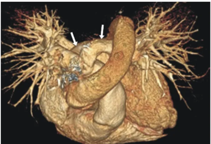
Fig. 2. Three-dimensional computed chest tomography showing the rise of an abnormal left pulmonary artery (arrow) from the posterior aspect of the right pulmonary artery and its passage along the right lateral wall of the trachea.
이와 같이 성인에서 발견되는 폐동맥 슬링은 매우 드물고 증상이 없거나 경미하며, 특별한 치료없이 예후가 양호하다.
저자들은 성인에서 발견된 무증상 폐동맥 슬링을 경험하였 기에 문헌 검토와 함께 이에 대해 보고하는 바이다.
증 례
38세 남자가 2주 전부터 발생한 운동 시 호흡곤란을 주소 로 내원하였다. 특별한 과거력이나 가족력은 없었고, 20년 간 하루 한 갑의 흡연력과 주1회 소주 1병을 마시는 음주력이 있었다. 현재 복용 중인 약제는 없었고, 최근 평소보다 담배를 많이 피웠다고 했다. 활력 징후는 혈압 130/90 mmHg, 맥박 수 60회/분, 호흡수 16회/분, 체온 36.6℃, 산소포화도 97%
로 정상이었고, 신체검진에서 천명 같은 이상 호흡음이나 심잡음은 없었으며, 양하지 부종도 관찰되지 않았다. 말초혈 액 검사에서 백혈구 5,820/mm3, 혈색소 14.6 g/dL, 헤마토크 리트 44.8%, 혈소판 247,000/mm3였고, 혈중요소질소/크레아 티닌 19/1.0 (mg/dL), 아스파르테이트아미노전이효소/알라닌 아미노전이효소 17/15 (IU/L), 총빌리루빈 0.5 mg/dL, 나트륨/
칼륨/염소 137/3.9/104 (mEq/L)로 모두 정상소견을 보였고, 소변검사도 이상소견은 없었다. 단순 흉부촬영에서 이상소 견은 없었으며, 심전도에서 정상 동율동을 보였다. 심혈관질 환 감별을 위하여 시행한 이차원 경흉부 심초음파에서 좌심 실 구혈률은 59%였고, 정상 이완기 심기능을 보였으며, 좌심 실 비대 소견은 관찰되지 않았다. 천식이나, 만성 폐쇄성 폐 질환 감별을 위해 시행한 폐기능 검사에서 기도 가역성은 없었고, 1초간 노력성 호기량의 노력성 폐활량에 대한 비 (forced expiratory volume in 1 second [FEV1]/forced vital capacity [FVC]) 0.72, 1초간 노력성 호기량(FEV1) 73%
(2.71 L), 노력성 폐활량(FVC) 81%(3.76 L), 노력성 호기 중간 유량(forced expiratory flow25-75%) 53%로 소기도 질환 의심 소견을 보였다. 또한 흉부 나선형 컴퓨터단층촬영을 시행하 였고, 조영증강 영상과 3차원 재건영상에서 좌폐동맥이 우 폐동맥의 후면에서 기시하여 기관과 식도 사이를 주행하는 폐동맥 슬링이 관찰되었다(Fig. 1, 2). 하지만 호흡곤란을 유 발할 수 있는 비정상적인 좌폐동맥의 기관 압박이나 기관고 리, 기관지 연화증 같은 선천성 기관-기관지 기형을 의심할 만한 소견은 발견되지 않았고, 검사를 진행하는 동안 특별한 치료 없이 자발적인 증상 호전을 보였다. 이를 통해 저자들은 환자가 호소했던 증상과 폐동맥 슬링의 연관성을 찾을 수 없었으므로, 무증상 폐동맥 슬링으로 보고 특별한 치료 없이 외래에서 경과관찰 중이다. 상기 환자는 입원 전 약 2주 간의
운동시 호흡곤란 외에 출생 시부터 현재까지 특별한 증상이 나 병력이 없었고, 퇴원 후 최근 3개월 동안 호흡곤란의 재발 이나 다른 증상은 없는 상태로 추후 지속적으로 외래에서 추적 관찰할 예정이다.
고 찰
폐동맥 슬링은 혈관륜(vascular ring)의 한 종류로, 발생학 적으로 왼쪽 6번째 대동맥궁의 왼쪽 근위부가 좌측 폐야와 정상적으로 연결되는 데 실패하고 우폐동맥의 평행부에서 측부 혈관들이 발달하여 좌측 폐야와 연결됨으로써 발생한다 [7]. 이는 1897년 Glaevecke와 Doehle에 의해 처음으로 발견
Asymptomatic pulmonary artery sling
YUJM VOLUME 31, NUMBER 2, DECEMBER 2014 111
되었고, 1958년 Contro 등에 의해 다른 혈관륜과 구분하기 위해 슬링이라는 용어를 처음으로 사용하기 시작했다[8,9].
폐동맥 슬링의 유병률에 대해서 정확히 알려진 바는 없다 [1,3]. 이는 폐동맥 슬링의 발생 자체가 드물고, 특별한 증상이 없는 무증상 폐동맥 슬링 환자에 대한 확인이 어렵기 때문이 다. 이와 같이 폐동맥 슬링의 정확한 유병률을 파악할 수는 없지만, 현재까지 보고된 증례들을 토대로 볼 때 무증상 폐동 맥 슬링은 전체 폐동맥 슬링 환자의 10%이며, 폐동맥 슬링의 남녀 성비는 3:2로 남자에서 조금 더 많이 발생한다[1,3].
이들은 선천성 혈관기형으로 50%에서 기관고리, 기관연 화증과 같은 선천성 기관-기관지 기형이 동반되고, 30%에서 동맥관개존, 심실중격결손, 심방중격결손 및 좌측상대정맥 과 같은 선천성 심장기형이 동반된다[1-4,10,11]. 그 외 소화 기계, 비뇨 생식기계 및 내분비계의 이상이 발견되는 경우도 있다[4,5]. 이렇게 동반된 선천성 기관-기관지 기형 유무에 따라 폐동맥 슬링은 동반된 기관-기관지 기형이 없는 1형과 완전 기관 고리와 같은 기관-기관지 기형을 동반한 2형으로 나눌 수 있다. 1형은 대부분 증상이 나타나지 않고 좋은 예후 를 보이며, 2형은 영아기부터 심한 호흡기 증상을 보이고 나쁜 예후를 보인다[4,12,13].
폐동맥 슬링은 비정상적인 좌폐동맥의 주행으로 인한 좌 폐동맥의 주위 구조물 압박 정도 또는 동반된 선천성 기형의 유무에 따라 증상의 정도가 다르다. 즉, 비정상적인 좌폐동맥 의 주행으로 좌폐동맥이 기도를 압박하거나 식도를 압박할 때 호흡곤란, 천명 및 청색증 등의 호흡기 증상이나 연하장애 와 같은 소화기 증상이 발생할 수 있고, 기관고리, 기관연화 증 같은 동반된 기관-기관지 기형에 의해서 천명, 호흡곤란 및 청색증 등의 호흡기 증상이 발생할 수 있다[1-4]. 또한 동반된 선천성 심장기형으로 호흡곤란, 청색증 및 부정맥 등이 발생할 수도 있다[14]. 하지만 비정상적인 좌폐동맥의 압박이 심하지 않거나 동반된 기관-기관지 기형 및 심장기형 이 없을 시에는 증상이 심하지 않거나 없을 수 있다[4,12,13].
본 증례의 무증상 폐동맥 슬링 환자도 시행한 검사를 통하여 비정상적인 좌폐동맥의 주위 구조물 압박이나 동반된 선천 성 기형이 동반되지 않았음을 확인할 수 있었다. 그러므로 호흡곤란이나 연하장애와 같은 증상이 발생하지 않았다고 추측해 볼 수 있다.
진단을 위해 흉부컴퓨터단층촬영, 흉부 자기공명영상, 심 장 초음파, 심혈관 조영술 및 바륨 식도 조영술을 시행할 수 있다[2,4,15]. 바륨 식도 조영술은 오랫동안 폐동맥 슬링 의 진단을 위해 사용되었던 검사로, 최근 흉부컴퓨터단층촬
영 및 기타 영상학적 진단 기술의 발달로 중요성이 감소하였 다. 경흉부 및 경식도 심장초음파는 폐동맥 슬링의 진단에 유용하고 동반된 심장기형의 진단에 도움을 줄 수 있지만, 종종 이상을 발견하지 못할 수 있다[3,4]. 따라서 정확한 해부 학적인 구조 및 주위 구조물 간의 관계를 정확히 보여줄 수 있는 흉부컴퓨터단층촬영과 흉부 자기공명영상이 가장 유용 한 검사이다[4,13,16].
증상이 있는 폐동맥 슬링 환자들은 수술적 치료가 필요하 다. 폐동맥 슬링은 매우 드물기 때문에 자연경과를 파악하기 는 어렵지만, 좌폐동맥의 주위 구조물 압박 정도 및 동반된 선천성 기형에 따라 증상이 심하고, 증상의 발현이 빠를수록 수술적 교정을 받지 않으면 나쁜 예후를 보이기 때문이다 [17]. 또한 1953년 Potts 등의 성공적인 수술적 교정 이후 지속적으로 수술적 치료에 대한 좋은 결과가 보고되고 있기 때문에, 증상이 심하고 증상의 발현이 빠른 폐동맥 슬링 환자 들에서는 적극적인 수술적 치료가 필요하다[14,18-21].
반면, 좌폐동맥의 주위 구조물 압박 정도가 심하지 않거나 선천성 기형이 동반되지 않은 환자들은 증상이 없거나 경미 하며 증상의 발현이 늦다. 이들은 대부분 나이가 들면서 증상 이 완화되고 특별한 치료 없이도 예후가 좋다[5,14]. 그러므 로 증상이 없거나 경미한 환자들은 특별한 치료 없이 임상경 과를 관찰하며 기다려보는 것이 좋다[1-4,12].
본 증례의 환자는 운동시 호흡곤란으로 내원하여 흉부 나선 형 컴퓨터단층촬영을 통해 폐동맥 슬링을 진단받았고, 특별 한 치료 없이 자발적으로 증상이 호전되어 현재 외래에서 호흡곤란의 재발 없이 경과관찰 중이다. 대부분의 폐동맥 슬링은 비정상적 좌폐동맥의 주위 압박이나 동반된 선천성 기관-기관지 기형 및 심장 기형으로 인해 호흡곤란 같은 호흡 기 증상이 발생하여 생후 1년 이내에 발견된다. 하지만 매우 드물게 특별한 증상 없이 성인에서 발견되기도 하는데, 이와 같은 무증상 폐동맥 슬링 환자들은 대부분 비정상적 좌폐동 맥의 압박이 심하지 않고 동반된 선천성 기형이 없다. 본 증례와 같은 성인의 무증상 폐동맥 슬링은 발견 자체가 드물 고 좌폐동맥의 주위 구조물 압박이나 동반 선천성 기형이 없는 증례는 아직 국내에서는 보고되지 않았기에, 문헌검토 와 함께 임상적 의미를 고찰하고 보고하는 바이다.
REFERENCES
1. Gundavaram MS, Gaba RC. Asymptomatic pulmonary artery
sling: a rare congenital vascular anomaly. Heart Lung Circ
2013;22:297-9.
Han Hee Chung et al.