143 http://www.jchestsurg.org
JCS
Journal of Chest SurgeryCase Report
Cor Triatriatum Associated with Total Anomalous Pulmonary Venous Connection: A Rare but Plausible Combination
Jun Oh Lee, M.D., Chun Soo Park, M.D., Ph.D.
Department of Thoracic and Cardiovascular Surgery, Asan Medical Center, University of Ulsan College of Medicine, Seoul, Korea
ARTICLE INFO Received May 8, 2020 Revised July 10, 2020 Accepted July 11, 2020 Corresponding author Chun Soo Park Tel 82-2-3010-3583 Fax 82-2-3010-6966
E-mail [email protected];
[email protected] ORCID
https://orcid.org/0000-0001-8718-8904
In a newborn in whom cor triatriatum was missed on echocardiography, infracardiac total anomalous pulmonary venous connection was successfully repaired with the aid of cardi- ac computed tomography (CT). In rare combinations, as in this case, an accurate diagnosis prior to surgery, which is of vital importance for successful repair, can be made through a high index of suspicion and the use of a supplemental imaging modality such as CT.
Keywords: Congenital heart disease, Cor triatriatum, Total anomalous pulmonary venous return
Copyright©The Korean Society for Thoracic and Cardiovascular Surgery. 2021. All right reserved.
This is an Open Access article distributed under the terms of the Creative Commons Attribution Non-Commercial License (http://creativecommons.org/licenses/
by-nc/4.0) which permits unrestricted non-commercial use, distribution, and reproduction in any medium, provided the original work is properly cited.
Case report
Although the association of total anomalous pulmonary venous connection (TAPVC) with cor triatriatum seems implausible, this rare combination has been convincingly documented and embryologically rational explanations have been proposed [1,2]. The rarity of this combination means that cardiologists or cardiac surgeons might not clearly identify and elaborate the anatomic details before surgery unless they have a high index of suspicion. Howev- er, complementary to echocardiography, additional imag- ing modalities such as cardiac computed tomography (CT) can provide critical information for making an accurate diagnosis. Here, we report the case of a baby born with this rare combination that was successfully repaired, and we present 1-year follow-up imaging.
An 8-day-old newborn was transferred to Asan Medical Center with severe respiratory distress and hypoxia requir- ing immediate mechanical ventilatory support. Transtho- racic echocardiography (TTE) demonstrated TAPVC, which drained into the portal vein through the vertical vein without flow disturbance. A common pulmonary ve- nous cham ber accommodating all the pulmonary venous blood was identified, but it was not thought based on TTE that a part of the common pulmonary venous chamber
was the proximal chamber of the left atrium (LA).
A cardiac CT scan clearly demonstrated infracardiac TAPVC, which was compatible with the TTE findings. In contrast to TTE, the CT scan also revealed a round cham- ber communicating with all the pulmonary veins that mimicked the LA, which had collapsed (Fig. 1). This find- ing suggested an obstructed cor triatriatum associated with infracardiac TAPVC. The descending vertical vein drained into the portal vein and there was no evidence of stenosis or narrowing.
At 12 days of age, the patient underwent urgent repair under moderate hypothermic cardiopulmonary bypass. An incision was made in the midline of the sternum and per- fusion cannulas were inserted through the aorta, right atri- al appendage, and inferior vena cava. After commencing cardiopulmonary bypass, 4 individual pulmonary veins, a posteriorly located pulmonary venous confluence, and the descending vertical vein were clearly identified. Under car- dioplegic arrest, the right atrium opened and a thick mus- cular membrane was seen posteriorly through the widely opened atrial septal defect (ASD). The membrane was in- cised at the center and widely resected to establish free communication between the proximal and distal (accessory and true) LA chambers. To enhance pulmonary venous in- flow, an incision on the posterior wall of the proximal LA
https://doi.org/10.5090/jcs.20.038 pISSN: 2765-1606 eISSN: 2765-1614 J Chest Surg. 2021;54(2):143-145
144
https://doi.org/10.5090/jcs.20.038
http://www.jchestsurg.org
JCS
chamber and a corresponding incision on the anterior wall of the pulmonary venous confluence were anastomosed with 8-0 Prolene sutures in an interlocked continuous run- ning fashion. The ASD was closed with bovine pericardi- um. The right atrial incision was repaired, and the de- scending vertical vein was divided. Convalescence from cardiopulmonary bypass was uneventful, and the patient was transferred to the pediatric intensive care unit with the sternum remaining open. The sternum was closed on post- operative day 2. The patient was taken off of mechanical ventilator support on postoperative day 13, transferred to the general ward on postoperative day 15, and discharged on postoperative day 31.
Follow-up TTE at discharge revealed no individual pul- monary vein stenosis, no flow disturbance in the LA, no evidence of pulmonary hypertension, and normal ventric- ular function. At the 1-year follow-up, the patient was do-
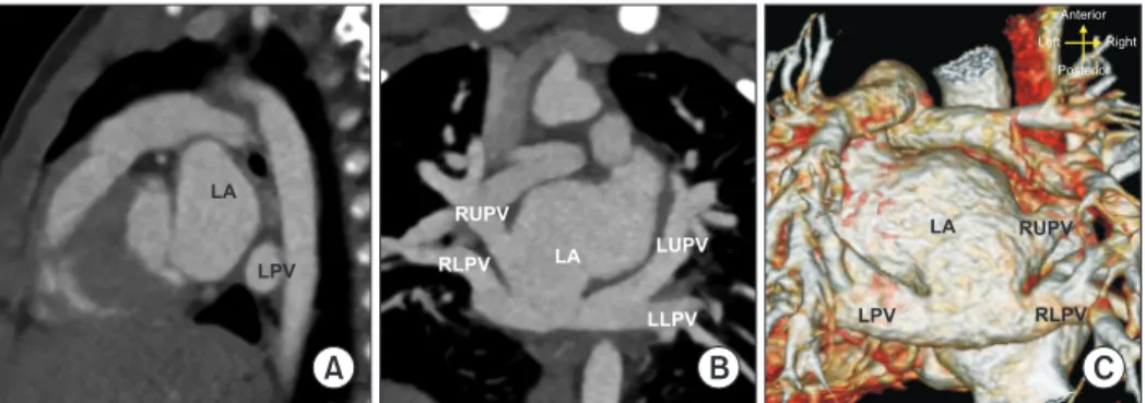
ing well without medication, an electrocardiogram was unremarkable, and unobstructed individual pulmonary veins and LA were demonstrated on a CT scan (Fig. 2).
The patient’s caregiver provided informed consent for publication of this research including medical records and clinical images on the line.
Discussion
Cor triatriatum is a rare congenital cardiac defect ac- counting for 0.1%–0.4% of all cases of congenital heart dis- ease. In cor triatriatum, the LA is divided into 2 chambers by an abnormal membranous structure, which can hinder the inflow toward the left ventricle (LV) and consequently function as a pulmonary venous obstruction resulting in pulmonary hypertension and cardiac failure. TAPVC is a well-known pulmonary venous anomaly that is also fre-
Fig. 1. (A) A preoperative simple chest Xray shows multifocal dependent opacities in both lungs due to pulmonary congestion. (B, C) The thick membrane between the 2 left atrial (A) chambers (arrow) bulged toward the anteriorly positioned collapsed true left atrium (DC) due to cor triatriatum (PC) in (B) sagittal and (C) coronal section CT images. (D) In the volumerendered CT image, the extent of incisions of the PVC and proximal left atrium can be delineated (red dotted line). DC, distal chamber; PC, proximal chamber; CT, computed to
mography; LV, left ventricle; LPV, left pulmonary vein; LLPV, left lower pulmonary vein; RLPV, right lower pulmonary vein; RUPV, right upper pulmonary vein; LUPV, left upper pulmonary vein; PVC, pulmonary venous confluence; VV, vertical vein.
A B C D
R
DC
PC
LV LPV
DC
LLPV LUP RUPV
RLPV PC
LV
PVC
VV RLPV
DC RUPV
PC
Front out SAL
IPR
Cranial Right Caual Left
Fig. 2. Oneyear followup computed tomography scan. (A) No residual membranous structures were identified in the left atrial cham
ber. (B, C) Individual pulmonary veins and the junction between the pulmonary venous confluence and LA were unobstructed in (B) the coronal section and (C) volumerendered images. LA, left atrium; LPV, left pulmonary vein; LLPV, left lower pulmonary vein; RLPV, right lower pulmonary vein; RUPV, right upper pulmonary vein; LUPV, left upper pulmonary vein.
C
A B
LA
LPV LA
LLPV LUPV RUPV
RLPV
LA
LPV RLPV
RUPV
Anterior Right Posterior Left
145
Jun Oh Lee and Chun Soo Park. Cor Triatriatum Associated with Total Anomalous Pulmonary Venous Return
http://www.jchestsurg.org
JCS
quently accompanied by a pulmonary venous obstruction.
Therefore, without adequate surgical treatment, concurrent existence of both anomalies could lead to severe morbidity and even death despite its extremely rare incidence [3,4].
An accurate diagnosis prior to surgery is paramount for successful repair, but the co-existence of these 2 abnormal- ities is barely detectable using traditional TTE unless the physician has a high index of suspicion. In this case, the presence of associated cor triatriatum could not be identi- fied by TTE, but was finally detected on a CT scan, where- by the anatomical relationship between the individual pul- monary veins and proximal and distal chambers of the LA were clearly delineated.
The classification of cor triatriatum was first suggested by Loeffler [5] and subsequently elaborated by Niwayama [6], Grondin et al. [7], and Krabill and Lucas [8]. According to the classification of Lucas, our case was compatible with cor triatriatum type B2, where the accessory chamber re- ceives all pulmonary veins without communication be- tween the accessory and true LA chambers, and pulmo- nary venous blood drains into the systemic vein in the fashion of TAPVC [1].
Patients with cor triatriatum associated with TAPVC can present with low cardiac output and cardiogenic shock due to insufficient LV preload resulting from the limited size of the interchamber (proximal and distal LA) and interatrial communication, which is the sole channel toward the LV.
As medical treatment usually does not work well for pa- tients with mechanically obstructed pulmonary venous in- flow, timely surgical repair is crucial for a favorable out- come. Although surgery was delayed to ensure an accurate diagnosis, our patient was stably maintained with a low inotrope dosage and minimum respiratory support until repair, owing to the grossly unobstructed pulmonary ve- nous drainage. The surgical procedures are quite straight- forward: the membrane should be resected as much as possible and anomalously returned pulmonary veins should be widely connected to the LA. In this case, the membrane was widely resected, following standard prac- tice, and TAPVC was repaired in a manner similar to con- ventional repair, with the proximal left atrial chamber and pulmonary venous confluence anastomosed from the pul- monary venous opening in the proximal LA.
Pulmonary vein stenosis is the most frequent and fatal complication after TAPVC repair. Most cases of pulmo- nary vein stenosis occur within 1 year following TAPVC
repair. Fortunately, the 1-year follow up image in our case showed no evidence of pulmonary venous inflow obstruc- tion.
In conclusion, an accurate diagnosis prior to surgery, which is of vital importance for successful repair, can be established through a high index of suspicion and the use of a supplemental imaging modality such as CT for this rare combination of cor triatriatum and TAPVC. An accu- rate diagnosis prior to surgery can enable satisfactory out- comes to be achieved despite delayed referral and repair.
Conflict of interest
No potential conflict of interest relevant to this article was reported.
ORCID
Jun Oh Lee: https://orcid.org/0000-0003-3373-1144 Chun Soo Park: https://orcid.org/0000-0001-8718-8904
References
1. Herlong JR, Jaggers JJ, Ungerleider RM. Congenital Heart Surgery Nomenclature and Database Project: pulmonary venous anomalies.
Ann Thorac Surg 2000;69(4 Suppl):S56-69.
2. Man JB, Chan YN, Sam SO, et al. Cor triatriatum with infracardiac total anomalous pulmonary venous drainage. Korean J Thorac Car- diovasc Surg 2002;35:52-5.
3. Burroughs JT, Edwards JE. Total anomalous pulmonary venous con- nection. Am Heart J 1960;59:913-31.
4. Vouhe PR, Baillot-Vernant F, Fermont L, Bical O, Leca F, Neveux JY. Cor triatriatum and total anomalous pulmonary venous connec- tion: a rare, surgically correctable anomaly. J Thorac Cardiovasc Surg 1985;90:443-5.
5. Loeffler E. Unusual malformation of the left atrium: pulmonary si- nus. Arch Pathol (Chic) 1949;48:371-6.
6. Niwayama G. Cor triatriatum. Am Heart J 1960;59:291-317.
7. Grondin C, Leonard AS, Anderson RC, Amplatz KA, Edwards JE, Varco RL. Cor triatriatum: a diagnostic surgical enigma. J Thorac Cardiovasc Surg 1964;48:527-39.
8. Krabill KA, Lucas RV. Abnormal pulmonary venous connections. In:
Emmanoulides GC, Riemenschneider TA, Allen HD, Gutgesselle, editors. Heart disease in infants, children, and adolescents: including the fetus and young adult. Baltimore (MD): Williams and Wilkins;
1995. p. 838-74.