목 차
Ⅰ. 서 론
Ⅱ. 연구재료 및 연구방법
Ⅲ. 연구결과
Ⅳ. 고 찰
Ⅴ. 결 론 참고문헌 영문초록
Ⅰ. 서 론
손상 받은 근육은 세 가지의 기전 즉, 근육섬유의 수, 길이, 및 그 두께의 증가에 의해 손상이 치유된다
1,2). 근육조직이 손상을 받은 경우 근육위성세포는 분 열되고 근육세포로 분화하여 근육섬유를 형성한다.
다기능 간질세포는 계열유도 유전자에 의해서 근모 세포로 분화하고 많은 수로 분열하여 인접 세포끼리 융합하여 다수의 핵을 가진 근관들을 형성한다. 이 근 관들은 구조 단백들이 형성되고 수축기가 형성되면 서 성숙된 근육의 기본 단위인 근섬유로 분화한다3-5). 이 과정에서 근육 특정 유전자들의 각 단계에 따른 조절에 의하여 근육의 분화가 일어나는 것으로 알려 져 있다4).
근육분화의 분자생물학적 기전은 많은 연구가 시 행되었으며, 특히 근육에 특정한 조절인자인 MyoD, myogenin, Myf-5, 와 MRF-4(Herculin) 등에 대한 연구가 시행되어 왔다6-10). 이러한 근육인자는 하나의 basic helix-loop-helix(이하 bHLH) motif를 가지고
* 이 논문은 1999년도 전남대학교 병원 임상연구소 임상연구비의 지원에 의하여 연구하였음
있다. 그리고 이 motif의 위치는 E-box라고 불리는 특정한 DNA 배열과 결합되어야 한다. 근육 특정 유 전자의 전사과정에서 근육에 특징적인 bHLH 인자인 E2A 단백(E12 와 E47)은 E-box에 결합하며 근육특 정 유전자의 전사가 일어난다4,5,). 그러나 다른 bHLH 인자인 Id(inhibitor of DNA binding) 단백은 근육 특 정인자를 억제한다11,12). 근육에 특징적인 bHLH 인자 의 전사는 E12, E47과 같은 양성조절자와 Id-1, 2, 3 와 같은 음성조절자에 의해 조절된다2).
근육의 분화는 다양한 인자들에 의해 조절되는데, FGF(fibroblastic growth factor)13,14), IGF(insulin- like growth factor)15-18)와 EGF(epidermal growth factor)19,20)등과 같은 펩타이드성 성장인자들에 의해 서도 조절된다. 그리고 Transforming Growth Factor -β(이하 TGF-β) superfamily와 같은 성장인자들 도 간질세포에서 분화되는 근모세포의 분화를 조절 하는 역할을 한다5). TGF-βsuperfamily는 TGF-β family, inhibin family와 bone morphogenetic protein(이하 BMPs) family등으로 구성되며, 이 중 BMPs subfamily 인 BMP-2, 4, 12, 13 등은 근육분화 에 효과를 갖는데, 이중 BMP-2는 근모세포의 분화를 골세포성으로 전환시켜 근육내에 이식시 이상성 골 형성을 유도한다고 하였다21,22). 그리고 BMP-4, 12, 13등도 근육세포의 분화를 차단하나 골세포성 특징 은 발현하지 않았다고 하였다23). Bone morphogenetic protein-7(이하 BMP-7)이라고도 불리우는 osteo- genic protein-1(이하 OP-1)은 원시적인 골 선조세포 (progenitor cell)의 아군집을 연골내 골화로 유도하는 역할을 한다고 믿어져 왔으나 완성된 골세포를 연골 세포로 전환시키지 않는 것으로 보인다고 하였다. 또
근육세포 분화에 대한 TGF-β1과 OP-1의 억제 효과
전남대학교 치과대학 구강내과학 교실, 마취과학 교실*
김 병 국․정 성 수*
한 분화 초기에 OP-1을 저용량 처리하면 간질성 선 조세포를 지방세포로 유도하나 동일한 세포에 고용 량 처리하면 연골성 특성을 갖는다24,25)고 하였으나, 근육세포에 대한 효과는 보고된 바 없다. TGF-β family인 TGF-β1의 근육세포 분화에 대한 억제효 과는 태생기 백서의 대퇴근에서 클론된 L6A1 세포에 서 myogenin mRNA의 발현상승을 차단하는 성장인 자의 능력에 의한다26). 그리고 myogenin이 발현 vector에 의해 만들어졌을 때, TGF-β1은 DNA에의 결합능력에 영향받지 않고 myogenin의 전사활성을 억제하는 형태로 작용한다27)는 등의 연구들이 있으나 근육 특정 유전자와 관련한 TGF-β1의 근육분화 억 제효과에 대해서는 이해가 부족한 상태이다.
위와 같이 TGF-β superfamily는 여러 세포의 성 장과 분화에 다양한 활성을 보여주며, 골격성 간질세 포의 분화에 중요한 역할을 한다. 그러나 TGF-β1과 OP-1의 골격근 세포의 분화에 대한 억제효과에 대한 기전은 아직까지 명확하지 않으며 논의의 대상이 되 어왔다. 이에 저자는 근육세포가 분화하는 동안 수용 체와 리간드의 변화를 통해 완전히 성숙된 근육세포 에서 TGF-β1의 근육세포 분화 억제효과를 확인하 고, OP-1의 근육세포 분화억제 및 골세포성 유도 가 능성에 대해 연구하였다.
Ⅱ. 연구재료 및 연구방법
1. 성장인자(Growth Factor) : human recombi- nant osteogenic protein-1(이하 OP-1, Creative Biomolecule, Boston, USA)과 transforming growth factor β1(이하 TGF- β1, R & D, Minneapolis, USA)
2. 세포배양
백서의 근육세포인 C2C12 세포를 Dulbecco's modified eagle medium(이하 DMEM, GIBCO BRL., Grand Island, USA)에 10%의 fetal bovine serum(이 하 FBS, Atlanta Biologicals, Norcross, USA)와 항생 제 penicillin 100U, streptomycin 100㎎과 ampho- tericin B 0.25㎎을 첨가한 성장용 배양액에서 배양하 였다. 세포는 2 X 10⁴개/㎠으로 접종하고 성장용 배양 액에서 배양하였다. 근육세포 분화에 대한 OP-1과 TGF-β1의 효과를 관찰하기 위하여 배양접시에 세 포가 밀생의 90% 수준에 도달하였을 때, 성장용 배양
액를 5% FBS가 함유된 DMEM인 분화용 배양액으 로 교환하였다. Ascorbic acid 50㎍/㎖과 10mM의 β -glycerophosphate를 분화용 배양액에 첨가하였다.
성장인자는 OP-1을 50ng/㎖, 100ng/㎖, 300ng/㎖과 600ng/㎖으로 2일, 7일과 14일간, 그리고 TGF-β1은 5ng/㎖을 1일, 2일, 3일과 4일간 처리하였다. 세포의 형태학적 변화는 역위상차 현미경(Olympus, Tokyo, Japan)으로 관찰하고 현미경에 외장된 camera로 촬 영하였다.
3. Total RNA 추출과 Northern hybridization
세포성 total RNA는 지침서의 지시에 따라 TRIzol reagent(GIBCO BRL., Grand Island, USA)를 이용한 single step법으로 추출하였다. 추출한 total RNA는 formazol(Molecular research center, Cincinnati, USA)에 용해시켰다. Total RNA 10㎍을 form- aldehyde가 함유된 1% agarose gel에 전기 영동하였 다. 영동된 total RNA는 capillary blotting 방법으로 Nytran Plus membrane(Schleicher and Schuell, Keene, USA)으로 이송하였다. Total RNA가 이송된 membrane은 ultraviolet crosslinker를 이용하여 고정 하였다. Total RNA가 고정된 membrane은 42℃의 hybaid mini hybridization oven(Labnet, Woodbridge, USA)에서 hybridization하였다. Hybridization 후 blot 된 membrane을 세정하여, film cassette에 넣고 Kodak X-Omat film에 영하 70℃에서 노출하였다.
Northern blot 분석을 위해 사용한 cDNA probes는 DECA prime DNA labelling kit(Ambion Inc., Austin, USA)을 이용하여 [α-32P]-dCTP(>3,000 Ci/mmol) (DuPont NEN research products, Boston, USA)으로 label하였다. 필름에 나타난 mRNA band는 densito- meter(Model GS-700 Imaging Densitometer, Bio-Rad Lab., Hercules, USA)를 이용하여 정량화하 고 glyceraldehyde-6-phosphate dehydrogenase (GAPDH) mRNA의 양으로 보정하였다.
사용된 cDNA probes는 다음과 같다. 골세포성 표 현형의 발현 가능성을 관찰하기 위하여 초기 골지표 인 1.6kb의 rat type I collagen cDNA insert(gifted by Dr. D.W. Rowe, Farmington, USA), 1.4kb의 rat osteopontine(이하 OPN) cDNA insert(gifted by Dr.
B.B. Mukherjee, McGill University, Montreal, Canada), 1.16kb의 mouse osteonectine(이하 SPARC) cDNA insert(gifted by Dr. B.L.M. Hogan,
Vanderbilt University, Nashville, USA)과 후기 골지 표인 2.5kb의 rat alkaline phosphatase(이하 ALP) cDNA insert(gifted by Dr. G.A. Rodann, Merck Sharpe and Dohme research Lab., West Point, USA), 1.05kb의 rat bone sialoprotein(이하 BSP) cDNA insert(gifted by Dr. J. Sodek, University of Toronto, Toronto, Canada), 0.327kb의 rat osteocalcin(이하 OC) cDNA insert(gifted by Drs.
Gupta and J.E. Aubin, University of Toronto, Toronto, Canada)을 사용하였으며, 1.2kb의 rat glyceraldehyde-6- phosphate dehydrogenase(이하 GAPDH) cDNA insert(gifted by Dr. P. Bradford, SUNY at Buffalo, USA)는 대조용으로 사용하였다.
근육 특정 지표인 bHLH family에 대한 cDNA probes는 human myoD1, human myosin, human myogenin, Id-1, 2, 3(이상 ATCC)를 사용하였으며, TGF β-1, 2, 3 ligand, type I and type II TGF β receptor(이상 ATCC)를 이용하여 TGF β1과 OP-1 처리에 따른 수용체와 리간드의 발현을 관찰하였다.
4. 세포분열능 검사
C2C12세포를 24 well plates(Becton-Dickson Lab, Lincoln Park, USA)에 2 X 10⁴개/well의 세포를 접종 하고 상기 방법으로 배양하였다. 근육세포 분열에 대 한 OP-1과 TGF-β1의 효과를 알아보기 위하여 세 포를 2% FBS가 함유된 배양액으로 교환하고, 1일, 2 일, 4일과 6일간 OP-1 과 TGF-β1을 처리하였다. 각 실험일에 세포를 trypsin 처리후 Coulter Z1 cell
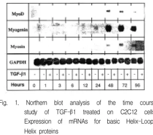
Fig. 1. Northern blot analysis of the time course study of TGF-β1 treated on C2C12 cells;
Expression of mRNAs for basic Helix-Loop- Helix proteins
counter(Coulter Corp., Miami, USA)를 이용하여 산 정하였다. 산정한 세포는 Microsoft Sigmastat을 이 용하여 ANOVA와 Duncan grouping을 시행하여 분 열능의 차이를 비교하였다.
Ⅲ. 연구결과
1. TGF-β1의 근육세포 분화에 대한 효과
1) basic HLH 단백 mRNAs
TGF-β1을 처리하지 않은 세포는 2일 째부터 myoD, myosin, myogenin mRNA 모두 발현하기 시 작하였으나, TGF-β1을 처리한 세포는 4일경과 후에 도 발현하지 않았다(Fig. 1).
2) Id 단백 mRNAs
TGF-β1을 처리하지 않은 세포에서는 시간 경과 에 따라 Id 단백 mRNAs의 발현은 차이가 없으나, TGF-β1을 처리한 세포는 처리 3시간 후부터 Id 단 백 mRNA의 발현이 감소하였다(Fig. 2).
3) TGF-β1 수용체 mRNAs
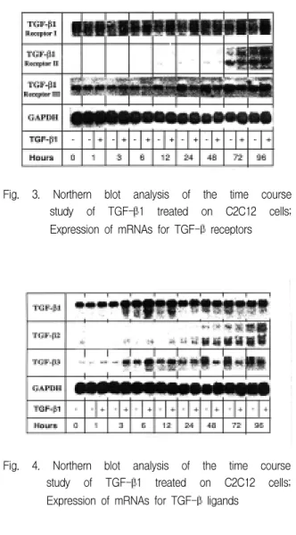
Type I 수용체 mRNA의 경우 TGF-β1을 처리한 세포가 처리하지 않은 세포에 비하여 강하게 발현하 였다. Type II 수용체 mRNA의 경우 TGF-β1을 처 리하지 않은 세포는 12시간 후부터 발현하기 시작하 여 증가 양상을 보이나, TGF-β1 처리한 세포는 24 시간까지 발현되지 않다가 72시간째부터 처리하지 않은 세포와 유사한 발현 양상을 보였다(Fig. 3).
Fig. 2. Northern blot analysis of the time course study of TGF-β1 treated on C2C12 cells;
Expression of mRNAs for ID proteins
Fig. 3. Northern blot analysis of the time course study of TGF-β1 treated on C2C12 cells;
Expression of mRNAs for TGF-β receptors
Fig. 4. Northern blot analysis of the time course study of TGF-β1 treated on C2C12 cells;
Expression of mRNAs for TGF-β ligands
Fig. 5. Northern blot analysis of the dose response study of OP-1 treated on C2C12 cells;
Expression of mRNAs for basic Helix-Loop- Helix proteins
4) TGF-β1 리간드 mRNAs
TGF-β1 mRNA의 경우 시간경과에 따라 두 군 모두 유사한 발현을 보였으며, TGF-β2 mRNA의 경 우 24시간까지 두 군 모두 유사한 발현을 보이나 48 시간 이후부터 TGF-β1을 처리하지 않은 세포에서 우세하게 발현하였다. TGF-β3 mRNA의 경우 12시 간 경과까지 TGF-β1을 처리한 세포에서 우세하였 으나 48시간 이후부터 발현이 역전되는 양상을 보였 다(Fig. 4).
2. OP-1의 근육세포 분화에 대한 효과
1) basic HLH 단백 mRNAs
MyoD와 myogenin mRNA 모두 OP-1을 처리하지 않고 β-glycerophosphate와 ascorbic acid만 처리한 세포에서 시간경과에 따라 발현이 감소하였으며, OP-1 50ng/㎖ 처리한 세포에서는 myoD와 myogenin mRNA 모두 2일과 7일에는 거의 발현하지 않다가 12 일째 뚜렷하게 발현하였다. OP-1 100ng/㎖ 이상 처리 한 세포에서는 전혀 발현하지 않았다(Fig. 5).
2) 골지표 단백 mRNAs
근육세포인 C2C12 세포에 OP-1의 용량에 따라 관 찰한 결과, type I collagen mRNA과 OPN mRNA의 경우 모두에서 조기에 발현하였다. ALP mRNA의 경 우 OP-1 100ng/㎖을 처리한 세포에서 7일째부터 발 현하였으며 300ng/㎖ 이상 처리한 세포에서는 2일째
Fig. 6. Northern blot analysis of the dose response study of OP-1 treated on C2C12 cells;
Expression of mRNAs for bone marker proteins
부터 조기 발현하였다. OC mRNA의 경우 OP-1 300ng/㎖ 이상 처리한 세포에서 14일째에 발현이 시 작하였다. BSP mRNA의 경우 OP-1 100ng/㎖ 이상 처리한 세포에서 14일째에 발현하였다(Fig. 6).
다. Id 단백 mRNAs
Id-1, 2, 3 mRNA 모두 OP-1 처리하지 않은 경우 시간경과에 따라 mRNA의 발현이 감소하였으나, OP-1 300ng/㎖ 이상 처리시 시간경과에 따라 유사하 게 증가되었다(Fig. 7).
Fig. 7. Northern blot analysis of the dose response study of OP-1 treated on C2C12 cells;
Expression of mRNAs for ID proteins
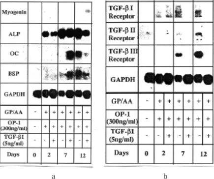
Fig. 8. Northern blot analysis of the study of combined OP-1 and TGF-β1 treated on C2C12 cells; a.
Expression of mRNAs for the myogenin and the bone marker proteins, b. Expression of mRNAs for the TGF-β receptors
a b
3. OP-1과 TGF-β1 동시처리시의 근육세포에 대 한 효과
1) 골 및 근육지표 단백 mRNAs
근육 분화의 지표인 myogenin mRNA의 경우 OP-1만 처리시 시간 경과에 따라 발현하지 않았으 나, 두 가지 동시에 처리한 경우 12일째에 발현하였 다. ALP mRNA의 경우 OP-1만 처리시 시간 경과에 따라 발현이 증가하나 두 가지 처리한 경우에는 7일 째 가장 많이 발현하였다가 12일째에는 감소하였다.
OC mRNA의 경우 OP-1만 처리시 12일째 발현하였 으나 두 가지 처리한 경우에 7일에 발현하여 12일째 에 감소하였다. BSP mRNA의 경우 OP-1만 처리시 7일에 시작하여 12일째에 발현하였으며, 두 가지 처 리한 경우 7일부터 발현하였으나 12일째에 감소하였 다(Fig. 8a).
2) TGF-β 수용체 mRNAs
Type I, II, III TGF-β 수용체 mRNA 모두에서 OP -1만 처리한 경우에 7일째부터 발현하였으며, 두 가지 모두 처리한 경우에는 type I TGF-β 수용체 mRNA 에서 시간경과에 따라 발현이 감소하였다(Fig. 8b).
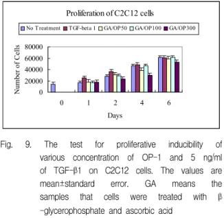
Fig. 9. The test for proliferative inducibility of various concentration of OP-1 and 5 ng/ml of TGF-β1 on C2C12 cells. The values are mean±standard error. GA means the samples that cells were treated with β -glycerophosphate and ascorbic acid
Proliferation of C2C12 cells
0 20000 40000 60000 80000
0 1 2 4 6
Days
Number of Cells
No Treatment TGF-beta 1 GA/OP50 GA/OP100 GA/OP300
4. TGF-β 1과 OP-1의 근육세포 분열능에 대한 효과
무 처리 배양 세포, OP-1으로 처리한 세포와 TGF-β1으로 처리한 세포 모두에서 시간 경과에 따 른 세포 수 증가 정도는 차이가 없었다(P>0.05)(Fig.
9).
Ⅳ. 고 찰
골격성 조직은 골모세포, 연골세포, 지방세포와 근 육세포 등의 다양한 형태의 간질세포들로 구성된다.
이 세포들은 골격성 간질세포라고 불리는 공통적인 간질세포에서 비롯되었다고 알려져 있다3,4). 이에 따 라 많은 연구자들이 골모세포, 연골세포, 지방세포와 근육세포 등으로 분화할 수 있는 다기능성 세포계에 대한 연구를 시행하였다28-31). 본 연구에서 사용한 C2C12 세포계는 성숙된 백서의 대퇴부 근육에서 클 론된 세포이다32). 이 세포계는 외부적 cDNA의 안정 된 transfection의 실험에 적합한 것으로 알려져 있으 며 근육세포를 코드화하는 실험에 추천되고 있다33). 이 세포는 정상 근육조직의 섬유를 따라 배열해 있으 며 근육위성세포에서 분화된 것으로 추정된다. 근육 조직이 손상을 받은 경우 근육위성세포는 분열되고 근육세포로 분화되어 근육섬유를 형성하게 된다1-5). C2C12 세포는 FBS 농도 10%에서 세포분열이 나 타나며, FBS 농도를 5%로 감소시키는 경우 또는 2%
말혈청으로 교환하는 경우 다핵성 근관으로 분화한
다32,33). 본 연구에서 세포의 수가 2배로 되는 데 소요
시간은 약 12시간이었으며, 5%의 FBS로 교환 후 2일 째부터 세포의 융합이 시작하여 4일 이후 다핵성 근 관들이 형성되기 시작하였으며 8일째부터는 자발적 인 수축을 시작하였다. MyoD의 발현능을 잃지 않게 분화용 배양액으로의 교체는 세포가 confluent상태의 90% 수준에 도달하였을 때 시행하였으며, 성장인자 처리도 동시 시행하였다.
본 연구에서 근육세포 분화에 대한 효과를 관찰하 고자 하는 TGF-β1은 사람의 혈소판, 태반과 소의 신장에서 분리될 수 있다. 이형체(isoforms)는 신장된 구상 입체구조를 갖는데, 각 단량체 사슬에서 9개의 cystein residue 중 8개가 TGF-β knot라고 불리는 사슬내 이황화 결합의 치밀한 형태를 갖는데 관여한
다34,35). TGF-β의 다형질 발현성 효과는 간질세포에
서 독립적 성장의 유도36), 지방세포 분화의 억제37), 교 원질과 fibronectin의 합성유도38), 상피분열 촉진과 상 피분화의 촉진39) 등으로 알려져 있다. TGF-β는 세 포의 형태에 따라 서로 다른 생물학적 활성을 가진다.
TGF-β는 슈반세포, 조골세포, 연골세포, 태생기 폐 의 섬유아세포등의 성장을 자극하나 대부분의 세포 에서 성장을 억제한다40,41).
OP-1은 BMP-7이라고도 불리우며 소의 골에서 고 순도 정제된 골형성 단백으로 18 kD의 분자량을 가 진 단량체성 기본단위와 이황화 결합을 하는 이형체 로 구성된다. 골형성 단백은 여러 다양한 포유류에서 분리한 골기질성 폴리펩티드의 계통으로, C-termini 에 잘 보존된 TGF-β의 7개의 cystein domain을 가 져 동물에서 골 선조세포의 아군집을 연골내 골화로 유도하는 역할을 한다고 하였으나 완성된 골세포는 연골세포로 바꾸지 않는 것으로 보인다24,25)고 하였다.
본 연구에서 TGF-β1은 수회의 실험결과 근육세 포 분화를 억제하는 적정 용량인 5ng/㎖로 선택하였 으며, 초기의 분화억제효과를 관찰하기 위하여 초기 4일간의 시간으로 본 연구를 시행하였다. OP-1의 경 우 각각의 농도에서 시간 경과에 따른 세포의 형태학 적 변화에 준하여 total RNA의 추출을 시행하였다.
각 농도에 따라 세포의 형태학적 변화는 1-2일 정도 의 오차가 있었다.
TGF-β1을 처리하지 않고 C2C12 세포를 배양했 을 때, 근육세포 분화과정에서 myoD와 myogenin mRNA의 발현은 2일째부터 나타나며 Id-1, 2, 3은 감 소하였다. TGF-β1을 처리한 세포에서는 4일까지 myoD와 myogenin mRNA의 발현을 억제하였으나,
Id-1, 2, 3은 감소하였다. 이는 TGF-β1의 경우 Id 단 백의 양성조절자와의 이성이형체 형성에 의한 것이 아닌 근육 특정 유전자의 발현 억제에 의한 것으로 보여진다. 다른 대표적인 분화 억제성 성장인자인 bFGF는 DNA binding domain에서 conserved protein kinase C의 인산화를 통해서 myogenin의 DNA 결합능력을 불활성시켜 근육세포의 분열을 촉 진시킨다고13,14) 알려져 있다. TGF-β1과 bFGF 또한 근육세포의 성숙세포로의 마지막 단계의 분화를 억 제하며 근육인자 mRNA의 발현을 억제한다. 그러나
bFGF13,14)와는 반대로, TGF-β1은 DNA 결합능과
Id-1 유도와는 독립적인 기전을 통해서 세포분열을 촉진하지 않고 근육분화 인자의 활성을 억제하는 즉, MyoD family의 DNA 결합능은 변화시키지 않는 것 으로 보여진다. 본 연구에서 OP-1은 100ng/㎖ 이상 의 용량에서 근육 특정 유전자의 발현을 강하게 억제 하였으며, 용량 증가에 따라 Id 단백의 발현을 증가시 켰다. 이는 OP-1 300ng/㎖ 이상에서 Id 단백이 강하 게 발현함으로써 근육분화와 관련하는 bHLH 단백 인자의 발현수준을 조절하여 근육세포분화를 방해한 것으로 보여진다. 또한 이는 C2C12 세포에서 bHLH 단백과 E12/E47사이의 이성이형체 형성을 방해함으 로서 근육분화를 억제한다는 Robert등의 보고36)와 일 치하는 것이다. OP-1에 의해 Id 단백의 발현이 증가 되면서 myoD와 myogenin의 발현도 억제되는 것은 OP-1이 TGF-β1과 다르게 근육 특정 유전자의 전 사와 DNA 결합능을 변화시킨 결과로 사료된다.
BMP-2의 근육세포 분화에 대한 효과를 연구한 Katagiri등21)도 BMP-2가 근육 특정 유전자의 발현을 억제하고 Id 단백 발현을 증가시킴으로서 근육의 분 화를 억제시킨다라는 본 연구의 OP-1의 경우와 유사 한 결과를 보고하여, BMP-2와 OP-1은 유사한 기전 으로 근육의 분화를 조절하는 것으로 추정된다. 위 내 용으로 보아, 양성과 음성조절자의 협동적인 조절이 근육분화 억제에 중요한 역할을 하는 것으로 보인다.
근육 인자들이 일반적인 coregulator와 관련되었는 지와 이러한 coregulator의 활성이 성장인자들의 세 포내 신호전달에 의해 조절되는지의 여부 등을 설명 하기 위한 계속적인 연구가 필요할 것으로 사료된다.
본 연구에서 TGF-β1 처리시 20ng/㎖ 이상의 용 량에서도 골지표 단백은 발현하지 않았으나, OP-1 처리시 100ng/㎖ 이상의 고농도에서 골지표 단백의 mRNAs가 발현되었다. 이러한 결과는 Murray등22), Yamaguchi등42)과 Katagiri등21)의 C2C12세포 분화에
대한 BMP-2의 효과에서의 결과와 유사한 양상을 보 인다. BMP-2와 OP-1의 분자량 및 역가의 차이에 의 해 효과를 비교하기 어려우나 BMP-2는 300ng/㎖ 이 상의 용량에서 OP-1보다 빠른 시간내에 골지표 단백 의 발현을 유도하였다. 이 결과로 보아 TGF-β1은 C2C12세포의 근육으로의 분화를 억제하나 이는 일시 적이고 가역적인 억제로 보여지며, BMP-2와 OP-1 은 C2C12세포의 근육으로의 분화를 억제하고 C2C12 세포를 골세포성 계열로 전환시키는 것으로 사료된 다. Murray등22)은 L6 세포계에서 BMPs는 근관의 형 성은 억제하였으나 골성 표현형의 발현은 유도하지 못하였으며, BMP-2의 효과는 C2C12 세포와 같은 근 육위성세포에서만 적용되는 것으로 제안하였다42). 즉 이는 잠재적인 선조세포에서는 BMP-2가 골세포성 계열로 분화를 전환시킬 수 있으나 보다 근육세포로 의 분화가 완성되어 있는 L6 세포같은 세포계에서는 분화과정을 변화시킬 수 있는 잠재력을 잃는 것으로 보여진다. 본 연구에서 비슷한 결과를 보인 OP-1도 근육위성세포에서만 효과를 보이지 않을 가 추정된 다.
TGF-β1 5ng/㎖과 OP-1 300ng/㎖을 동시에 처리 하였을 때, myogenin 발현이 초기에 완전히 억제되 었다가 두 가지 모두 처리한 세포에서 12일 째에 발 현하였으며, 골지표 단백의 경우 OP-1과 GP/AA만 처리한 경우 정상적인 발현을 보였으나, 두 가지를 모 두 처리한 세포에서는 7일째에 강하게 조기 발현하여 12일째에는 감소하는 양상을 보였다. Katagiri등21)과 Florini등35)의 BMP-2를 이용한 연구들에서도 본 연 구에서와 유사하게 TGF-β1은 BMP-2에 의해 유도 된 ALP와 OC 활성을 감소시켰다. 또한 TGF-β1은 다른 종류의 골성 세포에서도 ALP와 OC의 활성을 감소시켰으며, 생체에서 골막에 주사하였을 때 이소 성 신생골 형성을 유도하였으나 근육조직 내에 매식 하였을 때는 이소성 골형성이 나타나지 않았다38) 는 보고에 비추어 본 연구의 경우도 TGF-β1이 OP-1 의 효과를 가리며, 두 가지 동시 처리시 TGF-β1이 보다 강력한 조절자로 작용하는 것으로 사료되며, OP-1의 근육조직에 대한 연구도 병행되어야 할 것으 로 사료된다. 두 가지 동시 처리시 7일째의 골지표 단 백의 발현은 두 가지 성장인자의 상승효과에 의한 것 이며 이는 수용체와 신호전달 체계의 차이에 의한 것 으로 추정되나 이를 구명하기 위한 연구가 지속되어 야 할 것으로 사료된다.
TGF-β superfamily member의 세포내 신호전달
은 type I과 type II라는 두가지 형태의 특정한 serine/threonine kinase 수용체를 통해서 전달된다
43,44)
. Type I 과 type II TGF-β 수용체는 신호전달 에 있어 필수적인 것이며 리간드와 결합하여 이형성 복합체를 형성한다. TGF-β1은 세포표면에서 type II 수용체와 결합하나 이러한 결합이 전사나 성장억 제 작용을 유도하지는 않으며, Type I 수용체의 결합 특이성은 동시에 발현하는 특정 type II 수용체에 의 해 결정된다43-46). 본 연구에서 TGF-β1의 근육세포 분화억제에 있어서 type I 수용체가 초기 4일 간 상향 조절됨으로써 또 OP-1과 TGF-β1을 동시에 처리시 시간 경과에 따라 하강 조절됨으로써 근육 특정 유전 자의 발현 억제와 맞추어 음성 신호전달(negative signalling)에 주된 역할을 담당하는 것으로 사료된 다. TGF-β1 단독 처리시 리간드의 변화는 초기 분 화를 시작하는 48시간부터 처리하지 않은 세포에서 우세하게 발현함으로서 또 두 가지를 동시에 처리한 경우 발현이 증가되거나 발현 양상에 차이를 보이지 않음으로 보아 TGF-β2와 TGF-β3은 근육세포의 분화에 크게 간여하지 않는 것으로 사료된다.
본 연구에서는 시행되지 않았으나 OP-1과 BMP의 공통적인 분화능력은 같은 종류의 수용체와 결합에 의한다는 연구들이 있다. BMPR-IA와 BMPR-1B 수 용체가 BMP-2, 4와 OP-1을 위한 type I 수용체로 밝
혀졌다4,47,48). C2C12 세포에서도 BMPR-1A가 발현되
므로서 BMPR-1A가 BMP-2에 의해 유도되는 근육 분화의 음성 신호전달(negative signalling)에 관련한 다는 것이 제안되었다49). OP-1도 BMP-2와 같은 형 태의 수용체를 통해서 신호전달을 이루는 것으로 보 아 비슷한 기전으로 근육세포를 골세포성 계열로 전 환시킬 것으로 보여지며, 이를 확인하기 위해 근육세 포에서 OP-1에 대한 BMPR의 발현여부가 연구되어 져야 할 것이다.
위의 내용에 비추어 OP-1은 근육세포를 골세포성 계열로 세포를 전환시키고, TGF-β1는 가역적인 일 시적 근육 분화 억제효과를 가지는 것으로 사료되며, 두 가지 성장인자는 서로 다른 기전으로 근육세포 분 화를 억제할 것으로 추정된다. 보다 자세한 기전을 파 악하기 위하여 펩타이드성 성장인자들의 근육 분화 조절에 있어 다른 coregulator의 작용과 함께 세포 내 와 핵에서 이루어지는 신호전달 체계의 차이에 관한 심도있는 연구가 지속적으로 이루어져야 할 것으로 사료된다.
Ⅴ. 결 론
TGF-β1과 OP-1이 근육세포 분화에 미치는 영향 을 관찰하기 위하여, TGF-β1과 OP-1을 C2C12 세 포에 처리하여 형태학적 변화를 관찰하고, total RNA 를 추출하여 골표지 단백, 근육특절 조절성 단백, TGF-β수용체 및 리간드의 cDNA probe에 대한 mRNA의 발현을 Northern blot analysis를 이용하여 알아보고, 두가지 변형성장인자의 세포분열능에 대한 효과를 산정하였다.
C2C12 세포는 무처리 분화시 2일 째부터 세포들이 융합하기 시작하여 4일째부터 다핵성 근관들을 형성 하고 8일이상 경과시 자가 수축을 시작하였다. TGF- β1을 처리하여 배양하면 4일째까지 근육세포의 분 화가 억제되며 8일째부터 세포의 융합이 시작되었다.
C2C12 세포를 5ng/㎖의 TGF-β1을 처리하여 배양 한 4일까지 다핵성 근관 형성을 억제하며 Id 단백의 발현을 감소시켰다. 그러나 골세포성 표현형은 유도 되지 않았다. Type I TGF-β 수용체의 mRNA 발현 은 배양 48시간 이후 발현이 역전되었다. 이로 보아 Type I TGF-β 수용체는 근육분화를 억제하는데 음 성 신호전달에 역할을 하는 것으로 보여진다. OP-1 은 용량 의존성으로 100ng/㎖ 이상의 농도에서 ALP, OPN, BSP 발현을 유도하고, 300ng/㎖ 이상에서 OC 형성을 유도하였다. 이러한 골세포성 표현형을 발현 시키는데 필요한 OP-1의 농도는 근관 형성을 억제시 키는데 필요한 용량과 비슷하였다. OP-1을 100ng/㎖
이상으로 처리한 경우 OP-1은 처리 2일 후부터 근육 조절인자 mRNA의 발현을 억제하였다. OP-1 300ng/
㎖ 이상의 농도에서 Id-1, 2, 3 mRNA의 발현이 촉진 되었다. 두 가지 성장인자를 동시에 처리하였을 때, TGF-β1은 12일째에 OP-1의 분화억제를 감약시켰 으며 myogenin mRNA도 재 발현하였다. 그리고 TGF-β1은 처리 12일째에 OP-1에 의해 유도된 OC 와 BSP 형성을 감소시켰다. 세포 분열능에 있어서 TGF-β1과 OP-1 모두 분열촉진 효과는 나타내지 않았다. 이러한 결과로 보아 OP-1은 C2C12 근육세포 의 분화과정을 골세포성 계열로 전환시키나, TGF- β1은 골세포성 분화를 유도하지 않으며 가역적인 분 화억제 효과를 가지는 것으로 사료된다. 두가지의 성 장인자는 근육 분화의 억제에 있어서 서로 다른 수용 체를 통하는 서로 다른 기전에 의해 작용하는 것으로 추정된다.
참 고 문 헌
1. Bischoff, R. : Proliferation of muscle satellite cells on intact myofibers in culture. Dev. Biol., 115:129-139, 1986
2. Olsen, E.N. : Interplay between proliferation and differentiation within the myogenic lineage. Dev.
Biol., 154:261-272, 1992
3. Weintraub, H.: The MyoD family and myogenesis : redundancy, networks and thresholds. Cell 75:1241-1244,1994.
4. Yamaguchi, A.: Regulation of differentiation pathway of skeletal mesenchymal cells in cell lines by transforming growth factor(TGF-β) superfamily.
Seminar in Cell Biology, 6:165-173, 1995.
5. Dias, P., Dilling, M. and Houghton, P. : The molecular basis of skeletal muscle differentiation. Seminars in Diagnostic Pathology, 11:3-14, 1994.
6. Li, L. and Olson, E.N. : Regulation of muscle cell growth and differentiation by the MyoD family of helix-loop-helix proteins. Adv. Cancer Res., 58:95- 119, 1992.
7. Davis, R.L., Weintraub, H. and Lassar, A.B.:
Expression of a single transfected cDNA converts fibroblasts to myoblasts. Cell, 51:987-1000, 1987.
8. Weintraub, H., Davis, R. and Tapscott, S. : The MyoD gene family: Nodal point during specification of the muscle cell lineage. Science, 251:761-766, 1991.
9. Wright, W.E., Sassoon, D.A., and Lin, V.K. : Myogenin, a factor regulating myogenesis, has a domain homologous to MyoD. Cell 56:607-617, 1989.
10. Rhodes, S.J. and Konieczny, S.F. : Identification of MRF-4: A new member of the muscle regulatory factor gene family. Genes Dev. 3:2050-2061, 1989.
11. Davis, R.L. and Weintraub, H.: Acquisition of myogenic specificity by replacement of three amino acids residues from MyoD into E12. Science, 256:1027-1030, 1992.
12. Benezra, R., Davis, R.L., Lockshon, D., and Lassar, A.B.: The protein Id: A negative regulator of helix-loop-helix DNA binding proteins. Cell, 61:49- 59, 1990.
13. Li, L., Zhou, J., and James, G.: FGF inactivates myogenic helix-loop-helix proteins through phosphorylation of a conserved protein kinase C site in their DNA binding domains. Cell, 71: 1181-1194, 1992.
14. Vaydia, T.B., Rhodes, S.J., Taparowsky, E.J., and Konieczny, S.F.: Fibroblast growth factor and
transforming growth factor beta repress transcription of the myogenic regulatory gene MyoD1. Mol. Cell Biol., 9:3576-3579, 1989.
15. Florini, J.R., Ewton, D.J., Falen, S.L. and Van Wyk, J.J.: Biphasic concentration dependency of the stimulation of myoblast differentiation by somatomedins. Am. J. Physiol., 250:771-776, 1986.
16. Florini, J.R. and Magri, K.A.: Effects of growth factors on myogenic differentiation. Am. J. Physiol., 256:C701-C707, 1989.
17. Kiess, W., Haskell, J.F., Greenstein, L.A., Miller, B.E., Aarons, A.L., Rechler, M.M. and Nisley, S.P.: An antibody that blocks insulin-like growth factor binding to type II IGF receptor is neither an agonist nor an inhibitor of IGF -stimulated biologic responses in L6 myoblasts. J. Biol. Chem., 262:12745-12749, 1987.
18. Tollefsen, S.E., Lajara, R., Mckusker, R.H., Clemmons, D.R, and Rotwein, P.: IGF in muscle development.
expression of IGF-I, the IGF-I receptor, and an IGF binding protein during myoblast differentiation. J.
Biol. Chem., 264:13810-13813, 1989.
19. Meyer, S.D., Clair, J.H. and Ham, R.G.: EGF is a mitogen for human skeletal muscle satellite cells but does not block their differentiation. J. Cell Biol., 109:305a, 1989.
20. Olwin, B.B. and Hauschika, S.D.: Cell surface fibrobalst growth factor and epidermal growth factors are permanently lost during skeletal muscle terminal differentiation in culture. J. Cell Biol., 107:761-769, 1988.
21. Katakiri K., Yamauchi A., Komachi M., Abe E., Takahashi M., Ikeda T., Rosen V., Woozney Z.M. and Suda D.: Bone morphogenetic protein-2 converts the differentiation pathway of C2C12 myoblasts into the osteoblastic lineage. J. Cell Biol., 127:1755-1766. 1994.
22. Murray, SS., Murray, J.B., Glackin, C.A. and Urist, M.R.: Bone morphogenetic protein inhibits differentiation and affects expression of helix loop helix regulatory molecules in myoblastic cells. J. Cell.
Biochem. 53:51-60,1993.
23. Inada, M.. katagiri, T., Akiyama, S., Namiki, M.
Komaki, M. Yamaguchi, A., kamoi, K., Rosen, V., and Suda, T.: Bone morphogenetic protein-12 and -13 inhibit terminal differentiation of myoblasts, but do not induce their differentiation into osteoblasts.
Biochem. Biophys. Res. Commun., 222:317-322, 1996.
24. Cook, S. and Rueger, D.C.: Ostegenic Protein-1.
Clinical orthopedics and related research. 324:29-38,
1996.
25. Sampath, T.K., Maliakal, J.C., Hsuschika, T., Jones, W.K., Sak, H., Tucker, R.F., Couchlin, J.E., Tucker, M.M., Corbett, C., Orkaynak, E., Opermann, H., and Rueger, D.C.: Recombinant human osteogenic protein-1 induces new bone formation in vitro with specific activity compatible with natural bovin osteognic protein and stimulates osteoblast proliferation and differentiation in vitro. J. Biol. Chem.
267:20352-20362, 1992.
26. Brennan, T.J., Edmondson, D.G. and Li, L.: TGF-β represses the actions of myogenin through a mechanism independant of DNA binding. Proc. Natl.
Aca. Sci. USA 88:382-386,1991.
27. Massague, J., Cheifetz, S., Endo, T. and Nadal-Ginard, B.: Type β transforming growth factor is an inhibitor of myogenic differentiation. Proc. Natl.
Acad. Sci. USA 83:8206-8210, 1986.
28. Barr, E. and Leiden, J.M.: Systemic delivery of recombinant proteins by genetically modified myoblasts. Science, 254:1507-1509, 1991.
29. Silberstein, L., Webster, S.G., Travis, M. and Blaum, H.M.: Developmental progression of myosin gene expression in culture od muscle cells. Cell, 46:1075-1081, 1986.
30. Grigoriadis, A.E., Heersche, J.N.M. and Aubin, J.E.:
Differentiation of muscle, fat, cartilage, and bone from progenitor cells present in a bone-derived clonal cell population: effect of dexamethasone. J Cell. Biol., 106:2139-2151, 1988.
31. Yamauchi, A. and Kahn, AJ.: Clonal osteogenic cell lines express myogenic and adipocytic developmental potential. Calcif. Tissue Int., 49:221-225, 1991.
32. Yaffe, D. and Saxel, O.: Serial passaging and differentiation myogenic cell isolated from dystrophic mouse muscle. Nature Lond., 270:725-727, 1977.
33. McMahon, D.K., Anderson, P.A.W., Nassar, R., Bunting, J.B., Saba, Z., Oakley, A.E and Malouf, N.N.:
C2C12 cells : biophysical, biochemical, and immunocytochemical properties. Am. J. Physiol., 266:C1795-C1802, 1994.
34. Lawrence, D.A.: Transforming growth factor-β: a general review. Eur. Cytokine Netw., 7:363-374, 1996.
35. Florini, J.R., Roberts, A.B., Ewton, D.Z., Falen, S.L., Flander, K.C., and Sporn, M.B.: Transforming growth factor-β, A very potent inhibitor of myogenic differentiation, identical to the differentiation inhibitor secreted by buffalo rat liver cells. J. Biol. Chem.
261:16509-16513, 1986.
36. Robert, AB., Anzano, M.A., Wakefield, L.M., Roche, N.S., Stern, D.F. and Sporn, M.B.: Type β Transforming growth factor: a bifunctional regulator of cellular growth. Proc. Natl. Acad. Sci. USA 82:119-123, 1985.
37. Ignotz, RA. and Massague, J.: Type β transforming growth factor controls the adipogenic differentiation of 3T3 fibroblast. Proc. Natl. Acad. Sci. USA 82:530-534, 1985.
38. Heino, J. and Massague, J.: Cell adhesion to collagen and decreased myogenic gene expression implicated in the control of myogenesis by transforming growth factor beta. J. Biol. Chem., 265: 10181-10184, 1990.
39. Masui, T., Wakefield, L.M., Lechner, J.F., LaVeck, M.A., Sporn, M.B., and Harris, C.C.: Type β transforming growth factor is the primary differentiation inducing factor for normal human brochial epithelial cells. J. Cell. Biochem. 53:51-60, 1993.
40. Raynal, S. and Lawrence, D.A.: Differential effect of transforming growth factor β1 on protein levels of p21 WAF and cdk-2 and on cdk-4 kinase activity in human RD and CCL 64 mink lung cells, Inter. J.
Oncol., 7:337-343, 1995.
41. Benzakour, O., Merzak, A., Dooge, Y., Pironin, M., Lawrence, D.A. and Vigier, P.: Transforming growth factor beta stimulates mitogenically mouse NIH3T3 fibroblasts and those cells transformed by the EJ-H-ras oncogene. Growth Factors, 6:265-269, 1992.
42. Yamauchi, A., Ishzuya, T., Kintou, N., Wada, Y., Katagiri, T., Woozney, J.M., Rosen, V. and Yoshiki, S.: Bone morphogenetic protein-2 inhibits terminal differentiation of myogenic cells by suppressing the transcriptional activity of MyoD and Myogenin.
Biochem. Biophys. Res. Commun., 220:366-371, 1996.
43. Derynk, R.: TGF-beta receptor mediated signalling.
Trends Biochem. Science, 19:548-552, 1994.
44. Kawabata, M., Chytil, A., and Moses, H.L.: Cloning of a novel type II serine/threonine kinase receptor through interaction with the type I transforming growth factor beta receptor. J. Biol. Chem., 270:5625-5629, 1995.
45. Wrana, J.L., Attisano, L., Wieser, R., Ventura, F. and Massague, J.: Mechanism of activation of the TGF-beta receptor. Nature, 370:341-346, 1994.
46. Chen, R.H., Moses, H.L., Maruoka, E.M., Derynck, R.
and Kawabata, M.: Phosphorylation-dependent interaction of the cytoplasmic domains of the type I and type II transforming growth factor beta receptor.
J. Biol. Chem., 270:12235-12241, 1995.
47. ten Dijke, P., Yamashita, H., Ichijo, H., Franzen, P., Laiho, M., Miyazono, K. and Heldin, C.:
Characterization of Type I receptors for transforming growth factor-β and activin. Science, 264:101-104, 1994.
48. ten Dijke, P., Yamashita, H., Sampath, T.K., Redii, A.H., Estevez, M., Riddle, D.L., Ichijo, H., Heldin, C.
and Miyazono, K.: Identification of type I receptors for osteogenic protein-1 and bone morphogenic protein-4. J. Biol. Chem. 269:16985-16988, 1994.
49. Mishina, Y., Suzuki, A., Ueno, N., and Behringer, R.R.:
BMPR encodes a type I bone morphogenetic protein receptor that is essential for gastrulation during embryogenesis. Genes Dev., 9:3027-3037, 1995.
- ABSTRACT -
The Inhibitory Effect of TGF-β1 and OP-1 onto the Myogenic Differentiation
Byung-Gook Kim, DDS., PhD., Sung-Su Jung, MD., PhD*
Department of Oral Medicine, Anesthesiology*, School of Dentistry, Chonnam National University
In order to investigate the effect of Transforming growth factor β1(below TGF-β1) and osteogenic protein-1(below OP-1) onto the myogenic differentiation, C2C12 satellite myoblastic cell line was cultured and treated with both growth factors. At first morphological changes with microscopical examination were examined, and isolated total RNA to analyse mRNA expression of bone marker proteins, muscle regulatory proteins, TGF-β receptor and their ligands by Northern blot analysis. And cellular proliferative inducibility of both growth factors was also tested to C2C12 cells.
Incubating the cell with 5 ng/㎖ of TGF-β1 until 4 days almost inhibited multinucleated myotube formation expressing muscular regulatory proteins, and induced decreasing Id proteins. However, no osteoblastic phenotypes was induced by TGF-β1 in C2C12 cells. The mRNA expression of TGF-β receptors with TGF-β1 was conversed after 48 hours cultured. Type I TGF-β receptor was seemed to play a role in negative signalling for inhibition of myogenic differentiation. OP-1 dose dependently induced ALP activity, osteopontine production and bone sialoprotein production at concentrations above 100ng/㎖ and osteocalcin production at concentrations above 300ng/㎖. The concentration of OP-1 required to induce these osteoblastic phenotypes was the same as that required to almost completely inhibit myotube formation. Incubation with above 100ng/㎖ OP-1 suppressed the expression of mRNA for muscular regulatory proteins from 2 days after incubation. Expression of Id-1, 2, 3 mRNA were stimulated by OP-1 at concentration above 300ng/㎖.
When C2C12 cells were treated with both growth factors, TGF-β1 potentiated the inhibitory effect of OP-1 on myotube formation and expression of mRNA for myogenin at 12 days. And TGF-β1 reduced osteocalcin and bone sialoprotein production induced by OP-1 at 12 days in C2C12 cells. Both growth factor had no mitogenic effect. These results indicate that OP-1 converts the differentiation pathway of C2C12 myoblasts into that of osteoblastic lineage cells and it's not heritable, but TGF-β1 does not and has reversible inhibitory activity on the myogenic differentiation. TGF-β1 and OP-1 play a role in myogenic differentiation via different mechanism between them.