INTRODUCTION
Scrotal leiomyomas are uncommon tumors that arise from the subcutaneous tissue, or dartos muscle (1). The number of reported cases has recently been increasing (2). As in uter- ine leiomyomas, bizarre nuclei rarely occur in those of the genitourinary tract (3-8). Neither malignant transformation nor recurrences of scrotal bizarre leiomyoma has been report- ed. Awareness of scrotal leiomyoma with bizarre nuclei is critical not to misdiagnose as leiomyosarcoma because its surgical extent and the follow up are different.
CASE REPORT
A 65-yr-old man was admitted for transurethral resection of known urothelial carcinoma of the bladder. Physical exami- nation revealed an elastic, firm, nontender mass of 1.0 cm in the greatest dimension in the left scrotum, and it was imper- vious to light. He was not aware of the mass before the physi- cal examination. The serum level of some tumor markers such as alpha fetoprotein and human chorionic gonadotropin- beta was normal. Percutaneous mass excision was performed.
It was easily dissected from the tunica dartos.
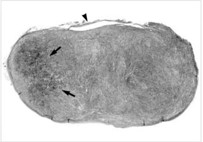
Grossly, the tumor was a well-circumscribed, 1.0 1.0 0.8 cm-sized, oval mass that originated from the tunica dar- tos, which was independent of the testis, epididymis, and funiculus spermaticus. Microscopically, it consisted of inter- lacing fascicles of spindle-shaped cells, and some of the tumor cells had pleomorphic nuclei and showed focally increased
cellularity (Fig. 1). The nuclei were large and multilobulated with hyperchromatic chromatin and macronucleoli (Fig. 2A).
Intranuclear cytoplasmic invagination producing eosinophilic pseudoinclusions was frequently observed (Fig. 2B). The tumor cells revealed no mitosis. Immunohistochemically, the tumor cells expressed vimentin (Zymed, San Francisco, CA, U.S.A., 1:50 dilution), desmin (Zymed, 1:50 dilution), and smooth muscle actin (Zymed, 1:100 dilution), but not cytokeratin (AE1/AE3, Zymed, 1:50 dilution), epithelial membrane antigen (Zymed, 1:50 dilution), HMB-45 (Zymed, 1:50 dilution), glial fibrillary acidic protein (Dako, Glostrup, Denmark, 1:350 dilution), or S-100 protein (Zymed, 1:100 dilution). The tumor cells showed a negative reaction for p53 protein (Zymed, 1:100 dilution), and less than 0.1% of the tumor cell nuclei showed Ki-67 labeling index (Zymed, 1:50 dilution). The tumor cells were stained red with Mas- son-Trichrome.
Resection of the bladder tumor by transurethral cystoscopy revealed noninvasive superficial papillary urothelial carcino- ma (G1, Ta). The postoperative course was uneventful for seven months after the operation, and no recurrence has been recognized.
DISCUSSION
Scrotal bizarre leiomyomas have been rarely reported with various names, i.e. symplastic, pleomorphic, bizarre or atypi- cal leiomyoma, and eight such cases could be retrieved in the world literature (3-8). All cases presented with an asymp-
*
*
Department of Pathology, Kangnam General Hospital Public Corporation, Seoul; *Department of Pathology, Samsung Medical Center, Sungkyunkwan University School of Medicine, Seoul, Korea
Received : 8 July 2002 Accepted : 31 July 2002
Joungho Han, M.D.
Department of Pathology, Samsung Medical Center, 50 Ilwon-dong, Kangnam-gu, Seoul 135-710, Korea Tel : +82.2-3410-2765, Fax : +82.2-3410-0025 E-mail : [email protected]
452 J Korean Med Sci 2003; 18: 452-4
ISSN 1011-8934
Copyright The Korean Academy of Medical Sciences
Scrotal leiomyomas with atypical bizarre nuclei are rare, which might be misdi- agnosed as malignant tumor. We describe a case of scrotal bizarre leiomyoma in a 65-yr-old man. The tumor was a 1 cm-sized, well circumscribed, oval mass arising from the tunica dartos muscle. Histologically, it was formed by whorling bundles of fusiform cells with occasional atypical, pleomorphic nuclei and pseudo- inclusions. Mitosis was not found. Although morphologically atypical, scrotal bizarre leiomyomas take on a biologic behavior not different from that of conven- tional leiomyoma, they should be distinguished from leiomyosarcoma to avoid unnecessary treatment.
Key Words :Neoplasms, Muscle tissue; Leiomyoma; Scrotum; Tunica dartos
Bizarre Leiomyoma of the Scrotum 453
tomatic, pedunculated tumor or ulcerative lesion. Scrotal leiomyomas are rare by themselves, and less than fifty cases have been reported (1, 2, 9). They rarely exhibit pleomor- phic nuclear changes analogous to their uterine counterparts.
In uterine leiomyoma, symplastic (atypical, bizarre, or pleo- morphic) uterine leiomyoma is a term reserved for that with giant cells, nuclear atypism, and minimal mitotic activity (up to 10/10 high-power fields) (10). Eosinophilic cytoplas- mic globules as observed in the present case which corre- sponded well to aggregates of intermediate filaments, are commonly not seen in leiomyomas with bizarre nuclei (5, 6).
Atypical cells were suggested to have resulted as a conse- quence of synthetic progestin treatment like those in uter-
ine leiomyomas. Unfortunately, there is no known relation- ship of atypical cells to preoperative hormonal treatment even in uterine leiomyomas. Bizarre cells are also regarded as degenerative changes, similar to that of ancient schwan- noma, ancient melanocytic nevus, or some dermal tumors (11). In contrast to the smooth muscle tumors of the uterus, the histologic criteria for the diagnosis of scrotal leiomyosar- comas have not yet been established. In the review of New- man and Fletcher, the presence of any mitotic activity was advocated as the criterion of potential malignancy (8). In our opinion, bizarre leiomyoma rather than symplastic or atypical variant is the appropriate term for the scrotal smooth muscle tumors with bizarre nuclei because the former can imply such lesions regardless of mitotic activity.
The smooth muscle of the tunica dartos is regarded as the cellular origin of scrotal leiomyoma. Clinically, scrotal leiomy- oma appears as a small, firm, nontender, slowly growing mass. It may present as a pedunculated lesion with ulcera- tion, needing to be differentiated from squamous carcinoma of the scrotum (12, 13). The painless nature of scrotal leiomy- omas corresponds well to the slow growing tumor pushing the nerve trunks outward instead of compressing them. It occasionally presents with a large pedunculated mass or with an ulcerative mass (2).
Simple excision is indicated to achieve cure of scrotal bizarre leiomyoma. Irradiation should be avoided because radiation may induce malignant transformation of the tumor. Long- term urologic follow-up, however, is indicated because of the rarity of scrotal bizarre leiomyomas and lack of evidence whether the lesions are progressive or regressing. It is impor- tant that both pathologists and urologists should not equate
Fig. 1.A well-circumscribed oval mass is contiguous to the tunica dartos (arrowhead). Arrows indicate focal areas with increased cellularity (H&E, original magnification).
Fig. 2.(A) Tumor cells show enlarged nuclei and multinucleation with fibrillary cytoplasm (H&E, 200). (B) Intranuclear eosinophilic pseu- doinclusions and multinucleated giant cells are seen (H&E, 400).
A B
454 N.R. Kim, C.O. Sung, J. Han
bizarre leiomyoma with leiomyosarcoma.
REFERENCES
1. Siegal GP, Gaffey TA. Solitary leiomyomas arising from the tunica dartos scroti. J Urol 1976; 116: 69-71.
2. Das AK, Bolick D, Little NA, Walther PJ. Pedunculated scrotal mass: leiomyoma of scrotum. Urology 1992; 39: 376-9.
3. Herbert M, Segal M, Hermann G, Sandbank J. Pleomorphic leimy- oma of the scrotum: immunohistochemical stains. Isr Med Assoc J 2001; 3: 543-4.
4. Rodriguez-Parets JO, Silva Abuin J, Abad Hernandez M, Tinajas Saldana A, Martin Rodriguez A, Garcia Macias C, Bullon Sopelana A. Atypical scrotal leiomyoma. A case report. Actas Urol Esp 1998;
22: 613-5 (Spanish).
5. Slone S, O’Connor D. Scrotal leiomyomas with bizarre nuclei: a report of three cases. Mod Pathol 1998; 11: 282-7.
6. De Rosa G, Boscaino A, Giordano G, Donofrio V, Staibano S, Maio
C, Iandolo M. Symplastic leiomyoma of the scrotum. A case report.
Pathologica 1996; 88: 55-7.
7. Nishiyama N, Hibi H, Yanaoka M, Naide Y. A case of bizarre leiomy- oma of the scrotum. Hinyokika Kiyo 1987; 33: 961-3 (Jpn).
8. Newman PL, Fletcher CD. Smooth muscle tumours of the external genitalia: clinicopathological analysis of a series. Histopathology 1991; 18: 523-9.
9. Ohtake N, Maeda S, Kanzaki T, Shimoinaba K. Leiomyoma of the scrotum. Dermatology 1997; 194: 299-301.
10. Rollason TP, Wilkinson N. Non-neoplastic conditions of the myometri- um and pure mesenchymal tumors of the uterus. In: Fox H, Wells M, editor, Hanes and Taylor Obstetrical and gynecological pathology.
5th ed. New York: Churchil Livingstone 2003; 497-548.
11. Kerl H, Soyer HP, Cerroni L, Wolf IH, Ackerman AB. Ancient melanocytic nevus. Semin Diagn Pathol 1998; 15: 210-5.
12. Chang SG, Lee SC, Park YK, Chai SE. Pedunculated leiomyoma of scrotum. J Korean Med Sci 1991; 6: 284-6.
13. Walser AC, Klotz T, Schoenenberger AJ. Ulcerating scrotal leiomy- oma. Urologe A 1999; 38: 370-1 (German).