This is an Open Access article distributed under the terms of the Creative Commons Attribution Non-Commercial License (http://creativecommons.org/licenses/by-nc/4.0/) which permits unrestricted non-commercial use, distribution, and reproduction in any medium, provided the original work is properly cited.
Copyright © 2015. Anatomy & Cell Biology
relation with the common bile duct and hepatic artery were usually included in the diagrams. However, the present anomaly would suggest that a fate of the left vitelline vein is still obscure and it may be out of the final hepatoduodenal ligament (Fig. 1) [4].
Case Report
Materials and methods
The study was performed in accordance with the pro- visions of the Declaration of Helsinki 1995 (as revised in Edinburgh 2000). In the large collection kept at the Embryology Institute, Universidad Complutense, Madrid, we described a portal vein anomaly using a series of serial sagittal sections stained with hematoxylin and eosin (5 mm in thickness) of a fetus on week 9 (crown-rump length, 36 mm).
The specimens in the collection were products of miscarriages and ectopic pregnancies managed at the Department of Obstetrics at the University. The study protocol was approved
Introduction
Collardeau-Frachon and Scoazec [1] summarized a role of communications between the left and right vitelline veins in development of the extrahepatic portal vein: (1) the inferior intervitelline anastomosis, (2) the middle intervitelline anastomosis, and (3) the superior or subhepatic anastomosis.
To explain anomalous portal vein courses, many researchers postulated diagrams showing fetal communications between the left and right vitelline veins especially of the chronological changes [2, 3]. All diagrams seemed to be considered on an assumption that the bilateral vitelline veins pass through the final hepatoduodenal ligament since the topographical
Case Report
http://dx.doi.org/10.5115/acb.2015.48.3.218 pISSN 2093-3665 eISSN 2093-3673
Corresponding author:
Ji Hyun Kim
Department of Anatomy, Chonbuk National University Medical School, 20 Geonji-ro, Deokjin-gu, Jeonju 54907, Korea
Tel: +82-63-250-1570, Fax: +82-63-271-6197, E-mail: [email protected]
An anomalous portal vein crossing the lesser sac and ending at the upper part of ductus venosus
Hee Chul Yu
1, Ji Hyun Kim
2, Gen Murakami
3, José Francisco Rodríguez-Vázquez
4, Baik Hwan Cho
11Department of Surgery and Biomedical Research Institute, Chonbuk National University Hospital, 2Department of Anatomy, Chonbuk National University Medical School, Jeonju, Korea, 3Division of Internal Medicine, Iwamizawa Asuka Hospital, Iwamizawa, Japan, 4Institute of Embryology, Universidad Complutense, Madrid, Spain
Abstract: In serial sagittal sections of a fetus on week 9 (crown-rump length, 36 mm), we incidentally found absence of the usual portal vein through the hepatoduodenal ligament. Instead, an anomalous portal vein originated behind the pancreatic body, crossed the lesser sac and merged with the upper part of the ductus venosus. During the course across the lesser sac, the vein provided a deep notch of the liver caudate lobe (Spiegel’s lobe). The hepatoduodenal ligament contained the hepatic artery, the common bile duct and, at the right posterior margin of the ligament, and a branch of the anomalous portal vein which communicated with the usual right branch of the portal vein at the hepatic hilum. The umbilical portion of the portal vein took a usual morphology and received the umbilical vein and gave off the ductus venosus. Although it seemed not to be described yet, the present anomalous portal vein was likely to be a persistent left vitelline vein. The hepatoduodenal ligament was unlikely to include the left vitelline vein in contrast to the usual concept.
Key words: Portal vein anomaly, Peritoneal cavity, Ductus venosus, Vitelline vein, Human fetus Received February 10, 2015; Revised March 16, 2015; Accepted May 26, 2015
An anomalous portal vein
http://dx.doi.org/10.5115/acb.2015.48.3.218
Anat Cell Biol 2015;48:218-221
219
www.acbjournal.org
by the University Ethics Committee (No. B08/374).
Results
The most striking feature of the specimen was found in a thick vein crossing the lesser sac from a site near the celiac arterial origin to merge with the upper part of the ductus venosus (Fig. 2). Since a major tributary of this anomalous vein was the superior mesenteric vein, we regarded it as a portal vein anomaly. Notably, the anomalous portal vein separated and far distant from the hepatoduodenal ligament, stomach and duodenum by the large liver caudate lobe (Spiegel’s lobe). The caudate lobe protruded leftward and occupied in the lesser sac.
During the course across the lesser sac (Fig. 2A-D), the vein provided a deep notch on the inferior aspect of the liver caudate lobe. In spite of the notch, the inferior vena cava was covered from the dorsal side by the liver parenchyma of the caudate lobe (Fig. 2H). The anomalous vein issued a branch when it took off the upper margin of the pancreatic body. The hepatoduodenal ligament contained (1) the hepatic artery, (2) the common bile duct and, at the right posterior margin of the ligament, and (3) a branch of the anomalous portal vein which communicated with the right branch of the portal vein at the hepatic hilum. Herein, the right branch of the portal vein was identified as a part from which the anterior and posterior sectorial trunks originated because the hilar bifurcation was absent. After merging with the branch, the right branch of the portal vein received a thin vein from the
pancreatic head. The umbilical portion of the portal vein appeared normal and it received the umbilical vein and gave off the ductus venosus. We did not find any anomalies in the intrahepatic vascular configuration as well as the extrahepatic course of the bile duct. We were able to identify three major hepatic veins as well as segmental portal vein branches. The cardiac anatomy including the great vessels to and from the heart was also normal.
Discussion
In spite of no hilar bifurcation of the portal vein, we hypo- thesized the site of the usual bifurcation according to anatomy of the portal vein branches. Thus, the present anomalous portal vein was connected with not only the ductus venosus but also the right branch of the portal vein. The both con- nections seemed to be remnants of the left vitelline vein due to the venous course between the retropancreatic area and the left end of the hepatic hilum. Conversely, we considered that, in the usual development, the left vitelline vein is not included in the hepatoduodenal ligament but regresses to disappear. Yi et al. [5] and Dighe and Vaidya [6] reported double portal veins running through the hepatoduodenal ligament: one of the duplicated portal veins might correspond to the present anomalous portal vein or a remnant of the left vitelline vein. However, the peritoneal attachment to the vein or the configuration of gastric mesenteries were unclear in the reports.
Rt. common cardinal vein Sinus venosus
Rt. vitelline vein
Rt. umbilical vein
Gut
Anastomosis
between vitelline veins
Sinus venosus
LHV
RHV
Ductus venosus
IVC
Gut
Vitelline veins
Rt. umbilical vein
Lt. umbilical vein
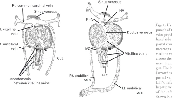
Fig. 1. Usual diagrams showing develo
pment of the portal vein. The vitelline veins provide sinusoids in the liver (left
hand side panel) and the extrahepatic portal vein course uses some of commu
nications between the left and right vitelline veins: the final portal vein first crosses the ventral aspect of the gut and, next, it crosses the dorsal aspect of the gut. The left vitelline vein near the liver (arrowhead) is not used for the final portal vein. IVC, inferior vena cava;
LHV, left hepatic vein; RHV, right hepatic vein. This figure omits details of the inferior vena cava development shown in our previous study [4].
Anat Cell Biol 2015;48:218-221 Hee Chul Yu, et al
220
www.acbjournal.org http://dx.doi.org/10.5115/acb.2015.48.3.218
A A A
A BBBB CCCC
D D D
D EEEE FFFF
G G G G
H
H II
J J
LHV LHV
ES ES
CL CL
ST ST
P P CACA
TC TC
DJ DJ
IVC IVC
DI DI
CL CL P2
P2 P3 P3
CL CL
STST AOAO P
P CACA SMV SMV
DI DI
CL Ductus CL
venosus Ductus
venosus
P2 P2
PV PV
ST ST QL
QL PP SMVSMV
SMA TC SMA
TC
Heart Heart
IVC IVC
DI DI
LPV
LPV CLCL
ST ST
P P QL
QL TC TC
SMV SMV DJ DJ
LPV
LPV CLCL
DU DU
P P
TC
TC DJDJ
P4 P4
PV PV
QL
QL DUDU P P
DJ DJ
UV UV
P4 P4
QL
QL DUDU PP
IVC IVC
CL CL RHV RHV RPV RPV
UV UV
CL
CL CLCL
P P
DJ DJ TC TC
CBD CBD
DU DU
RHV RHV
PST PST
AD AD
AST AST
GB GB
RK RK IVC
IVC DUDU
UP UP
UV UV QLQL
RPV RPV SMV SMV IVC IVC Ductus
venosus Ductus
venosus
Fig. 2. Anomalous portal vein seen in a 36mm fetus. Sagittal sections. Hematoxylin and eosin staining. Panel (A) (or panel I) displays the most left (or right) side of the figure. The lesser sac contains liver caudate lobe (CL; panels A-E). An anomalous portal vein (arrows) provides a peritoneal fold crossing the lesser sac. The vein is originated from the superior mesenteric vein (SMV) (D) and ends at the ductus venosus (B). A branch of the anomalous portal vein joins the usual right portal vein (RPV) in panel (F). In panel (F), PV indicates the usual site of the hilar bifurcation of the portal vein. The right portal vein issues branches to the pancreas (P) and segment 6 of the liver (arrowheads in panel G) and it divides into the right anterior and posterior sectorial trunks (AST, PST) of the liver (I). Panel (H) includes the usual longitudinal course of the inferior vena cava (IVC).
Panel (J) exhibits a schematic representation of the portal vein configuration: dotted line with star indicates the usual portal vein course. Intervals between panels are 0.2 mm (A-B), 0.3 mm (B-C), 0.2 mm (C-D), 0.3 mm (D-E), 0.2 mm (E-F, F-G), 0.4 mm (G-H), and 0.6 mm (H-I), respectively. Panels (A), (B), (C), (E), (F), and (G) or panels (D) and (H) are prepared at the same magnification. Scale bars=1 mm. AD, right adrenal; AO, aorta; CA, celiac artery; CBD, common bile duct; DI, diaphragm; DJ, duodenojejunal junction; DU, duodenum; ES, esophagus;
GB, gall bladder; LHV, left hepatic vein; LPV, left branch of the portal vein; P2, P3, and P4, portal vein branches to segment 2-4, respectively; QL, quadrate lobe of the liver; RHV, right hepatic vein; RK, right kidney; SMA, superior mesenteric artery; ST, stomach; TC, transverse colon; UP, umbilical portion of the portal vein; UV, umbilical vein.
An anomalous portal vein
http://dx.doi.org/10.5115/acb.2015.48.3.218
Anat Cell Biol 2015;48:218-221
221
www.acbjournal.org
In contrast to the adult morphology, the liver caudate lobe (Spiegel’s lobe) was located closely to the abdominal esophagus. This was consistent with our previous description about the fetal liver caudate lobe extending into the lesser sac [7]. A deep notch (the so-called caudate notch) is often seen on the inferior surface of the adult caudate lobe [8, 9]. The notch seems to be sculptured by the common hepatic artery or a hepatic artery variant coming from the left gastric artery [7]. However, the present anomaly suggested a rare possibility that an anomalous portal vein is likely to make a deep notch of the caudate lobe. The artery-derived notch is unlikely to correspond to a border between the left and right portal vein territories in the caudate lobe. Although the pathogenesis involves the portal vein development, the branching pattern seemed to be normal in the present anomaly (Fig.
3). Therefore, at any situation for providing the caudate notch, the left/right border in the caudate lobe is unlikely to correspond to the notch.
Consequently, in contrast to the usual diagram (Fig. 1), the present anomaly exhibited a possibility that the left vitelline vein does not accompany the hepatic artery and bile duct in the hepatoduodenal ligament but it is likely to run
outside of the ligament. A re-examination of the portal vein development seemed to be needed especially for describing the topographical relation to mesenteries.
Acknowledgements
This study was supported by a grant (0620220-1) from the National R & D Program for Cancer Control, Ministry of Health & Welfare, Republic of Korea.
References
1. Collardeau-Frachon S, Scoazec JY. Vascular development and differentiation during human liver organogenesis. Anat Rec (Hoboken) 2008;291:614-27.
2. Inoue M, Taenaka N, Nishimura S, Kawamura T, Aki T, Yamaki K, Enomoto H, Kosaka K, Yoshikawa K. Prepancreatic postduodenal portal vein: report of a case. Surg Today 2003;
33:956-9.
3. Tomizawa N, Akai H, Akahane M, Ino K, Kiryu S, Ohtomo K. Prepancreatic postduodenal portal vein: a new hypothesis for the development of the portal venous system. Jpn J Radiol 2010;28:157-61.
4. Jin ZW, Cho BH, Murakami G, Fujimiya M, Kimura W, Yu HC.
Fetal development of the retrohepatic inferior vena cava and accessory hepatic veins: Re-evaluation of the Alexander Barry's hypothesis. Clin Anat 2010;23:297-303.
5. Yi SQ, Tanaka S, Tanaka A, Shimokawa T, Ru F, Nakatani T. An extremely rare inversion of the preduodenal portal vein and common bile duct associated with multiple malformations.
Report of an adult cadaver case with a brief review of the literature. Anat Embryol (Berl) 2004;208:87-96.
6. Dighe M, Vaidya S. Case report. Duplication of the portal vein: a rare congenital anomaly. Br J Radiol 2009;82:e32-4.
7. Hwang SE, Cho BH, Hirai I, Kim HT, Kim JH, Fujimiya M, Murakami G, Kimura W. Topographical anatomy of Spiegel's lobe and its adjacent organs in mid-term fetuses: its implication on the development of the lesser sac and adult morphology of the upper abdomen. Clin Anat 2010;23:712-9.
8. Murakami G, Hata F. Human liver caudate lobe and liver segment. Anat Sci Int 2002;77:211-24.
9. Kanamura T, Murakami G, Ko S, Hirai I, Hata F, Nakajima Y.
Evaluating the hilar bifurcation territory in the human liver caudate lobe to obtain critical information for delimiting reliable margins during caudate lobe surgery: anatomic study of livers with and without the external caudate notch. World J Surg 2003;27:284-8.
IVC
IVC
Lt. umbilical vein Ductus venosus
Coronary sinus
Closed
SPV Closed IMV
Duodenum SMV
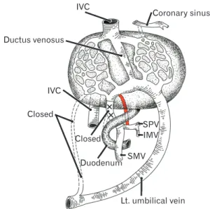
Fig. 3. Understanding of the present anomaly as a left vitelline vein.
The usual portal vein course behind the duodenum is closed and, in
stead, the portal vein used a part of the left vitelline vein course (see also Fig. 1). IMV, inferior mesenteric vein; IVC, inferior vena cava;
SMV, superior mesenteric vein; SPV, splenic vein. The closure of the right umbilical vein is normal.