Notes Bull. Korean Chem. Soc. 2013, Vol. 34, No. 6 1867 http://dx.doi.org/10.5012/bkcs.2013.34.6.1867
Study on Long-Term Stability of Non-Biofouling
Poly[(3-(methacryloylamino)propyl)-dimethyl(3-sulfopropyl)ammonium hydroxide] Films Under Biologically Relevant
Conditions
Baekhap Choi, Daewha Hong, Juno Lee, Sung Min Kang,† Young Hwan Jung,‡ Eun Hyea Ko, Yongseong Kim,§ Insung S. Choi,* and Jungkyu K. Lee#,* Department of Chemistry, KAIST, Daejeon 305-701, Korea. *E-mail: [email protected]
†Department of Marine Bio-Materials & Aquaculture, Pukyong National University, Busan 608-737, Korea ‡Department of BioNanomaterials, Bio-Campus of Korea Polytechnic, Chung-nam 320-905, Korea
§Department of Science Education, Kyungnam University, Masan 631-701, Korea
#Department of Chemistry, Kyungpook National University, Daegu 701-702, Korea. *E-mail: [email protected] Received January 28, 2013, Accepted February 24, 2013
Key Words : Cell chips, Non-biofouling, Polymer grafting, Poly(MPDSAH), Stability
In the fabrication of biomedical micro- and nano-systems, including cell chips and prosthetic devices, is used inten-sively polymer grafting, such as surface-initiated polymeri-zation, as an interface controller to preferentially inhibit or promote cell adherence and growth with the molecular-level control.1-7 For example, we reported that the film of a zwitterionic polymer, poly[(3-(methacryloylamino)propyl)-dimethyl(3-sulfopropyl)ammonium hydroxide] [poly-(MPDSAH)], possessing both anionic sulfonate and cationic quaternary amine, effectively prevented the non-specific ad-sorption of proteins and adherence of cells.8-10 In compari-son, a slight structural-variation to poly[(2-(methacryloyl-oxy)ethyl)-dimethyl(3-sulfopropyl)ammonium hydroxide] [poly(MEDSAH)] was found to sustain long-term growth of human embryonic stem cells.11 It is, however, the long-term film stability that should be considered more seriously for practical use of polymer coating in biomedical devices, besides the intrinsic cell-repelling or cell-attracting property of polymer films; the prolonged desired-functioning of a polymer film is determined absolutely by the long-term stability of the film.
The poly(MPDSAH) film was reported to possess the biopassive efficacy longer than the commonly used ethylene glycol-based polymer, poly(EG), against NIH 3T3 fibroblast cells: the cell micropatterns on the poly(MPDSAH)
sub-strate were maintained up to 20 days, while the integrity of the cell micropatterns on the poly(EG) substrate began to be deteriorated after 4 days of culture.9 In this previous work of ours, it was qualitatively observed that the loss of cell-repelling ability was connected with the film degradation. In other words, the previous work suggested that we should have both intrinsic non-biofouling property and chemical/ biological stability of films for long-term use of polymer coating. In this respect, studies on film (in)stability under biological conditions would contribute to molecular design for polymer grafting applied to biomedical devices, especial-ly to cell-based systems. In this work, we investigated the stability of poly(MPDSAH) films under various biologically relevant conditions for one month. The thickness decreases of the films under cell culture media and in protein solutions were obtained by ellipsometric measurements to get a mechanistic insight into film degradation.9 Because most of polymer films were known to be degraded to a certain extent even in pure water,12 herein we focused on the relative and comparative effects of biological entities, such as proteins, on the film degradation.
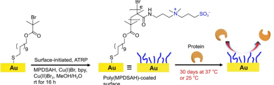
The poly(MPDSAH) films were grown from a gold-coat-ed substrate after formation of self-assemblgold-coat-ed monolayers of a polymerization initiator, 11,11'-dithiobis[1-(2-bromo-2-methylpropionyloxy)undecane] ([BrC(CH3)2C-OO(CH2)11
1868 Bull. Korean Chem. Soc. 2013, Vol. 34, No. 6 Notes
S]2)13 (Figure 1). We formed thick (> 20 nm) films for this
work, because the poly(MPDSAH) films less than about 3 nm in thickness lost their non-biofouling property,9 and the surface adsorption of biomolecules would interfere with the thickness measurement. Briefly, the surface-initiated, atom transfer radical polymerization (SI-ATRP)10,14-20 was perform-ed in the water-methanol solution of MPDSAH, bipyridine, Cu(I)Br, and Cu(II)Br2 at room temperature for 16 h under
an argon atmosphere. The resulting gold substrate was immersed in water at 40 oC for 4 days to remove the copper complexes and any remaining monomers.8 This removal step allowed us to obtain uniform films with 24.3 ± 1.5 nm in thickness. For the degradation studies, the widely-used cell culture medium− Gibco® Dulbecco’s modified eagle medium (DMEM) supplemented with 10% fetal bovine serum (FBS) and 1% antibiotics solution− and four repre-sentative proteins (bovine serum albumin, lysozyme, fibrino-gen, and RNase A) were employed. Regarding the proteins, their isoelectric points (pI), sizes, adsorption behaviors, layer formations, and degrees of denaturation on various surfaces have been well studied, and they are widely used for bio-medical devices.21 Bovine serum albumin (BSA, MW: 69 kDa, pI: ~4.8; molecular biology tested, Sigma) as a soft protein is relatively subjected to be easily denatured on adsorption,22,23 but lysozyme (MW: 14.6 kDa, pI: ~11.0; grade III from chicken egg white, Sigma) is a class of hard proteins, because it resists denaturation and exhibits concen-tration-dependent layer formation.24 Fibrinogen (MW: 340
kDa, pI: ~5.5; fraction I from human plasma, plasminogen-free, Sigma) is a large plasma glycoprotein with high surface affinity, which usually displaces pre-adsorbed proteins,25,26 and RNase A (MW: 13.7 kDa, pI: ~9.63; type III-A from bovine pancreas, Sigma) has widely been used as a classic model system to understand catalytic cleavage of single-stranded RNA.27 The poly(MPDSAH)-grafted substrate was immersed for 30 days in pure water, phosphate buffered saline (PBS) (10 mM, pH 7.4), the PBS solution of a protein (1 mg/mL), or cell culture medium, and the film thickness was measured every day after washing with pure water and drying under a stream of argon.
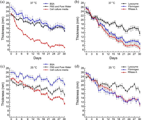
Figure 2 shows the graphs of film thickness versus immer-sion time under the different conditions. In the cell culture medium the film was degraded about 2 times faster than in the PBS solution or pure water at 37 °C, implying the participation of biological roles in film degradation: after 30 days, the thickness difference (initial thickness minus final thickness) was 15.2 nm for the cell culture medium and 7.2 nm for the PBS solution and pure water (Figure 2(a)). We observed no noticeable difference between PBS and pure water, excluding the active involvement of salts in film degradation. In the cell culture medium, DMEM contains glucose, amino acids, vitamins, and salts; FBS contains BSA, growth factors, antibodies, and others. The plasma protein, fibrinogen, is generally removed as fibrin from the serum. We, therefore, tested the effects of BSA and fibrino-gen on the film degradation along with two other
Notes Bull. Korean Chem. Soc. 2013, Vol. 34, No. 6 1869
sentative proteins, lysozyme and RNase A (Figure 2(a) and (b)). Because BSA in PBS in itself did not show additional and discernible degrading-effect (Figure 2(a)), we thought that the faster thickness decrease observed for the cell cul-ture medium was caused not by BSA, but by other biological components in the culture medium. Of interest, fibrinogen and RNase A, not present in cell culture medium, had a similar degradation-capability to the cell culture medium. Based on this result, it could be deduced that each protein has the ability of degrading polymer films with its own level; in addition, decreasing the immersion temperature from 37 °C to 25 °C led to the decrease in film degradation for protein solutions and cell culture medium, supporting for this hypothesis (Figure 2(c) and (d)). After 30 days, the difference of thickness-decrease between the two temper-atures is 3.5 nm for the cell culture media, 3.5 nm for BSA, 2.6 nm for fibrinogen, 4.1 nm for RNase A, or 3.2 nm for lysozyme. In contrast, the temperature effect was negligible for PBS and pure water (only 0.3-nm difference).
The degradation processes seemed to involve changes in the chemical compositions of polymer films, e.g., cleavage of side chains by proteins, inferred from data of water con-tact angles (Table 1). After 30 days at 37 °C, we observed a slight increase in the contact angles (from 11.6 ± 0.9° at Day 2 to about 20°). The contact angle changes could not be explained with simple oxidative desorption of thiolates from gold; the different contact angles also might indicate that different proteins and biomolecules had different cleavage characteristics in the degradation processes.
Experimental
Formation of Poly(MPDSAH) Films on Gold Substrates. The polymerization initiator, 11,11'- dithiolbis[1-(2-bromo-2-methylpropionyloxy)undecane] ([BrC(CH3)2COO(CH2)11
-S]2) was synthesized by following the reported procedure.13
A fresh bare gold substrate was immersed in a 1-mM ethanolic solution of the polymerization initiator for 18 h to form the self-assembled monolayers (SAMs) of the poly-merization initiator, washed with ethanol several times, and dried under a stream of argon. The surface-initiated, atom transfer radical polymerization from the initiator anchored onto the gold substrate was carried out for 18 h at room temperature under argon atmosphere. Briefly, MPDSAH (2.5 g, 8.55 mmol, 96%, Aldrich), CuBr2 (3.8 mg, 0.017 mmol,
99.999%, Aldrich), and 2,2'-bipyridyl (0.0667 g, 0.427 mmol, > 99%, Aldrich) were added to a mixture of pure water (4 mL) and methanol (1 mL) in a Schlenk flask. CuBr (0.0245 g, 0.171 mmol, 99.999%, Aldrich) was slowly added to the
reaction mixture, after the mixture was degassed for 4 h by passing a continuous stream of dry argon through the vigor-ously stirred solution. The formed poly(MPDSAH) film was washed with pure water and methanol several times and dried under a stream of argon. To remove the copper com-plexes and remaining monomers, the resulting substrate was immersed in water at 40 oC for 4 days, because the swollen polymer film could promote the diffusion of the impurities into the water.
Characterizations. The thickness of poly(MPDSAH) films was measured with Gaertner L116s ellipsometer (Gaertner Scientific Corporation, IL) equipped with a He-Ne laser (632.8 nm) at a 70o angle of incidence. A refractive index of 1.46 was used for all the samples. More than five different points on each sample were measured, and the average values were recorded. Contact angle measurements were performed using Phoenix 300 apparatus (Surface Electro Optics Co. Ltd, Korea) equipped with a video camera. The static contact angles of water drops of ~5 µL were measured at five different locations on each sample, and average values were recorded.
Acknowledgments. This work was supported by the Kyungpook National University Research Fund, 2012.
References
1. Noimark, S.; Dunnill, C. W.; Wilson, M.; Parkin, I. P. Chem. Soc. Rev. 2009, 38, 3435.
2. Chen, C. S.; Mrksich, M.; Huang, S.; Whitesides, G. M.; Ingber, D. E. Science 1997, 276, 1425.
3. Ma, H. W.; Hyun, J.; Zhang, Z. P.; Beebe, T. P.; Chilkoti, A. Adv. Funct. Mater. 2005, 15, 529.
4. Callow, J. A.; Callow, M. E. Nat. Commun. 2011, 2, 244. 5. Huang, N. P.; De Paul, S. M.; Textor, M. Biomacromolecules
2011, 12, 4213.
6. Gong, P.; Grainger, D. W. Method. Mol. Biol. 2007, 381, 59. 7. Ratner, B. D.; Bryant, S. J. Annu. Rev. Biomed. Eng. 2004, 6, 41. 8. Cho, W. K.; Kong, B.; Choi, I. S. Langmuir 2007, 23, 5678. 9. Cho, W. K.; Kong, B.; Park, H. J.; Kim, J.; Chegal, W.; Choi, J. S.;
Choi, I. S. Biomaterials 2010, 31, 9565.
10. Kim, J.; Hong, D.; Jeong, S.; Kong, B.; Kang, S. M.; Kim, Y. G.; Choi, I. S. Chem. Asian J. 2011, 6, 363.
11. Villa-Diaz, L. G.; Nandivada, H.; Ding, J.; Nogueira-De-Souza, N. C.; Krebsbach, P. H.; O'Shea, K. S.; Lahann, J.; Smith, G. D. Nat. Biotechnol. 2010, 28, 581.
12. Ostuni, E.; Chapman, R. G.; Liang, M. N.; Meluleni, G.; Pier, G.; Ingber, D. E.; Whitesides, G. M. Langmuir 2001, 17, 6336. 13. Shah, R. R.; Merreceyes, D.; Husemann, M.; Rees, I.; Abbott, N.
L.; Hawker, C. J.; Hedrick, J. L. Macromolecules 2000, 33, 597. 14. Ma, H. W.; Wells, M.; Beebe, T. P.; Chilkoti, A. Adv. Funct. Mater.
2006, 16, 640.
15. Kim, D. J.; Lee, K.-B.; Chi, Y. S.; Kim, W.-J.; Paik, H.-j.; Choi, I. Table 1. Static water contact angles of the poly(MPDSAH) films after 2 and 30 days at 37 °C
Contact
Angle (o) PBS
Cell Culture
Medium BSA
a Lysozymea Fibrinogena RNase Aa
After 2 days 13.1 11.0 11.2 10.6 11.5 12.0
After 30 days 19.7 19.0 18.0 11.0 18.2 34.8
1870 Bull. Korean Chem. Soc. 2013, Vol. 34, No. 6 Notes
S. Langmuir 2004, 20, 7904.
16. Lee, S. B.; Koepsel, R. R.; Morley, S. W.; Matyjaszewski, K.; Sun, Y. J.; Russell, A. J. Biomacromolecules 2004, 5, 877. 17. Lee, B. S.; Lee, J. K.; Kim, W.-J.; Jung, Y. H.; Sim, S. J.; Lee, J.;
Choi, I. S. Biomacromolecules 2007, 8, 744.
18. Lee, B. S.; Chi, Y. S.; Lee, K.-B.; Kim, Y.-G.; Choi, I. S. Bio-macromolecules 2007, 8, 3922.
19. Yoon, K. R.; Ramaraj, B.; Lee, S.; Yu, J. S.; Choi, I. S. J. Biomed. Mater. Res. A 2009, 88A, 735.
20. Azzaroni, O.; Brown, A. A.; Huck, W. T. S. Angew. Chem. Int. Ed. 2006, 45, 1770.
21. Kim, J.; Somorjai, G. A. J. Am. Chem. Soc. 2003, 125, 3150. 22. Norde, W.; Flavier, J. P. J. Colloid Interf. Sci. 1992, 64, 87. 23. Su, T. J.; Lu, J. R.; Thomas, R. K.; Cui, Z. F.; Penfold, J. J. Colloid
Interf. Sci. 1998, 203, 419.
24. Wertz, C. F.; Santore, M. M. Langmuir 2002, 18, 1190. 25. Schaaf, P.; Dejardin, P.; Schmit, A. Langmuir 1987, 23, 1131. 26. Wertz, C. F.; Santore, M. M. Langmuir 2002, 18, 706. 27. Raines, R. T. Chem. Rev. 1998, 98, 1045.
28. Castner, D. G.; Hinds, K.; Grainger, D. W. Langmuir 1996, 12, 5083.