Korean J Gastroenterol Vol. 68 No. 6, 331-333 https://doi.org/10.4166/kjg.2016.68.6.331 pISSN 1598-9992 eISSN 2233-6869
IMAGE OF THE MONTH
Korean J Gastroenterol, Vol. 68 No. 6, December 2016 www.kjg.or.kr
상피하 종양 형태로 나타난 심재성 낭성 위염
정진태
대구가톨릭대학교 의과대학 내과학교실
Gastritis Cystica Profunda Presented as a Subepithelial Tumor
Jin Tae Jung
Department of Internal Medicine, Catholic University of Daegu School of Medicine, Daegu, Korea
CC This is an open access article distributed under the terms of the Creative Commons Attribution Non-Commercial License (http://creativecommons.org/licenses/
by-nc/4.0) which permits unrestricted non-commercial use, distribution, and reproduction in any medium, provided the original work is properly cited.
Copyright © 2016. Korean Society of Gastroenterology.
교신저자: 정진태, 42472, 대구시 남구 두류공원로 17길 33, 대구가톨릭대학교 의과대학 내과학교실
Correspondence to: Jin Tae Jung, Department of Internal Medicine, Catholic University of Daegu School of Medicine, 33 Duryugongwon-ro 17-gil, Nam-gu, Daegu 42472, Korea. Tel: +82-53-650-4217, Fax: +82-53-624-3281, E-mail: [email protected]
Financial support: None. Conflict of interest: None.
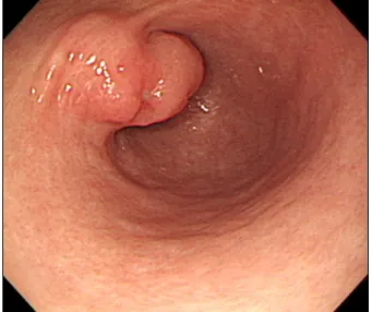
Fig. 1. Endoscopic finding. A 2×3 cm sized subepithelial lesion at the anterior wall of the distal antrum.
증례 1: 43세 여자가 건강검진 목적으로 개인병원에서 시 행한 상부위장관 내시경검사에서 위의 상피하 종양이 의심되 어 초음파 내시경을 위해서 전원되었다. 과거력 및 가족력에 서 특이사항은 없었다. 신체검진에서 혈압 120/80 mmHg, 맥 박수 분당 66회, 호흡수 분당 14회, 체온 36.5oC였으며 의식 은 명료하였고 전신상태는 양호하였다. 말초혈액검사에서 백 혈구 4,900/mm3, 혈색소 11.0 g/dL, 혈소판 199,000/mm3였 다. 간기능, 신장기능 및 혈청 전해질은 정상 범위였다. 본원 에서 시행한 상부위장관 내시경에서 전정부 원위부의 전벽에 발적 및 얕은 궤양을 가지고 있으며 정상 점막으로 덮여 있는 2×3 cm 크기의 상피하 종양이 관찰되었다(Fig. 1). 초음파 내시경에서 위의 2층과 3층에 위치하며 무음영의 낭성에코와 격막을 가지고 있는 것으로 관찰되었는데, 크기가 1.8×1.4 cm로 심재성 낭성 위염(gastritis cystica profunda, GCP)이 의심되어 내시경 점막하 박리술을 시행하였다(Fig. 2). 조직병 리 소견에서 다수의 낭종이 점막 고유근층 및 점막하층에서 관찰되었으며 저도 이형성증을 동반하는 GCP로 진단되었다 (Fig. 3). 2개월 후 추적내시경 검사에서 반흔 외에 다른 소견 은 없어 외래에서 추적관찰 중이다.
증례 2: 52세 남자가 건강검진 목적으로 개인병원에서 시 행한 상부위장관 내시경검사에서 위의 상피하 종양이 의심되 어 초음파 내시경을 위해서 전원되었다. 과거력 및 가족력에 서 특이사항은 없었다. 신체검진에서 혈압 110/70 mmHg, 맥
박수 분당 72회, 호흡수 분당 16회, 체온 36.5oC였으며 의식 은 명료하였고 전신상태는 양호하였다. 말초혈액검사에서 백 혈구 8,800/mm3, 혈색소 14.2 g/dL, 혈소판 245,000/mm3였 다. 간기능, 신장기능 및 혈청 전해질은 정상 범위였다. 본원 에서 시행한 상부위장관 내시경에서 위저부의 대만부에 정상 점막으로 덮여 있는 0.8 cm의 상피하 종양이 관찰되었으며
332
정진태. 상피하 종양 형태로 나타난 심재성 낭성 위염The Korean Journal of Gastroenterology
Fig. 2. Endoscopic ultrasound and endoscopic finding. (A) A 17.5×13.5 mm sized anechoic cystic region with septation at the 2nd and 3rd layers of the stomach. (B) A 3.6×2.4 cm sized endoscopic submucosal dissection specimen.
Fig. 3. Microscopic findings (H&E stain). (A) Normal mucosal gland and dilated glands in the submucosa (×100). (B) Cystically dilated glands with surround fibrous stroma in the submucosa (×400).
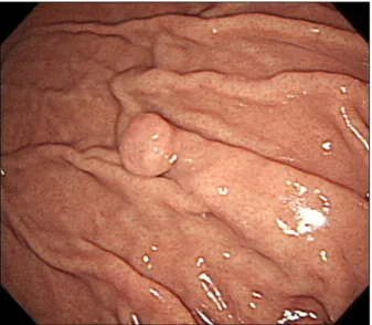
Fig. 4. Endoscopic finding. A 0.8 cm sized subepithelial lesion at the greater curvature of the fundus.
Fig. 5. Endoscopic ultrasound finding. A 7×5 mm sized hypoechoic homogenous region at the 2nd and 3rd layers of the stomach.
Jung JT. Gastritis Cystica Profunda Presenting with a Subepithelial Tumor
333
Vol. 68 No. 6, December 2016
생검 겸자로 눌렀을 때 비교적 단단하게 촉지되었다(Fig. 4).
초음파 내시경으로 관찰했을 때 위의 2층과 3층에 위치하고 저음영의 에코를 가지고 있었으며, 크기가 0.7×0.5 cm로 유 암종일 가능성을 완전히 배제할 수 없어 내시경 점막절제술을 시행하였다(Fig. 5). 조직병리검사 결과 GCP로 진단되었다.
진단: 심재성 낭성 위염
GCP는 위점막 심부 및 점막하층에 다수의 낭종을 형성하 는 비교적 드문 질환으로 조직학적으로 위선상피세포의 과형 성 및 낭성 확장을 특징으로 하고 있다.1 소화성 궤양으로 수 술을 받은 환자에서 위 공장 문합부에 발생한 용종성 병변을 발견하여 이를 GCP로 처음 보고하였으며 이후 위 수술병력이 있는 환자에서 GCP와 위암이 동반된 예가 보고되었다.2 그러 나 2001년 이후로 위 수술 병력이 없는 환자에서도 GCP가 발생한 보고가 있으며 대부분 양성 질환으로 간주하고 있다.3
GCP의 발생기전은 명확하지 않지만 수술 병력이 있는 경 우는 수술 자체에 의해 발생하거나 만성 허혈에 따른 위점막 의 미란, 봉합사 등의 이물질에 의해 발생할 수 있으며, 수술 병력이 없는 경우는 이전의 위궤양이나 선천적인 요인 등이 관여할 것으로 추정하고 있다.4,5 GCP의 임상 증상은 비특이 적으로 무증상부터 상복부 불쾌감, 복통, 출혈 등의 다양한 증상이 있을 수 있는데, 내시경 소견은 상피하 종양, 유경성 또는 무경성의 용종 형태로 주로 관찰되며 점막에 미란이나 궤양을 보이는 경우에는 위암을 동반할 가능성이 높은 것으로 보고하고 있다.6
GCP의 임상적 의의는 전암성 병변 혹은 암 주위 병변일 가능성이 많다는 것이다. 수술 병력이 있는 경우 위 소장 문합 부위에서 GCP가 위암과 함께 발생할 수 있는 합병증으로 보 는 의견이 있다.7 수술 병력이 없는 경우 선종 및 선암을 동반 한 GCP가 많이 보고되고 있는데, Lee 등8의 연구에 의하면 GCP의 약 반수에서 위암을 동반하였고 대부분 조기 위암이 었으며 위암이 동반되지 않은 환자군에서도 저도 이형성 및 고도 이형성이 다수 발견되어 GCP 환자들을 검사하거나 추 적할 때 이를 염두에 둘 것을 권유하였다.
GCP의 치료는 현재까지 명확하게 정립되어 있지 않지만 대부분 양성질환으로 간주하며 출혈이나 위장관 폐쇄 등의 합 병증이 있거나 궤양 및 선종이 있어 위암의 동반 가능성이 완전히 배제되지 않을 때 진단 및 치료 목적으로 내시경 절제 술이나 수술 등을 고려해 볼 수 있다.9 최근에는 내시경 술기 의 발전으로, 조기위암에 대해 내시경 점막하 박리술 후 시행 한 조직검사에서 점막하 조직에 위암과 동반된 GCP의 예를 드물지 않게 보고하고 있다. 이번 증례도 첫 번째 예는 점막 표면에 궤양을 동반하고 있었으며 두 번째 예는 유암종 등의 다른 종양과 감별이 되지 않아 진단 및 치료목적으로 내시경 절제술을 시행하였다.
REFERENCES
1. Koga S, Watanabe H, Enjoji M. Stomal polypoid hypertrophic gas- tritis: a polypoid gastric lesion at gastroenterostomy site. Cancer 1979;43:647-657.
2. Littler ER, Gleibermann E. Gastritis cystica polyposa. (Gastric mucosal prolapse at gastroenterostomy site, with cystic and in- filtrative epithelial hyperplasia). Cancer 1972;29:205-209.
3. Kim W, Park SC, Lee JY, et al. A case of gastric adenocarcinoma diagnosed after being followed up as submucosal tumor for 10 years. Korean J Gastroenterol 2001;37:291-295.
4. Fonde EC, Rodning CB. Gastritis cystica profunda. Am J Gastroenterol 1986;81:459-464.
5. Chakrovorty RC, Schatzki PF. Gastric cystic polyposis. Am J Dig Dis 1975;20:981-989.
6. Ozenç AM, Ruacan S, Aran O. Gastritis cystica polyposa. Arch Surg 1988;123:372-373.
7. Qizilbash AH. Gastritis cystica and carcinoma arising in old gas- trojejunostomy stoma. Can Med Assoc J 1975;112:1432-1433.
8. Lee HJ, Lee TH, Lee JU, et al. Clinical features of gastritis cystica profunda in patients without history of gastric surgery (Gastric Cancer Patients vs. Non-cancerous Patients). Korean J Med 2006;71:511-517.
9. Cho HJ, Kim JE, Jeong BJ, et al. A case of gastric adenocarcinoma arising from gastritis cystica profunda. Korean J Gastrointest Endosc 2004;28:237-241.