Korean J Gastroenterol Vol. 64 No. 6, 387-389 http://dx.doi.org/10.4166/kjg.2014.64.6.387 pISSN 1598-9992 eISSN 2233-6869
IMAGE OF THE MONTH
Korean J Gastroenterol, Vol. 64 No. 6, December 2014 www.kjg.or.kr
하부위장관 출혈을 유발한 속발성 유전분증
김승범, 김경옥
영남대학교 의과대학 내과학교실
Lower Gastrointestinal Bleeding Induced by Secondary Amyloidosis
Sung Bum Kim and Kyeong Ok Kim
Department of Internal Medicine, Yeungnam University College of Medicine, Daegu, Korea
CC This is an open access article distributed under the terms of the Creative Commons Attribution Non-Commercial License (http://creativecommons.org/licenses/
by-nc/3.0) which permits unrestricted non-commercial use, distribution, and reproduction in any medium, provided the original work is properly cited.
교신저자: 김경옥, 705-703, 대구시 남구 현충로 170, 영남대학교 의과대학 내과학교실 소화기분과
Correspondence to: Kyeong Ok Kim, Division of Gastroenterology and Hepatology, Department of Internal Medicine, Yeungnam University College of Medicine, 170 Hyeonchung-ro, Nam-gu, Daegu 705-703, Korea. Tel: +82-53-620-3835, Fax: +82-53-623-8038, E-mail: [email protected]
Financial support: None. Conflict of interest: None.
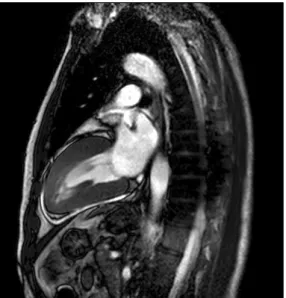
증례: 54세 남자 환자가 3일 전부터 발생한 혈변과 복통을 주소로 응급실에 내원하였다. 혈변은 매회 소량으로 하루 3-4 회 발생하였고 복통은 하복부에 우리한 양상을 보였으며 배변 후 호전되는 양상을 보였다. 환자는 내원 2개월 및 1개월 전 각각 혈변으로 2차 진료기관에 입원하여 대장염 진단하에 치 료를 받았으며, 보존적 치료로 증상이 호전되었다. 만성 B형 간염 보유자로 30년 동안 하루 반 갑의 흡연을 하였으며 음주 는 하지 않았다. 활력징후는 혈압 100/60 mmHg, 맥박수 75 회/분, 호흡수 20회/분, 체온 36.8oC로 측정되었고, 신체검사 에서 결막은 창백하지 않았고 흉부 청진에서 심, 폐 잡음은 들리지 않았으며 복부 촉진에서 경미한 하복부 압통이 관찰되 었다. 혈액검사에서 백혈구 10,040/mm3, 혈색소 11.9 g/dL, 혈소판 223,000/mm3였고, C 반응성 단백 2.467 mg/dL로 상 승되어 있었다. 총 단백질은 5.68 g/dL, 알부민은 3.92 g/dL 였으며 간 및 신장기능에 이상 소견은 보이지 않았다. 대장 내시경검사에서 대장 전장에 걸쳐 거친 점막이 관찰되었으며, 하행 및 구불 결장에 미만성 궤양에 동반된 출혈성 수포 병변 이 관찰되었다(Fig. 1). 환자의 증상과 내시경 병변 부위 및 소견을 바탕으로 허혈성 장염을 의심하였으며, 이와 관련된 기저 질환 및 위험인자를 평가하기 위해 심초음파 검사를 시 행하였다. 검사 결과 비후성 심근병증 또는 침윤성 심근병증 을 시사하는 비대칭 심실중격비대 소견이 관찰되었으며, 심장 자기공명영상 검사에서 미만성 좌심실벽 비후가 관찰되었다
(Fig. 2). 심장 외 타장기 침윤성 병변 유무를 확인하기 위해 이전 내시경검사에서 얻은 조직에 대한 특수 염색을 의뢰하였 다.
진단: 허혈성 장염이 동반된 유전분증
하행 및 구불 결장의 조직검사에서 만성 염증 소견과 함께 점막하 혈관 주위로 비후 소견이 관찰되었으며, Congo red 특수염색에서 혈관벽 내로 염색되는 물질이 관찰되어 유전분 증으로 진단되었다(Fig. 3). 혈액과 소변 단백전기영동 검사에 서 M 단백질은 관찰되지 않았고 kappa and lambda chain 조직염색은 음성 소견을 보여 속발성 유전분증으로 진단되었 다. 보존적인 치료에도 소량씩 간헐적인 혈변이 관찰되었고 추적 결장경 검사에서도 병변은 이전에 비해 호전이 없었다.
이후로도 혈변이 지속되어 외과와 의논하여 수술을 계획하였 으며, 환자는 좌측 반결장절제술을 시행받았다. 절제된 좌측 대장은 육안적으로 전반에 걸쳐 궤양과 출혈이 관찰되었으며, 장막하 지방으로도 출혈이 관찰되었다(Fig. 4). 조직검사에서 유전분증과 동반된 허혈성 장염으로 인한 다발성 궤양이 관찰 되었고, 절제 부위의 양측 변연에서도 유전분증의 침범이 관 찰되었다.
유전분증은 비정상적으로 접지된 단백질이 생성되어 불 용성 섬유상으로 침착하는 질환이다.1 유전분증의 경우 원발 성 또는 속발성으로 나누며, 원발성 유전분증은 가장 흔한 아
388
김승범, 김경옥. 하부위장관 출혈을 유발한 속발성 유전분증The Korean Journal of Gastroenterology
Fig. 1. Endoscopic findings at initial visit. (A) Coarse mucosa with diminished colonic vasculature is noted throughout the colon. (B) Diffuse mucosal ulceration with necrotic tissue and hemorrhagic bullae are seen on descending and sigmoid colon.
Fig. 2. Cardiac MRI. Diffuse left ventricular wall thickening is noted on vertical long axis veiw.
형으로 면역글로불린 경쇄의 침착으로 발생하며 뇌 이외의 모 든 장기에 침착하여 증상을 유발할 수 있다. 속발성 유전분증 의 경우는 유전분 A 단백질이 침착하여 발생하며 염증성 관 절염, 만성 감염, 면역 결핍성 질환, 염증성 장질환, 종양 또는 전신성 혈관염이 원인이 되어 나타날 수 있다. 60%의 반응성 유전분증 환자에서 소화기계 침범이 보고되었으며,2 소화기계 침범 시 대장 및 직장의 침범은 90% 이상에서 관찰된다.3
대장 침범의 경우 유전분 섬유상의 장 신경 침범으로 운동 장애를 유발하여 변비 또는 설사로 나타날 수 있으며,4 점막 침범으로 인해 출혈, 협착, 장 폐쇄, 또는 천공이 합병될 수
있다. 유전분증의 침범으로 인한 장출혈의 알려진 기전으로는 장벽 전 층 또는 장에 혈액을 공급하는 작은 혈관벽에 유전분이 침착하여 이로 인해 궤양이 발생하거나, 유전분이 장막 간 또는 점막하 혈관벽에 침착하여 나타나는 혈관 변화 등이 있다.5-7 유 전분의 혈관 또는 장벽으로의 침범으로 허헐성 장염이 발생할 수 있으며, 이로 인해 미만성 점막 출혈이 발생할 수 있다.
유전분증의 경우 구강부터 항문까지 위장관계 전체를 침범 할 수 있으나 비특이적인 소견을 보여 다른 질환과의 감별이 어려우며 확진은 조직검사를 통해서만 할 수 있다. 위장관을 침범하는 경우 직장에 대한 무작위 조직검사가 가장 추천되는 데 이는 접근이 쉽고 75-94%의 비교적 높은 조직진단 민감도 를 보이기 때문이다.8 특징적으로 Congo red 염색에 반응하 며, 편광현미경으로 관찰 시 초록색의 이중 굴절상이 관찰된 다.9
유전분증으로 인한 위장관 증상에 대한 특별한 치료 방법 은 없는 실정이며, 대개 보존적인 치료를 시행한다. 설사에 대해서는 dimethyl sulfoxide 또는 somatostatin analogue 등을 사용해 볼 수 있다. 위장관 출혈의 지혈에 있어 내시경의 역할은 미미하며, 혈관 조영술을 이용하여 출혈 혈관이 관찰 되는 경우 색전술을 시행할 수 있다.10 증례의 환자도 혈관 조영술을 시행하였으나 출혈혈관은 관찰되지 않아 색전술을 시행할 수는 없었다. 수술의 경우 국소적인 유전분증으로 인 한 출혈인 경우 고려할 수 있으나, 유전분증의 심장 또는 신장 으로의 침범이 있는 경우 마취 및 술 후 합병증의 위험이 높아 수술 전 충분한 평가가 통해 수술에 따른 이득과 위험을 고려 해야 한다.11,12 이 증례의 경우도 심장 침범으로 인한 위험이 있었으나 보존적인 치료로 출혈이 조절되지 않았으며 병변이 전
Kim SB and Kim KO. Amyloidosis Induced Lower GI Bleeding
389
Vol. 64 No. 6, December 2014
Fig. 3. Microscopic findings of colon tissue. (A) Amorphous pinkish hyaline deposits are seen in vessel wall (H&E, ×200). (B) Congo red staining shows amorphous hyaline deposits in vessel walls (×200).
Fig. 4. Gross findings of surgically resected specimen. Mucosal ulcerations and hemorrhage at subserosal fat are noted without any mass-like lesions.
혀 호전을 보이지 않아 수술적 절제를 고려할 수 밖에 없었다.
하혈을 주소로 내원한 환자에서 심한 염증성 장질환 등이 배제되었으나 점막 병변이 2주 이상 지속되는 경우 유전분증 등의 다른 기저질환의 동반 유무에 대한 면밀한 검사가 고려 되어야 하겠다.
REFERENCES
1. Pepys MB. Amyloidosis. Annu Rev Med 2006;57:223-241.
2. Okuda Y, Takasugi K, Oyama T, Onuma M, Oyama H. Amyloidosis in rheumatoid arthritis--clinical study of 124 histologically pro- ven cases. Ryumachi 1994;34:939-946.
3. Tada S, Iida M, Iwashita A, et al. Endoscopic and biopsy findings of the upper digestive tract in patients with amyloidosis. Gastro- intest Endosc 1990;36:10-14.
4. Ito T, Sakakibara R, Ito S, et al. Mechanism of constipation in familial amyloid polyneuropathy: a case report. Intern Med 2006;45:1173-1175.
5. Mallory A, Struthers JE Jr, Kern F Jr. Persistent hypotension and intestinal infarction in a patient with primary amyloidosis.
Gastroenterology 1975;68:1587-1592.
6. Jarnum S. Gastrointestinal haemorrhage and protein loss in pri- mary amyloidosis. Gut 1965;6:14-18.
7. Chang SS, Lu CL, Tsay SH, Chang FY, Lee SD. Amyloidosis-in- duced gastrointestinal bleeding in a patient with multiple myeloma. J Clin Gastroenterol 2001;32:161-163.
8. Ebert EC, Nagar M. Gastrointestinal manifestations of amyloi- dosis. Am J Gastroenterol 2008;103:776-787.
9. Parsons J. Identification of crystalline material from lung biopsy by x-ray diffraction. Henry Ford Hosp Med Bull 1962;10:355-357.
10. Maeshima E, Yamada Y, Yukawa S. Massive gastrointestinal hemorrhage in a case of amyloidosis secondary to rheumatoid arthritis. Scand J Rheumatol 1999;28:262-264.
11. Mardinger O, Rotenberg L, Chaushu G, Taicher S. Surgical man- agement of macroglossia due to primary amyloidosis. Int J Oral Maxillofac Surg 1999;28:129-131.
12. Sattianayagam PT, Hawkins PN, Gillmore JD. Systemic amyloi- dosis and the gastrointestinal tract. Nat Rev Gastroenterol Hepatol 2009;6:608-617.